Thoracic Cancer
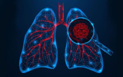
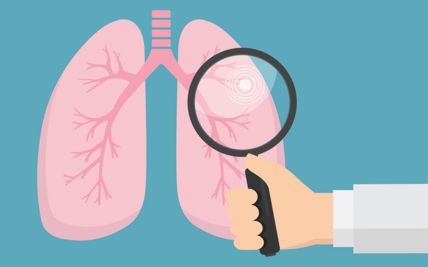
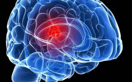
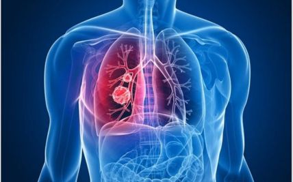
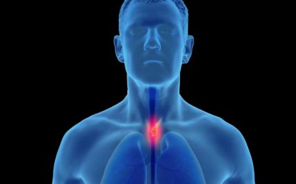
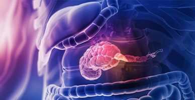
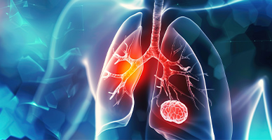
Thoracic cancer refers to cancers that develop within the chest. These may arise in the lungs, airways, pleura, thymus, mediastinum, chest wall, or esophagus. Lung cancer is the most common thoracic cancer, but each of these diseases has distinct biological features and treatment requirements.
Thoracic cancers can be difficult to detect early because their symptoms may resemble common respiratory or digestive conditions. Their location near the heart, major blood vessels, spinal cord, and healthy lung tissue can also make diagnosis and treatment complex.
Care may involve thoracic surgeons, pulmonologists, medical oncologists, radiation oncologists, gastroenterologists, pathologists, radiologists, nuclear medicine specialists, and reconstructive surgeons. The treatment plan is based on the cancer type and stage, molecular findings, lung and heart function, overall health, and individual needs.
Types of Thoracic Cancer
Thoracic cancer is not a single disease. Identifying where the cancer began and the type of cell involved is essential for selecting treatment.
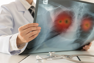
Non-small cell lung cancer, or NSCLC, accounts for most lung cancers. It generally grows and spreads more slowly than small cell lung cancer, although its behaviour varies considerably.
The main subtypes are:
Adenocarcinoma: Usually develops in the outer regions of the lungs. It is the most common lung cancer in people who have never smoked, although it also affects people with a smoking history.
Squamous cell carcinoma: Commonly begins in the larger airways and is strongly associated with tobacco exposure.
Large cell and other poorly differentiated carcinomas: Less common cancers that may develop in any part of the lung.
Molecular testing is important in many NSCLCs because specific genetic changes may make the tumour eligible for targeted treatment.
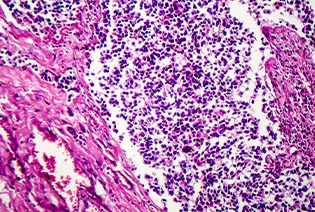
Small cell lung cancer, or SCLC, is a fast-growing cancer that is strongly associated with tobacco use. It tends to spread earlier than NSCLC and is often treated with chemotherapy and immunotherapy, with radiation therapy used in selected stages. Although SCLC may initially respond well to treatment, it has a relatively high risk of recurrence.
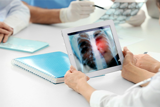
These tumours arise from neuroendocrine cells in the lungs and airways. They include typical carcinoid, atypical carcinoid, large-cell neuroendocrine carcinoma, and small cell lung cancer. Typical and atypical carcinoids often grow more slowly than high-grade neuroendocrine cancers. Surgery is commonly used for localised carcinoid tumours, while high-grade cancers usually require systemic treatment.
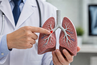
Pleural mesothelioma begins in the thin tissue lining the lungs and chest cavity. It is strongly associated with previous asbestos exposure, although symptoms may not develop until decades after exposure. The cancer can spread along the pleural surface and restrict lung expansion. Treatment may include surgery, medicines, radiation therapy, pleural procedures, or a combination of approaches.
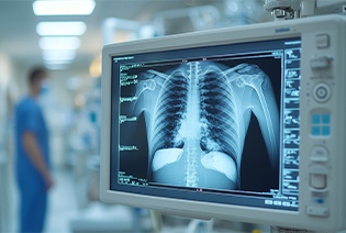
These cancers begin in the thymus, a small organ in the upper chest. Thymoma may be associated with autoimmune conditions, particularly myasthenia gravis. Thymic carcinoma is generally more aggressive and more likely to spread. Surgery is the principal treatment when the tumour can be removed completely. Locally advanced disease may require chemotherapy and radiation therapy in addition to surgery.

These rare cancers develop in the trachea or major bronchi. They may cause breathlessness, wheezing, coughing, coughing up blood, or recurrent chest infections. Treatment depends on the tumour type, length of airway involved, and whether it has spread. Selected tumours may be treated with airway-preserving surgery, radiation therapy, endobronchial procedures, or systemic treatment.
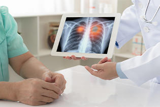
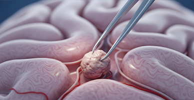
The mediastinum is the space between the lungs containing the heart, major blood vessels, thymus, lymph nodes, nerves, and other tissues. Cancers in this area include thymic tumours, lymphomas, germ-cell tumours, and sarcomas. A biopsy is usually needed because different mediastinal tumours may appear similar on imaging but require very different treatments.
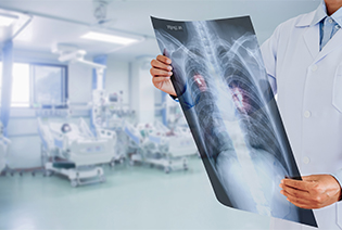
Chest wall cancers may arise from the ribs, sternum, cartilage, muscles, or soft tissues. They include chondrosarcoma, Ewing sarcoma, osteosarcoma, and soft tissue sarcoma. Treatment often involves wide surgical removal. Reconstruction may be needed to restore chest stability, protect the lungs and heart, and preserve breathing function.
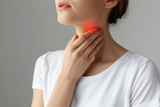
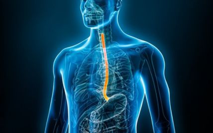
Esophageal cancer begins in the food pipe connecting the throat to the stomach. The main types are squamous cell carcinoma and adenocarcinoma. Depending on its location and the organisation of the clinical programme, esophageal cancer may be managed by a thoracic or gastrointestinal cancer team. Treatment can include endoscopic procedures, surgery, chemotherapy, immunotherapy, radiation therapy, or combined chemoradiation.
Risk factors differ between lung, pleural, thymic, airway, chest wall, and esophageal cancers. Some people develop thoracic cancer without an identifiable risk factor.
Modifiable Risk Factors
Smoking cigarettes, bidis, cigars, pipes, or other tobacco products
Exposure to second-hand tobacco smoke
Occupational exposure to asbestos, silica, diesel exhaust, arsenic, chromium, nickel, or other carcinogens
Indoor exposure to radon
Air pollution
Heavy alcohol consumption, particularly when combined with tobacco use, for esophageal squamous cell carcinoma
Obesity and chronic acid reflux, which increase the risk of esophageal adenocarcinoma
Inadequate workplace protection when handling hazardous substances
Non-Modifiable Risk Factors
Increasing age
A personal or family history of lung or another thoracic cancer
Previous radiation therapy involving the chest
Long-standing lung disease, including pulmonary fibrosis
Previous tuberculosis or other conditions that cause lung scarring
Inherited cancer-predisposition syndromes
Barrett’s esophagus
Reduced immunity
Certain autoimmune conditions associated with thymoma
Genetic changes acquired within tumour cells
Smoking is the leading cause of lung cancer, but lung cancer can also occur in people who have never smoked. Persistent symptoms therefore require evaluation regardless of smoking history.
Signs and Symptoms
Symptoms depend on the cancer’s location, size, and extent. Some early thoracic cancers are found incidentally during imaging performed for another reason.
Possible signs and symptoms include:
These symptoms frequently have non-cancerous causes. However, a persistent or worsening symptom should be assessed. Coughing up more than a small streak of blood, severe breathlessness, chest pain, confusion, or swelling of the face and neck requires urgent medical attention.
Diagnosis begins with a review of symptoms, tobacco and occupational exposure, previous illnesses, and family history. The doctor examines the chest and lymph nodes and may assess oxygen levels, breathing, and general health. Imaging is used to locate the abnormality and determine its extent, while a biopsy is usually needed to confirm the cancer type. The safest biopsy route is selected according to the tumour’s location. Whenever possible, enough tissue is obtained for both pathological diagnosis and biomarker testing.
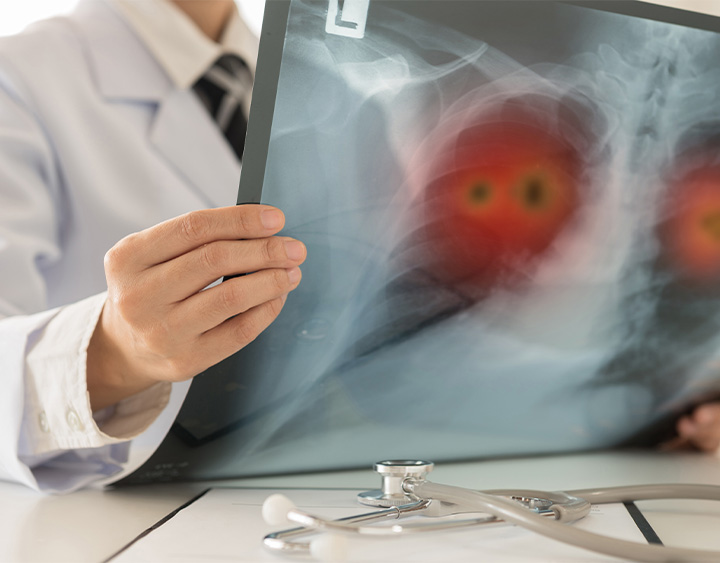
A chest X-ray may be the first test performed for a persistent cough, breathlessness, chest pain, or suspected infection. It can show a lung mass, collapse of part of the lung, or fluid around the lung. A normal chest X-ray does not rule out lung cancer. Further imaging may be required when symptoms or clinical findings remain concerning.
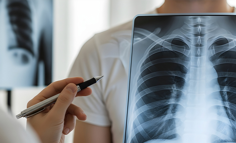
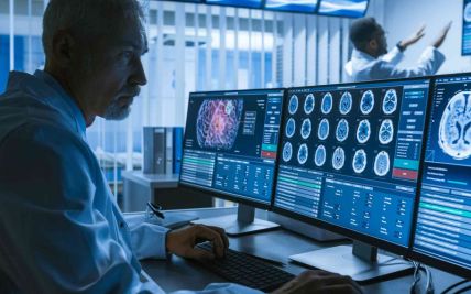
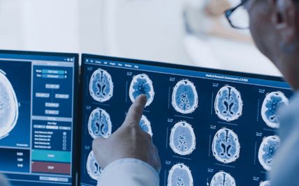
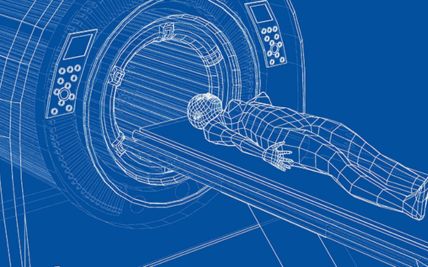
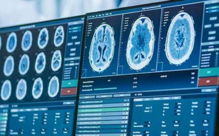
A CT scan provides detailed images of the lungs, airways, pleura, mediastinum, chest wall, and nearby lymph nodes. It helps determine the tumour’s size and location and whether nearby structures are involved. CT imaging of the chest and upper abdomen is commonly used when lung or another thoracic cancer is suspected. It can also guide a needle biopsy.
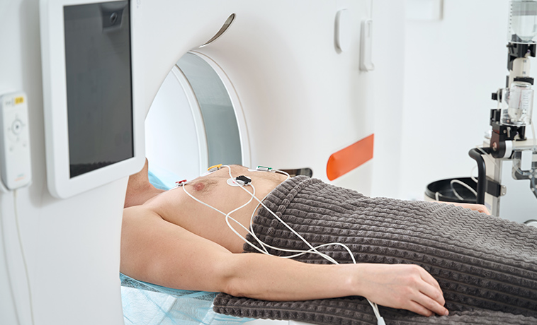
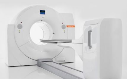
PET-CT identifies areas with increased metabolic activity and combines them with detailed anatomical images. It can help evaluate lymph nodes, identify distant disease, select an appropriate biopsy site, and plan treatment. Inflammation and infection can also appear active on PET-CT. Suspicious findings may therefore require tissue confirmation.
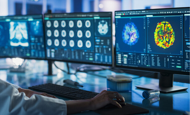
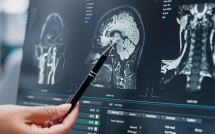
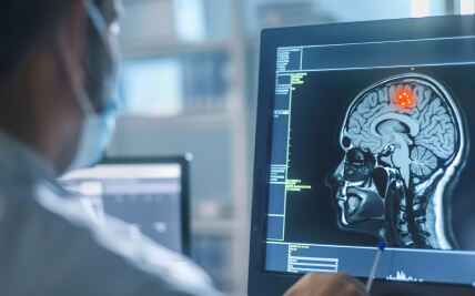
MRI provides detailed images without ionising radiation. It may be used to assess the brain, spinal cord, chest wall, major nerves, heart, or blood vessels. Brain MRI is commonly considered for selected lung cancers where there is a meaningful risk of brain spread or when neurological symptoms are present.
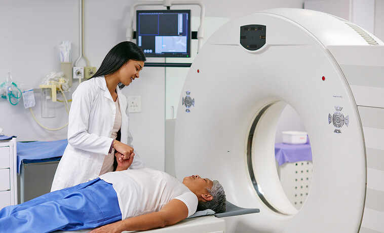
Endobronchial ultrasound, or EBUS, combines bronchoscopy with ultrasound to examine structures beside the airways. It is particularly useful for evaluating and sampling lymph nodes in the mediastinum. Endoscopic ultrasound, or EUS, is performed through the esophagus and can assess lymph nodes and tumours close to the esophageal wall. EBUS and EUS can reduce the need for a more invasive surgical biopsy.


A bone scan may be used in selected cases to look for cancer in the bones. PET-CT has replaced it in many clinical situations, but it may still be recommended when PET-CT is unavailable or when particular bone findings require assessment.
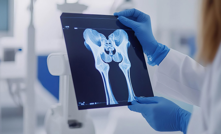
A bronchoscope is passed through the nose or mouth into the airways. The doctor can examine the trachea and bronchi and collect tissue using forceps, brushes, needles, or lavage. Advanced navigation techniques may help reach selected small or peripheral lung nodules.
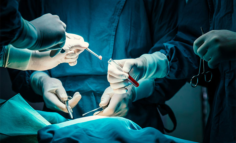
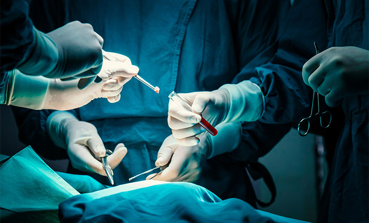
A needle is passed through the airway or esophageal wall under ultrasound guidance to sample lymph nodes or centrally located tumours These procedures can confirm the diagnosis and establish lymph-node involvement during the same examination.
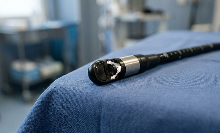
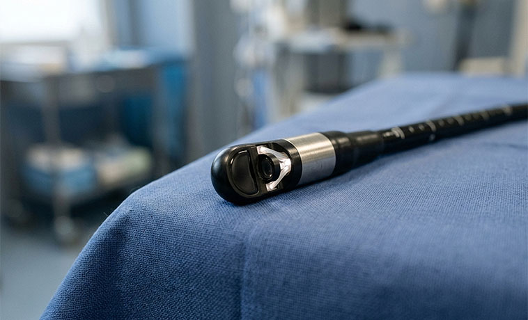
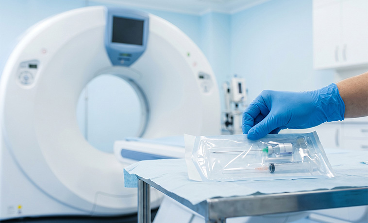
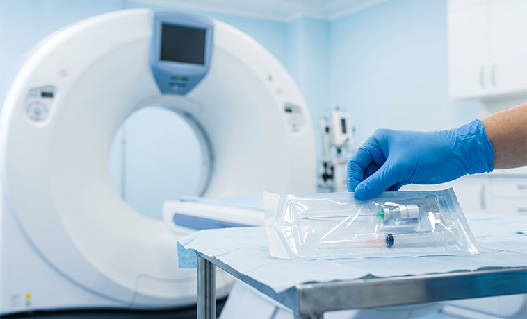
A needle is passed through the chest wall into the tumour under CT guidance. It is commonly used for lesions near the outer part of the lung, pleura, or chest wall. The procedure may cause bleeding or a pneumothorax, in which air collects around the lung. The patient is monitored for these complications.
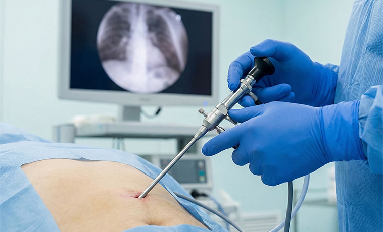
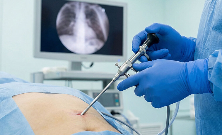
Thoracoscopy allows the pleural surface, lung, and chest cavity to be examined through small incisions. Video-assisted thoracoscopic surgery, or VATS, may be used when a larger tissue sample is needed or when less invasive tests have not established the diagnosis. It is particularly useful for suspected mesothelioma, pleural tumours, or difficult-to-reach lung lesions.
Fluid around the lung can be removed with a needle and examined for cancer cells. This can help diagnose lung cancer, mesothelioma, or another cancer involving the pleura. A negative fluid result does not always exclude cancer. A pleural biopsy may be required if suspicion remains high.
Mediastinoscopy is a surgical procedure used to sample lymph nodes and other tissues in the central chest. It may be recommended when EBUS or another less invasive procedure cannot provide adequate tissue or when additional confirmation is required before treatment.
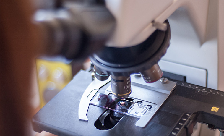
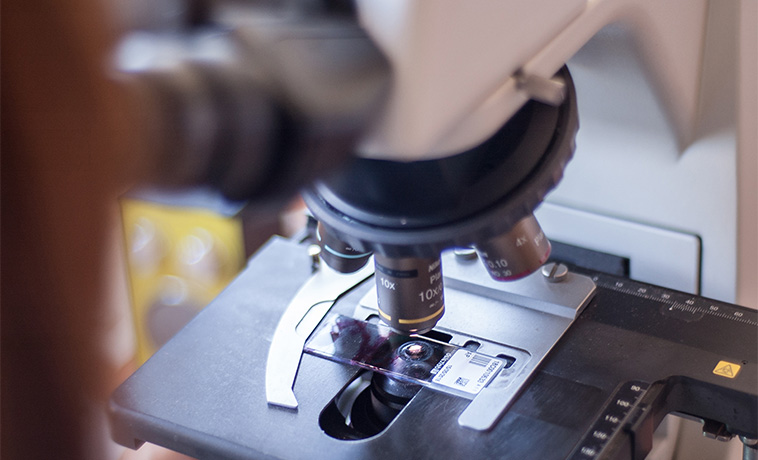
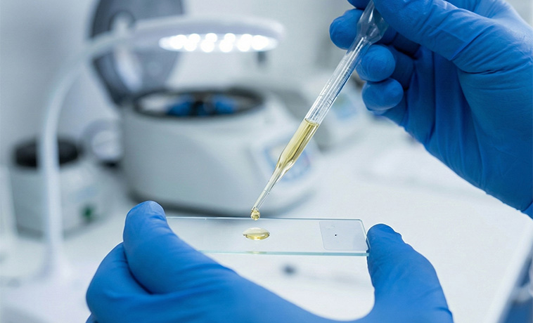
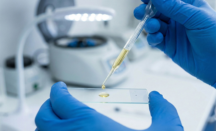
A specialist pathologist examines biopsy tissue to determine whether cancer is present and identify its type and subtype. Immunohistochemistry helps distinguish adenocarcinoma, squamous cell carcinoma, small cell cancer, mesothelioma, thymic cancer, lymphoma, and metastatic cancer from another organ.
For eligible NSCLC, molecular testing may assess changes involving genes such as EGFR, ALK, ROS1, BRAF, KRAS, MET, RET, NTRK, and HER2. The exact panel depends on the cancer subtype, stage, available treatments, and clinical guidelines.
PD-L1 testing may help guide immunotherapy. However, it is considered alongside the cancer stage, molecular findings, previous treatment, and overall clinical situation.
A liquid biopsy examines tumour-related DNA circulating in the blood. It may be useful when adequate tissue cannot be obtained or when rapid molecular information is required. A negative liquid-biopsy result does not exclude an actionable alteration, and tissue testing may still be needed.
Routine blood tests assess blood counts, kidney and liver function, calcium, nutritional status, and fitness for treatment. Tumour markers alone cannot diagnose or screen for lung cancer.


Different thoracic cancers use different staging systems. The stages below describe non-small cell lung cancer, the most common thoracic cancer. Mesothelioma, thymic cancer, esophageal cancer, sarcoma, and other thoracic cancers require their own disease-specific staging.
Stage 0: Abnormal cells are confined to the lining of the airways and have not invaded deeper tissue. This is also called carcinoma in situ.
Stage I: The cancer is confined to the lung and has not spread to lymph nodes. Stage I is divided according to the tumour’s size and local features.
Stage II: The cancer may be larger, may involve a nearby structure, or may have spread to lymph nodes within the lung or near the main airway on the same side of the chest.
Stage III: The cancer has spread more extensively within the chest. It may involve mediastinal or supraclavicular lymph nodes, lymph nodes on the opposite side, or nearby structures such as the chest wall, diaphragm, heart covering, major vessels, trachea, or esophagus.
Stage IV: The cancer has spread beyond the original lung. This may include a tumour in the opposite lung, cancerous nodules on the pleura or pericardium, malignant fluid around the lung or heart, or spread to organs such as the brain, liver, bones, or adrenal glands.
A wedge resection removes the tumour with a small surrounding portion of lung. It may be considered for selected small, peripheral tumours or for people who cannot safely undergo a larger operation. Lymph-node sampling or removal is generally performed when the procedure is being used to treat lung cancer.
A segmentectomy removes an anatomical segment of the lung, including its bronchus and blood supply. It preserves more lung tissue than a lobectomy and may be suitable for selected small, early-stage tumours. The decision depends on tumour size and location, lymph-node findings, surgical margins, and lung function.
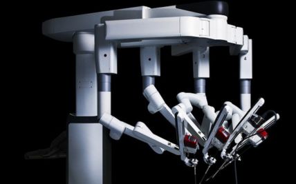
A lobectomy removes one lobe of the lung and is a common operation for operable NSCLC. Nearby lymph nodes are also removed or sampled for accurate staging. The procedure may be performed through open surgery, VATS, or robotic-assisted thoracic surgery, depending on the tumour and individual clinical situation.
A sleeve resection removes a section of an involved airway, sometimes with part of the lung, and reconnects the remaining airway. It may allow the surgeon to remove a centrally located tumour without removing the entire lung. Suitability depends on the tumour’s relationship to the airway, blood vessels, and lymph nodes.
A pneumonectomy removes an entire lung. It may be required for selected central tumours that cannot be completely removed with a smaller operation. Because the procedure places greater demands on the heart and remaining lung, detailed breathing and cardiac assessments are required beforehand.
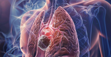
Thymectomy removes the thymus and surrounding tissue and is the principal treatment for a resectable thymoma or thymic carcinoma. Larger tumours may require removal and reconstruction of involved pericardium, lung, nerves, or blood vessels. Other mediastinal tumours require treatment based on their pathology. Surgery is not appropriate for every mediastinal mass, particularly lymphomas that are primarily treated with medicines.
Selected people with pleural mesothelioma may undergo surgery to remove visible disease from the pleura and other involved tissues. Procedures vary in extent and are considered as part of a multidisciplinary treatment plan. Extensive surgery is suitable only for carefully selected patients after assessment of disease extent, lung function, heart function, and expected benefit.
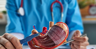
Esophagectomy removes part or most of the esophagus together with nearby lymph nodes. The stomach or, less commonly, part of the intestine is used to restore continuity of the digestive tract. Surgery may be performed through open, minimally invasive, robotic-assisted, or combined approaches according to the tumour’s location and extent.
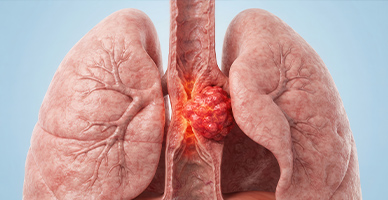
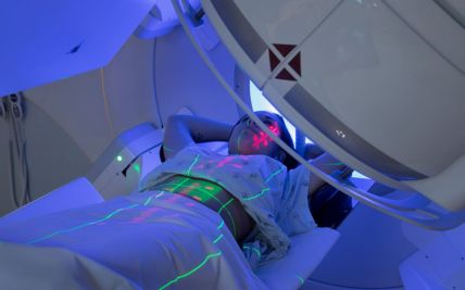
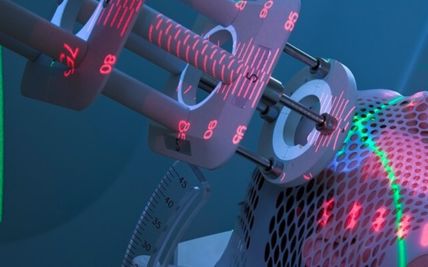
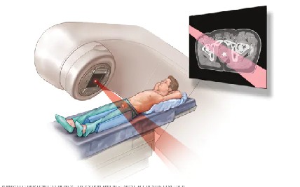
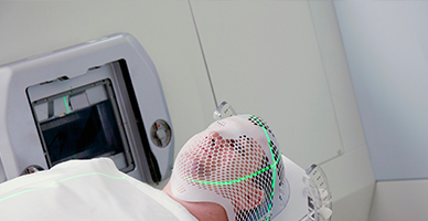
External beam radiation directs high-energy beams at the tumour and nearby areas at risk. It may be used for lung, pleural, thymic, mediastinal, airway, chest wall, or oesophageal cancers. The dose and number of sessions depend on the cancer type, stage, treatment purpose, and nearby organs.
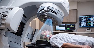
Intensity-modulated radiation therapy shapes and adjusts the radiation dose around the tumour. Image-guided radiation therapy uses imaging during treatment to confirm tumour and patient positioning. These techniques help improve accuracy when treating moving or irregularly shaped tumours in the chest.
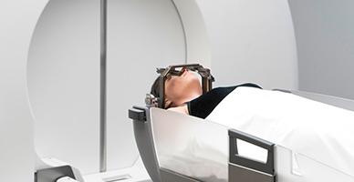
Stereotactic body radiation therapy, or SBRT, delivers highly focused radiation over a limited number of sessions. It is commonly considered for selected early-stage lung cancers when surgery is unsuitable and for carefully chosen metastatic tumours. The tumour’s location, movement with breathing, and proximity to major airways or other critical structures influence whether SBRT is appropriate.
Chemotherapy and radiation therapy may be given during the same period to increase their combined effect. This is an established treatment for many unresectable Stage III NSCLCs, limited-stage SCLCs, and selected oesophageal or other thoracic cancers. Because combined treatment can cause more side effects, patients require careful assessment, planning, and supportive care.
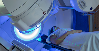
Proton therapy uses proton beams that release most of their radiation within the treatment area, with little dose continuing beyond it. In selected chest tumours, this may reduce radiation exposure to the heart, healthy lungs, esophagus, or spinal cord.
It may be considered when treatment planning demonstrates a meaningful advantage over advanced photon techniques, including for selected locally advanced, recurrent, paediatric, or complex thoracic cancers. Proton therapy is not routinely required for every patient, and suitability is determined through individual plan comparison.
Chemotherapy may be given before surgery to shrink a tumour, after surgery to reduce recurrence risk, or as the principal treatment for advanced disease. It is central to the treatment of small cell lung cancer and is also used for NSCLC, mesothelioma, thymic cancer, oesophageal cancer, and other thoracic malignancies. It may be combined with radiation therapy or immunotherapy.

Targeted medicines act on specific molecular changes within cancer cells. They are particularly important for eligible NSCLCs with alterations such as EGFR, ALK, ROS1, BRAF, KRAS G12C, MET, RET, NTRK, or HER2. Because molecular findings can change treatment substantially, adequate biomarker testing should be completed when clinically appropriate.
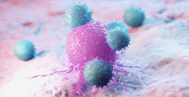
Immunotherapy helps the immune system recognise and attack cancer cells. It may be used alone or with chemotherapy for selected NSCLCs and small cell lung cancers. It may also be considered before or after surgery in certain stages and is used for selected mesotheliomas, oesophageal cancers, and other thoracic cancers. Suitability depends on the diagnosis, stage, biomarkers, autoimmune history, and previous treatment.

Antibody-drug conjugates combine an antibody directed at a cancer-related target with a cancer-killing medicine. They may be used for selected advanced thoracic cancers with an appropriate biomarker. Their role depends on the tumour type, molecular findings, previous treatment, and regulatory approval.

For symptom relief and select therapeutic purposes, our thoracic oncology team also offers image-guided interventions such as radiofrequency ablation and pleural procedures to manage fluid build-up around the lungs.
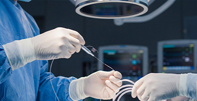
After removing a tumour from the trachea or bronchus, the remaining airway may be reconnected. This can preserve functioning lung tissue and maintain airflow in appropriately selected patients.
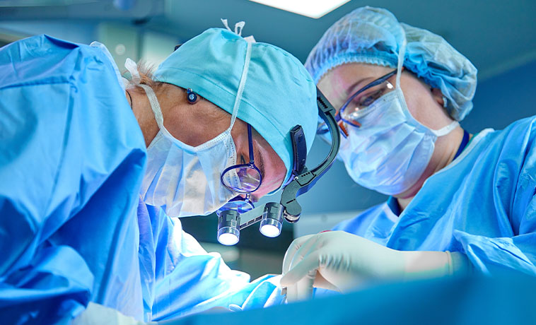
Removal of a chest wall tumour may leave a defect involving the ribs, sternum, muscles, or skin. Mesh, plates, biological materials, or transferred tissue may be used to restore stability, protect the organs within the chest, and support breathing.
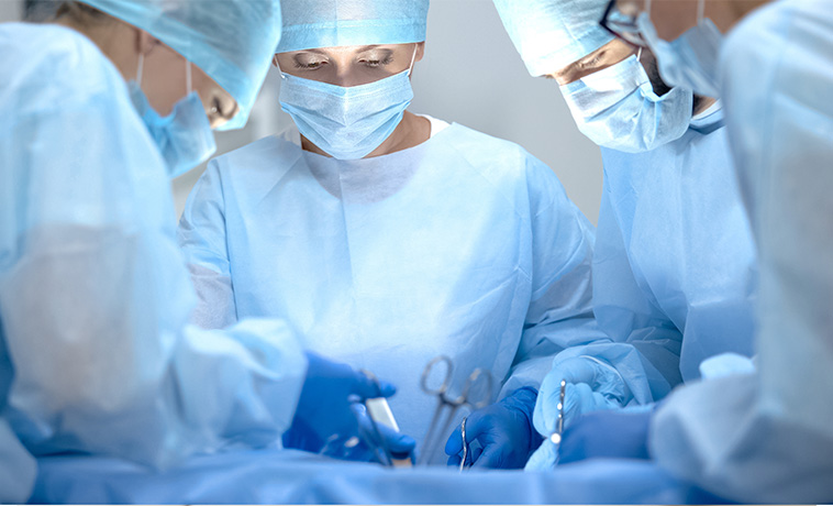
Following esophagectomy, the stomach is commonly reshaped and brought up to reconnect the digestive tract. A segment of the intestine may be used when the stomach is unsuitable.
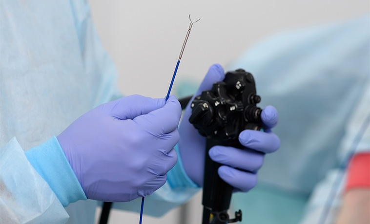
Large defects involving the skin or muscles of the chest may be covered using local, regional, or free-tissue flaps. Reconstruction is planned to protect underlying structures, close the wound securely, and support subsequent treatment.
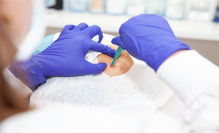
Tumours narrowing a major airway may be treated through a bronchoscope using selected techniques such as mechanical removal, laser, electrocautery, argon plasma coagulation, cryotherapy, or airway stenting. These procedures can help improve breathing, control bleeding, or reopen an obstructed airway. They may be combined with cancer-directed treatment.
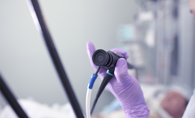
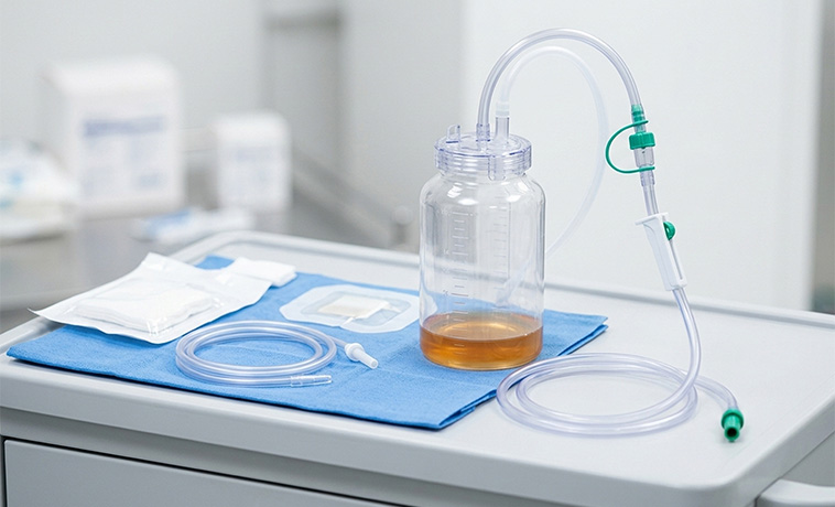
Fluid around the lung can cause breathlessness and chest discomfort. It may be removed through a needle, drainage tube, or indwelling pleural catheter. Pleurodesis uses a medicine to help the pleural layers adhere and reduce recurrent fluid accumulation. The appropriate procedure depends on how quickly the fluid returns and whether the lung can re-expand.
Radiofrequency, microwave, or cryoablation destroys tumour tissue using heat or cold delivered through a probe. It may be considered for selected small lung tumours or limited metastatic disease when surgery is unsuitable. Ablation is not appropriate for every tumour and requires evaluation of its size, location, and relationship to major airways and blood vessels.
Prognosis for Thoracic Cancer
The outlook for thoracic cancer varies considerably. Some early-stage tumours can be treated with curative intent, while advanced cancers may require long-term treatment to control the disease, relieve symptoms, and maintain quality of life.
Factors that influence prognosis include:
Cancer type and pathological subtype
Stage at diagnosis
Whether the tumour can be completely removed
Lymph-node and distant-organ involvement
Molecular and immune-marker findings
Response to treatment
Lung and heart function
Overall health and nutritional status
Tobacco use during and after treatment
Development of recurrent disease
Stage alone does not determine an individual’s outcome. People with the same stage may respond differently depending on the cancer’s biology, available treatment options, and general health.


Screening for Thoracic Cancer
Screening is established primarily for lung cancer in people at high risk. There is no routine population-wide screening test for all thoracic cancers.
Low-Dose CT Screening
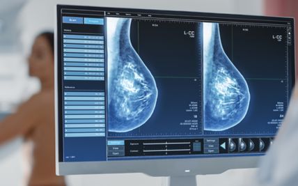
Low-dose CT can detect some lung cancers before symptoms develop. It may be recommended for people within a defined age range who have a substantial smoking history and currently smoke or stopped within a specified period.
Eligibility criteria vary between guidelines and countries. Screening should be offered through a structured programme that includes counselling, management of abnormal findings, and support for stopping tobacco.
Why Choose ACC for Thoracic Cancer Treatment
A multidisciplinary thoracic oncology team
Pulmonology, thoracic surgery, medical oncology, radiation oncology, pathology, and imaging expertise
Bronchoscopy, EBUS, image-guided biopsy, PET-CT, and molecular profiling
Open, VATS, and robotic-assisted thoracic surgical options
Precision radiation techniques, including SBRT and proton therapy for selected patients
Targeted therapy and immunotherapy guided by clinical and molecular findings
Complex airway, chest wall, and oesophageal reconstruction
Tumour Board review for coordinated, individualised treatment planning

Health Blogs