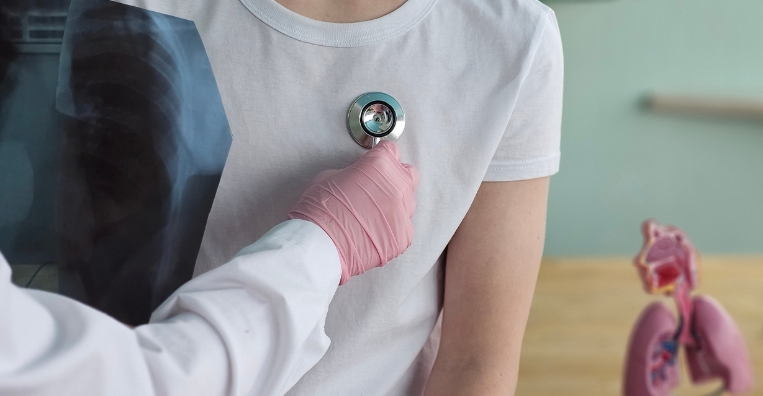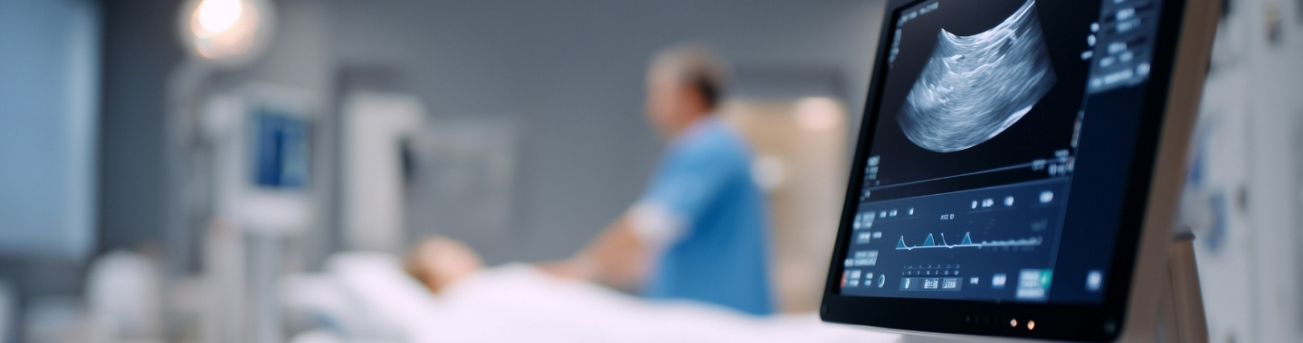
Endobronchial ultrasound (EBUS) combines bronchoscopy with ultrasound to examine structures beyond the walls of the trachea and bronchi. It is most commonly used to assess and sample lymph nodes or masses in the centre of the chest.
EBUS-guided transbronchial needle aspiration, or EBUS-TBNA, allows cells and small tissue samples to be collected without a surgical incision. A different form, called radial EBUS, may help locate selected abnormalities deeper in the lungs.


Role in Cancer Care


What to Expect
Before the Test
You will receive instructions about when to stop eating and drinking. Tell the clinical team about:
All medicines and supplements you take, particularly anticoagulants and antiplatelet medicines
Diabetes or medicines that may require adjustment while fasting
Bleeding disorders or significant heart or lung conditions
Any oxygen requirements
Allergies or previous reactions to sedatives, anesthetics or medicines
Pregnancy or the possibility of pregnancy
Dentures, removable dental work or loose teeth
Do not stop prescribed medicines unless instructed by your clinical team. Blood tests, breathing assessments or recent imaging may be reviewed before the procedure. If sedation or general anesthesia is planned, arrange for a responsible adult to take you home and follow the instructions about supervision afterwards.

During the Test
EBUS may be performed under moderate or deep sedation or general anesthesia, depending on the centre, the examination planned and your individual needs. Local anesthetic may be applied to the throat and airways, while your oxygen level, heart rate and blood pressure are monitored.
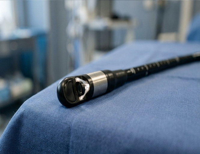
The bronchoscope is usually passed through the mouth and into the airways. Although coughing, pressure or a temporary sensation of breathlessness may occur, the scope does not usually prevent breathing.
The doctor examines the lymph nodes and nearby structures using ultrasound and guides a needle into selected areas. Several samples may be collected to improve the likelihood of obtaining enough diagnostic material. The procedure commonly takes approximately 30–60 minutes, although the duration can vary.

After the Test
You will be monitored until the sedation or anesthesia has worn off. Do not eat or drink until the throat numbness has resolved and the clinical team confirms that swallowing is safe. A temporary sore throat, hoarse voice, cough or small amount of blood-streaked phlegm may occur.
If you received sedation or general anesthesia, do not drive, operate machinery, drink alcohol, make important decisions or sign legal documents for the period specified by the clinical team. Arrange for someone to accompany you home and remain with you if advised.
The doctor may discuss the initial findings shortly after the procedure. Cytology, histopathology and molecular test results take longer and will be reviewed when the laboratory analysis is complete.
Seek urgent medical advice if you develop:
Increasing or severe breathlessness
Chest pain
Persistent fever or chills
Heavy or persistent bleeding when coughing
Fainting, confusion or any rapidly worsening symptoms

Potential Benefits
- Beyond Airways: It provides ultrasound images of lymph nodes, masses and other structures beyond the airway walls.
- Targeted Sampling: It allows selected abnormalities to be sampled under continuous ultrasound guidance.
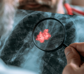
- Cancer Diagnosis: Samples collected through EBUS can help confirm whether cancer is present and identify its type.
- Nodal Staging: It can determine whether cancer has spread to accessible lymph nodes in the centre of the chest.
- Molecular Testing: Suitable samples may provide material for biomarker or molecular analysis that helps guide treatment.
- Treatment Planning: Its findings can support decisions about surgery, systemic therapy, radiotherapy and other treatments.
- Minimal Incisions: It can sample many central chest abnormalities without requiring an external surgical incision.
- Radiation-Free Guidance: The ultrasound component uses sound waves rather than ionising radiation to guide sampling.