




Radiation oncology is a key part of comprehensive cancer care. It uses carefully planned doses of ionising radiation to damage the DNA of cancer cells, stopping or slowing their growth. Radiation therapy is a local or regional treatment, which means it is directed at a specific area of the body.
Read MoreProton therapy is a form of external beam radiation therapy that uses protons rather than X-rays. The physical properties of protons allow the radiation dose to be shaped so that less radiation may continue beyond the target, potentially reducing exposure to selected nearby tissues. It may be considered when this dose distribution offers a meaningful clinical advantage, but it is not necessary or superior for every patient or every cancer.
Read More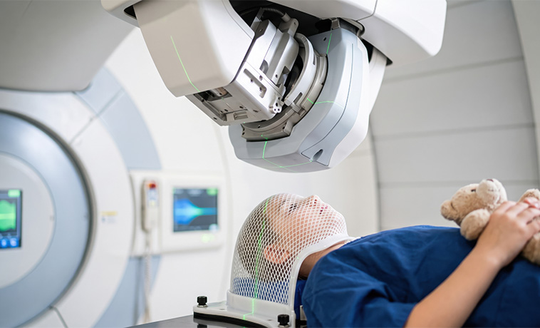
Zap-X is a dedicated platform used to deliver stereotactic radiosurgery to selected targets in the brain, head, and upper neck. It directs multiple precisely focused radiation beams at the target while limiting the dose to surrounding tissue. Despite its name, radiosurgery does not involve an incision; suitability depends on the diagnosis, size, number, and location of the lesions and any previous treatment.
Read More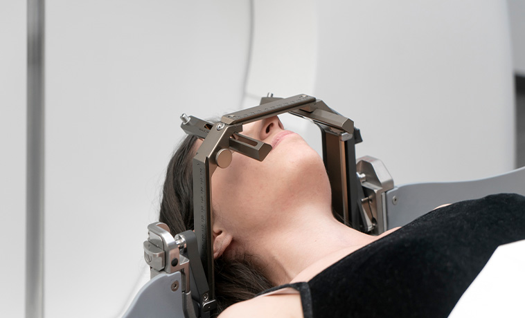
CyberKnife is a robotic radiation-delivery system used for stereotactic radiosurgery and stereotactic body radiation therapy. It delivers radiation from multiple angles and uses image guidance to track the target and adjust for positioning or certain types of movement during treatment. It may be used for selected tumours in the brain or body, but the technology is chosen only when clinically appropriate.
Read More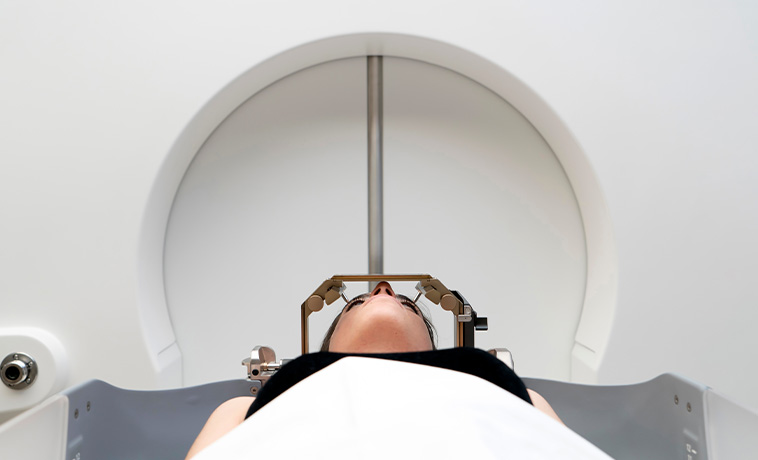
External beam radiation therapy delivers high-energy radiation from a machine outside the body. The machine directs radiation at a planned treatment area and does not touch the patient. Treatment is usually divided into sessions called fractions, although the number and schedule vary according to the diagnosis and treatment goal. Patients do not become radioactive after standard external beam treatment.
Read More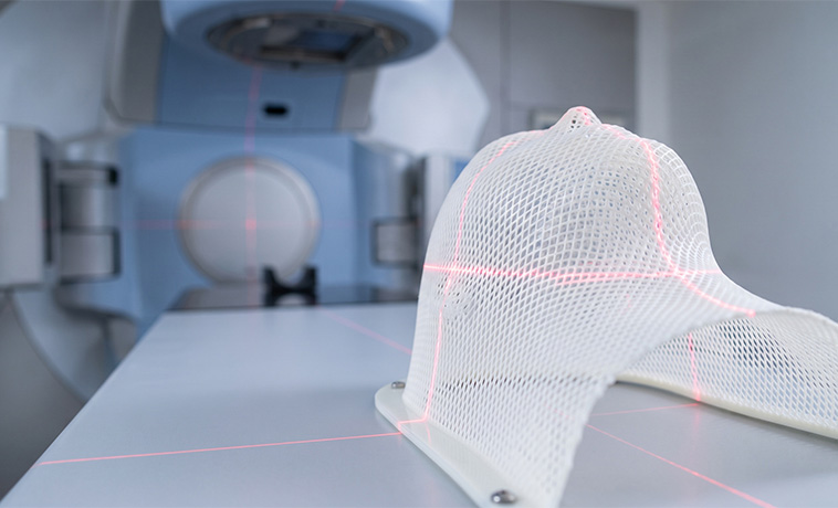
Three-dimensional conformal radiation therapy, or 3D-CRT, uses imaging and computerised planning to shape radiation beams to the three-dimensional outline of the target. Beams are delivered from different directions to treat the planned area while reducing unnecessary exposure to nearby tissues. It may be appropriate when the target and surrounding anatomy can be treated safely without more complex modulation.
Intensity-modulated radiation therapy, or IMRT, is an advanced form of external beam treatment that varies the intensity of radiation across multiple beams. This allows the dose to conform closely to complex target shapes and helps limit exposure to selected nearby organs. IMRT requires detailed planning and quality assurance before treatment begins.
Read More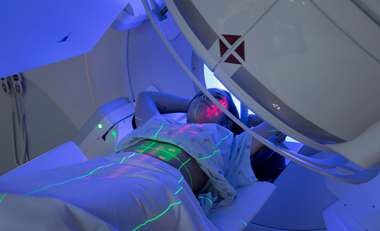
Volumetric modulated arc therapy, or VMAT, is a form of intensity-modulated radiation therapy delivered while the treatment machine rotates around the patient. The beam shape, intensity, and delivery rate can change during rotation, allowing a planned dose to be delivered efficiently to complex targets while limiting exposure to surrounding structures.
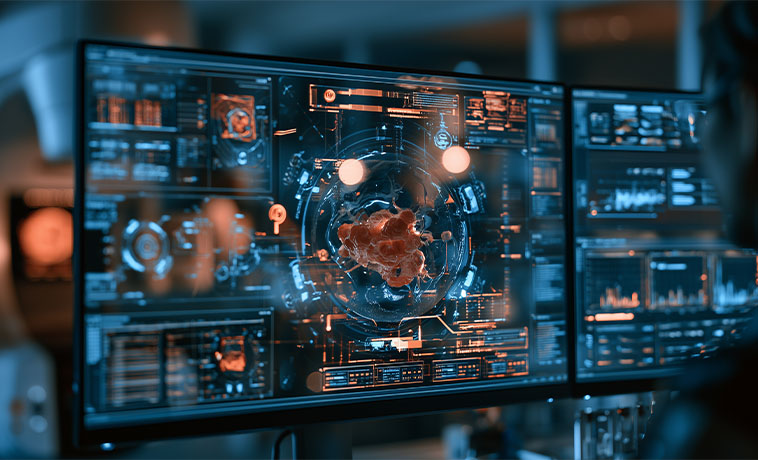
Image-guided radiation therapy, or IGRT, uses imaging before or during treatment to confirm the position of the patient and target. The team can make positioning corrections before delivering radiation, helping account for daily anatomical variation or movement. IGRT is commonly combined with techniques such as IMRT, VMAT, SBRT, and stereotactic radiosurgery rather than being a separate type of radiation dose.
Read More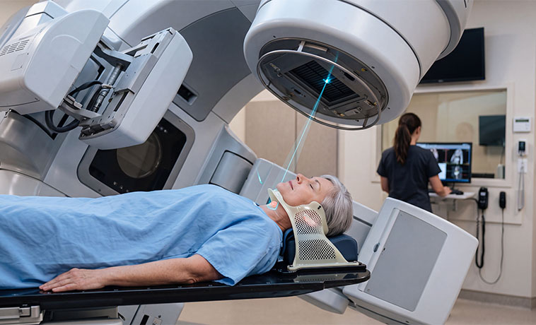
Stereotactic body radiation therapy, or SBRT, delivers highly focused radiation to selected targets outside the brain, usually in a small number of treatment sessions. It requires precise immobilisation, imaging, treatment planning, and motion management when the target moves with breathing. SBRT is suitable only for selected patients based on the size, number, and location of lesions and their proximity to sensitive organs.
Read More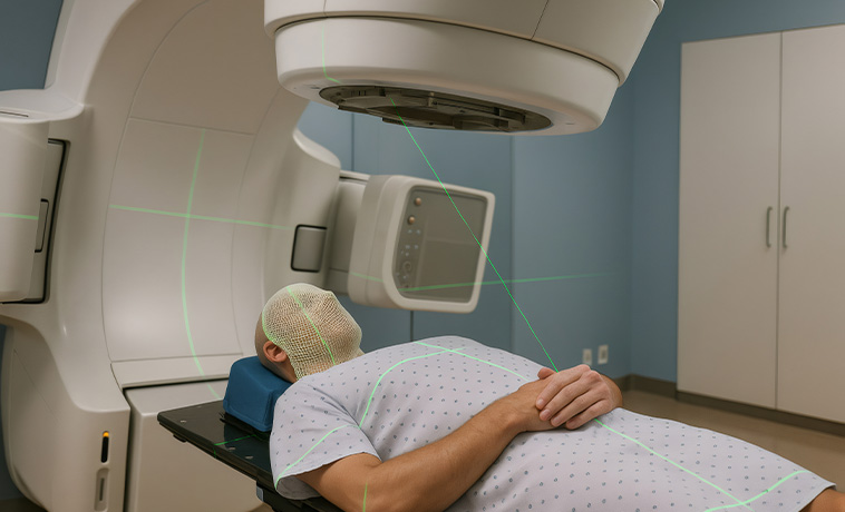
Stereotactic radiosurgery, or SRS, delivers a highly focused dose of radiation to selected targets in the brain or other intracranial structures, usually in one or a few sessions. It does not involve an incision and may be delivered using different treatment platforms. Suitability depends on the diagnosis, size, number, and location of the targets and their relationship to critical structures.
Read More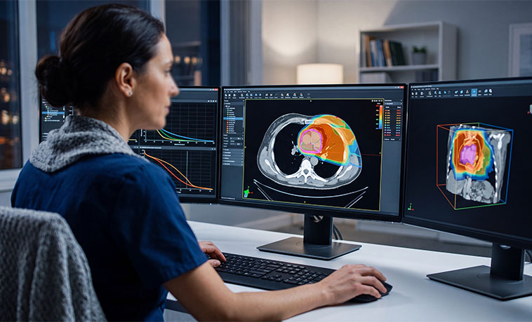
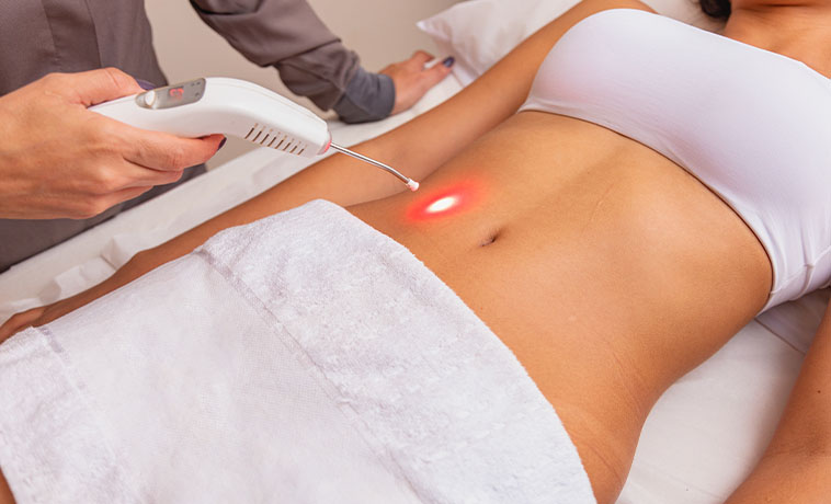
Brachytherapy is a form of internal radiation therapy in which a sealed radioactive source is placed inside or close to the treatment area. This allows a high radiation dose to be delivered over a short distance while limiting exposure to more distant tissues. Depending on the procedure, the source may be temporary or permanent, and specific radiation-safety instructions may be required.
Read More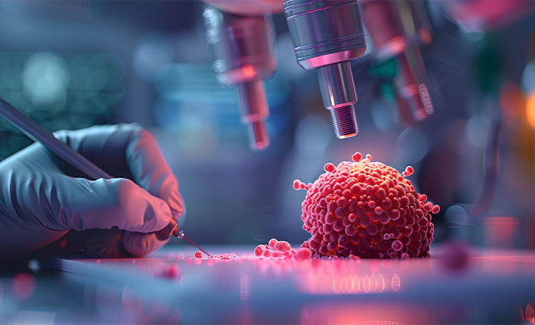
Total body irradiation, or TBI, delivers radiation to the whole body and may be used as part of the conditioning regimen before selected stem cell or bone marrow transplants. Its purpose may include treating remaining abnormal cells and suppressing the immune system to help donor cells establish themselves. TBI requires careful planning and close coordination between radiation oncology and the hemato-oncology and transplant teams.

How Radiation Oncology Fits into Your Overall Care Plan
Radiation therapy may be used as the main treatment or as one part of a broader cancer care plan. Its purpose may be to cure the cancer, reduce the risk of recurrence, control disease, prepare for another treatment, or relieve symptoms.
Radiation may be given before surgery to reduce the extent of disease, after surgery to treat possible remaining cancer cells, or alongside systemic therapy when a combined approach is appropriate. In selected situations, it may be used instead of surgery or to treat a limited area of recurrent or metastatic disease.
Palliative radiation therapy may be used to relieve symptoms such as pain, bleeding, obstruction, breathing difficulty, or pressure on nearby structures. Palliative treatment schedules are often shorter, but the appropriate schedule depends on the clinical situation.
Radiation therapy may also be coordinated with stem cell transplantation, nuclear medicine treatments, or image-guided interventional procedures. The sequence and combination of treatments are determined by the multidisciplinary team
What Patients Can Expect
Before Treatment
Your first consultation may include a review of your medical history, examination findings, biopsy or pathology reports, imaging, previous treatments, current medicines, and relevant health conditions. Your radiation oncologist will explain the aim of treatment, available techniques, likely number of sessions, potential side effects, and other treatment options.
If radiation therapy is recommended, you will usually attend a planning or simulation appointment. Imaging such as a CT scan is used to map the treatment position and define the target and nearby organs. A mask, mould, cushion, or other immobilisation device may be made to help you remain in the same position for every session. Small skin marks or tattoos may be used for positioning when necessary.
Your treatment does not usually begin on the day of simulation. The radiation oncologist, medical physicist, and planning team use the simulation images and other scans to design and verify an individual treatment plan.
Tell the team if you are pregnant or could be pregnant, have an implanted electronic device, have previously received radiation therapy, experience claustrophobia, or cannot remain comfortably in the planned position.
During Treatment
At each session, the radiation therapy team will position you carefully and may take images to confirm alignment. You will need to remain still, but you can breathe normally unless specific breathing instructions are provided. The treatment machine may move around you but will not touch you.
Radiation delivery is painless, and external beam treatment does not make you radioactive. The therapists leave the room while radiation is being delivered but can see, hear, and communicate with you throughout the session.
Side effects vary according to the area treated, radiation dose, treatment schedule, and whether other treatments are given at the same time. They may include fatigue, skin changes, hair loss limited to the treated area, or site-specific effects involving eating, swallowing, digestion, urinary function, or other organs. Your team will monitor you regularly and provide measures to prevent or manage side effects.
Brachytherapy may require an applicator or radioactive source to be placed in or near the treatment area. Depending on the procedure, anaesthesia, a short admission, or temporary radiation-safety precautions may be required.

After Treatment
Some effects improve within weeks after treatment, while others may take longer to resolve. Certain late effects can develop months or years later, depending on the treatment area and dose. Your team will explain which symptoms to monitor and when to seek medical advice.
Follow-up may include consultations, physical examinations, imaging, laboratory tests, rehabilitation, nutritional support, or other assessments. Imaging is generally scheduled at an appropriate interval after treatment because radiation-related inflammation can sometimes make early scans difficult to interpret.
The multidisciplinary team will assess recovery and disease response and determine whether further treatment, surveillance, or supportive care is required.



