




Interventional oncology is a specialised area of cancer care that uses imaging to guide minimally invasive procedures for diagnosing, treating, or relieving symptoms caused by cancer. Instead of making a large surgical incision, specialists generally reach the treatment area through a small skin puncture using needles, probes, or thin catheters.
Read MoreRadiofrequency ablation, or RFA, uses image guidance to position a needle-like probe within a tumour. Radiofrequency electrical energy generates heat at the probe tip, destroying tissue within a planned treatment area. RFA may be considered for selected, usually small or limited tumours and metastases, depending on their location and proximity to blood vessels, bile ducts, nerves, or other sensitive structures.
Read More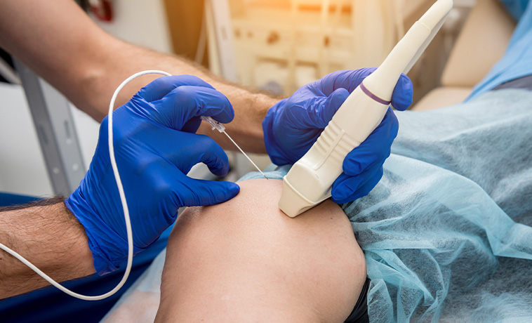
Microwave ablation, or MWA, uses image guidance to place one or more probes within a tumour. The probes produce an electromagnetic microwave field that rapidly generates heat and destroys tissue within the treatment area. MWA may be considered for selected tumours based on their size, number, location, surrounding anatomy, and the ability to create an adequate treatment margin safely.
Read More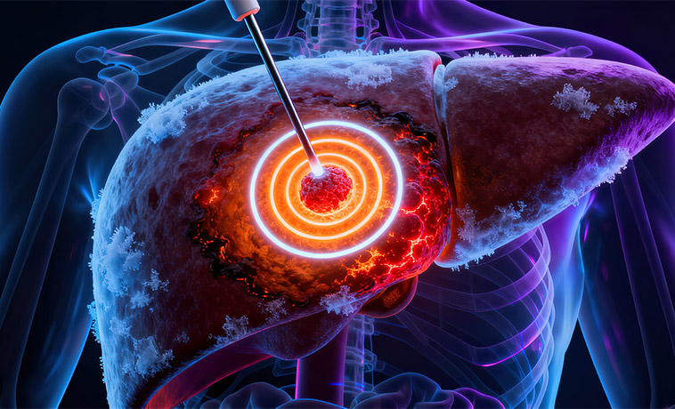
Cryoablation uses image-guided probes to freeze and destroy tumour tissue through repeated freezing and thawing cycles. Imaging allows the treatment team to monitor the developing area of frozen tissue during the procedure. Cryoablation may be considered for selected tumours, particularly when careful monitoring of the treatment zone is important or when another ablation method is less suitable.
Read More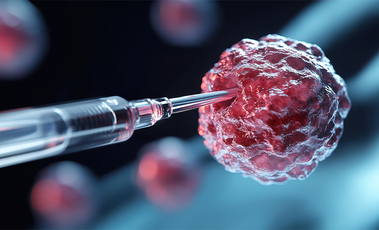
Embolisation uses a thin catheter inserted into a blood vessel to deliver particles or other materials that reduce or block blood flow to a tumour. In some procedures, embolisation is combined with locally delivered chemotherapy, known as chemoembolisation. It may be used to control selected tumours, reduce tumour-related bleeding, relieve symptoms, or support another treatment, depending on the organ involved and the tumour’s blood supply.
Read More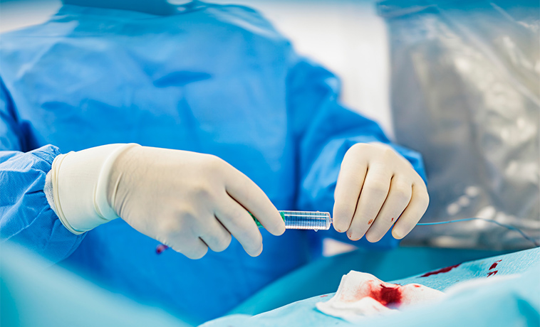
Radioembolisation is a targeted internal radiation treatment used for selected primary or metastatic tumours in the liver. A catheter is guided into the arteries supplying the tumour, and microscopic beads containing a radioactive isotope, usually yttrium-90, are delivered into the tumour’s blood supply. The procedure requires angiographic mapping, nuclear medicine assessment, radiation-dose planning, and coordinated evaluation by interventional radiology, nuclear medicine, medical physics, and the wider oncology team.
Read More

How Interventional Oncology Fits into Your Overall Care Plan
Interventional oncology may be used as the main local treatment for selected tumours or as one part of a broader cancer care plan. It is generally most suitable when the disease can be reached safely using image guidance and treated within an acceptable margin.
Ablation may be considered for selected primary tumours, a limited number of metastases, or residual or recurrent disease. It may also be used when surgery is not appropriate because of the tumour’s location, previous treatment, reduced organ reserve, or the patient’s overall health. Embolisation and radioembolisation may be used to control selected tumours supplied by identifiable blood vessels. Depending on the clinical situation, these procedures may help reduce tumour activity, manage bleeding or other symptoms, or support treatment before surgery, transplantation, radiation therapy, or systemic treatment.
These procedures treat visible and accessible areas of disease. They do not necessarily treat microscopic cancer cells or disease elsewhere in the body. Chemotherapy, targeted therapy, immunotherapy, radiation therapy, surgery, or another treatment may therefore still be required.
Your interventional oncologist works with the multidisciplinary team to determine the most appropriate procedure, timing, and combination of treatments.
What Patients Can Expect
Before Treatment
Your interventional oncology team will review your diagnosis, pathology reports, imaging, previous treatments, current medicines, allergies, and relevant health conditions.
Before the procedure, you may need:
Blood tests to assess blood counts, clotting, and organ function
Additional imaging to define the treatment area
Anesthesia or sedation assessment
Temporary adjustment of blood thinners or other medicines under medical guidance
Fasting for a specified period
Antibiotics, hydration, or other supportive medicines
Pregnancy screening when relevant
Angiographic mapping and nuclear medicine assessment before radioembolisation
Your specialist will discuss:
The aim of the procedure
Available alternatives
Expected benefits and limitations
Possible risks and side effects
The planned type of anaesthesia or sedation
Whether admission or overnight observation may be required
The possibility of further or repeat treatment
You will have an opportunity to ask questions before providing informed consent.

During Treatment
Depending on the procedure, the interventional oncologist will:
Insert a probe through the skin into the tumour for RFA, MWA, or cryoablation
Insert a catheter into a blood vessel for embolisation or radioembolisation
Use ultrasound, CT, MRI, or fluoroscopy to guide treatment
Monitor the treatment area and surrounding structures
Deliver heat, cold, embolic material, chemotherapy, or radioactive microspheres as planned
The procedure may be performed under local anaesthesia with sedation or under general anesthesia. Your heart rate, blood pressure, breathing, oxygen level, and overall condition will be monitored throughout. The duration depends on the treatment, tumour location, number of areas being treated, and procedural complexity.

After Treatment
After the procedure, you will be monitored in a recovery area. Some patients can return home the same day, while others may require overnight observation or a longer hospital stay.
You will receive instructions covering:
Pain relief and other medicines
Wound or puncture-site care
Hydration and diet
Activity restrictions
Return to work, driving, and exercise
Radiation-safety precautions after radioembolisation, when applicable
Follow-up appointments and imaging
Temporary effects may include pain, fatigue, nausea, mild fever, bruising, or soreness at the treatment site. Follow-up may include blood tests and imaging to assess the treated area and determine whether another procedure or additional cancer treatment is required.
Contact your care team promptly if you develop severe or worsening pain, heavy bleeding, increasing swelling at the puncture site, persistent vomiting, a high or persistent fever, breathing difficulty, chest pain, confusion, jaundice, markedly reduced urine output, or another symptom listed in your discharge instructions.



