Pediatric Cancer
Pediatric cancers are cancers that develop in children and adolescents. They differ from adult cancers in the cells they arise from, their biological behaviour, and the way they respond to treatment. Leukemia, brain and spinal tumours, lymphoma, neuroblastoma, Wilms tumour, retinoblastoma, and sarcomas are among the more common types.
Most pediatric cancers are not caused by lifestyle choices or anything a parent did or did not do. They usually develop because of genetic changes within growing cells, although only a minority are linked to an inherited cancer-predisposition syndrome.
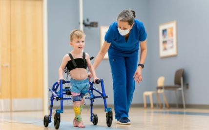
A child’s age, growth, development, fertility, education, and emotional well-being must be considered throughout treatment. Care often involves pediatric oncologists, pediatric surgeons, radiation oncologists, radiologists, pathologists, transplant physicians, genetic counsellors, and child-development specialists working closely with the child and family.
Types of Pediatric Cancer
Pediatric cancer is not a single disease. Each cancer has its own biological features, age pattern, staging system, and treatment pathway.
Leukemia begins in the blood-forming tissues, including the bone marrow. Abnormal blood cells multiply and interfere with the production of healthy red blood cells, white blood cells, and platelets. Acute lymphoblastic leukemia, or ALL, is the most common childhood cancer. Acute myeloid leukemia, or AML, is less common but generally progresses quickly. The cell type, genetic findings, organ involvement, and early response to treatment determine the treatment plan.
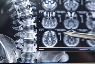
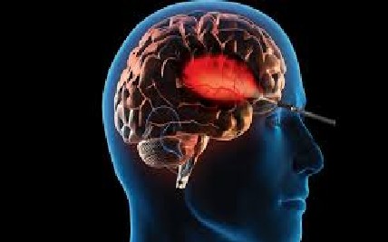
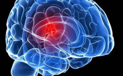
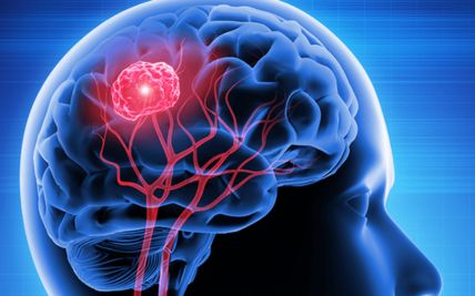
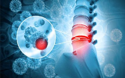
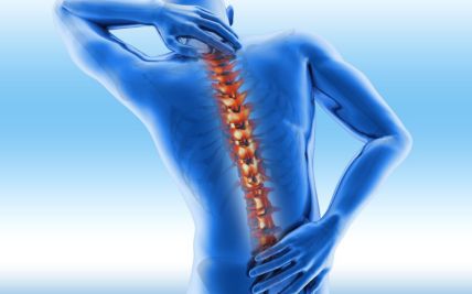
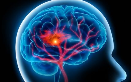
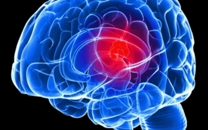
Brain and spinal tumours form a diverse group that includes low-grade gliomas, high-grade gliomas, medulloblastomas, ependymomas, germ-cell tumours, and other rare tumours. Symptoms and treatment depend on the tumour’s location, type, grade, and molecular features. Treatment must balance tumour control with the protection of neurological, cognitive, hormonal, visual, and developmental function.

Lymphoma develops from lymphocytes, which are part of the immune system. The two main groups are Hodgkin lymphoma and non-Hodgkin lymphoma. Pediatric non-Hodgkin lymphomas are often fast-growing but may respond well to appropriately planned treatment. The cancer subtype, stage, tumour burden, and organs involved determine therapy.
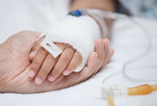
Neuroblastoma develops from immature nerve cells, most often in the adrenal glands or along nerve tissue beside the spine. It mainly affects infants and young children. Its behaviour varies widely. Some tumours may resolve or mature with limited treatment, while high-risk neuroblastoma can spread to the bone marrow, bones, lymph nodes, liver, or other organs and requires intensive combined treatment.
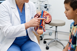
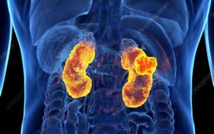
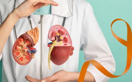
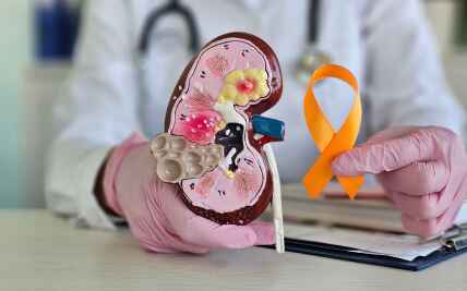
Wilms tumour, or nephroblastoma, is a kidney cancer that mainly affects young children. It may present as a painless abdominal swelling or be discovered during evaluation of abdominal symptoms. Treatment commonly combines surgery and chemotherapy, with radiation used in selected cases. The tumour’s stage, histological features, and involvement of one or both kidneys influence treatment.
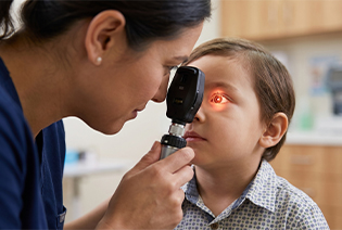
Retinoblastoma develops in the retina and primarily affects young children. A white reflection in the pupil, an abnormal appearance in photographs, or a new squint may be early signs. It may affect one or both eyes and can be hereditary. Treatment prioritises the child’s life, followed by preservation of the eye and useful vision wherever this can be achieved safely.
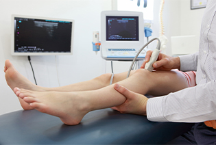
Osteosarcoma is a bone cancer that commonly develops near the knee or upper arm during periods of rapid growth. Persistent bone pain or swelling may be an early symptom. Treatment usually combines chemotherapy with surgery. Limb-sparing surgery is possible for many appropriately selected children, although the operation depends on the tumour’s location and involvement of nerves and blood vessels.
Ewing sarcoma can arise in bone or soft tissue and often affects children, adolescents, and young adults. It commonly develops in the pelvis, chest wall, or long bones. Treatment generally combines chemotherapy with surgery, radiation, or both. Molecular testing helps confirm the diagnosis.

Rhabdomyosarcoma is a soft-tissue sarcoma arising from cells related to skeletal-muscle development. It may occur in the head and neck, urinary or reproductive organs, limbs, or other sites. Treatment may include chemotherapy, surgery, and radiation. The tumour’s site, subtype, size, spread, and response determine the risk group and treatment approach.

Germ-cell tumours develop from cells that normally form eggs or sperm. They can occur in the ovaries or testicles or outside the reproductive organs, including the chest, abdomen, pelvis, brain, or lower spine. Some are non-cancerous, while others are malignant. Tumour-marker blood tests, imaging, pathology, age, and tumour location guide diagnosis and treatment.
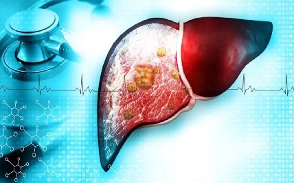
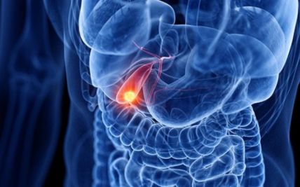
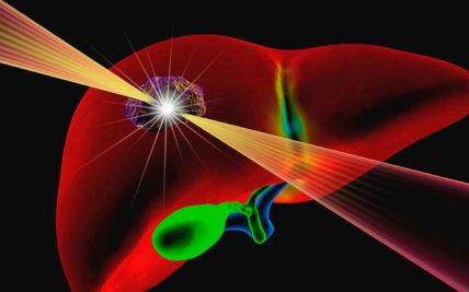
The main malignant liver tumours in children are hepatoblastoma and hepatocellular carcinoma. Hepatoblastoma mainly affects younger children, while hepatocellular carcinoma is more common in older children and adolescents. Treatment may include chemotherapy, liver surgery, or liver transplantation in carefully selected cases.
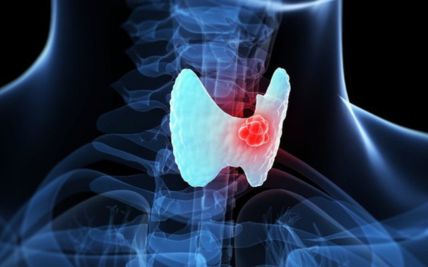
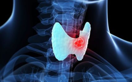
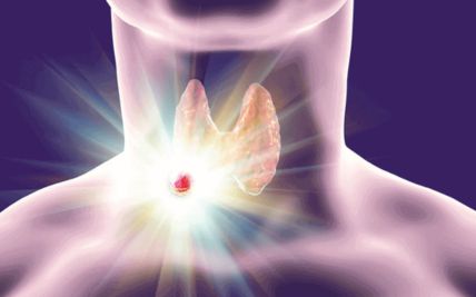
Children and adolescents may also develop thyroid cancer, melanoma, nasopharyngeal cancer, ovarian or testicular cancer, gastrointestinal tumours, and other rare cancers. These require age-appropriate treatment based on the specific cancer rather than a single pediatric cancer protocol.
The cause of most pediatric cancers is unknown. Unlike many adult cancers, childhood cancers are rarely linked to lifestyle or long-term environmental exposure.
Modifiable Risk Factors
There are no clearly established lifestyle measures that can prevent most pediatric cancers. Parents should not be made to feel that a child’s cancer resulted from food, everyday activities, minor illnesses, or routine household exposures.
Certain infections associated with selected cancers may be reduced through vaccination, infection prevention, and appropriate medical care. However, these infections account for only a small proportion of pediatric cancers.
Non-Modifiable Risk Factors
Inherited cancer-predisposition syndromes
Down syndrome, which increases the risk of particular leukemias
Li–Fraumeni syndrome
Neurofibromatosis type 1 or 2
Beckwith–Wiedemann syndrome
Constitutional mismatch-repair deficiency
Familial retinoblastoma caused by an inherited RB1 variant
Other inherited bone-marrow failure or tumour-predisposition conditions
Certain developmental or congenital conditions
A weakened immune system
Previous chemotherapy or radiation treatment
Previous organ or stem-cell transplantation
Certain infections associated with specific lymphomas or other cancers
Cancer-related genetic changes that develop within cells without being inherited
Most children diagnosed with cancer have no identifiable risk factor. Only a minority of pediatric cancers are directly caused by an inherited genetic variant.
Signs and Symptoms
The symptoms of pediatric cancer can resemble common childhood illnesses. The pattern, persistence, progression, and combination of symptoms are therefore important.
Possible signs and symptoms include:
Most children with these symptoms do not have cancer. However, symptoms that persist, recur, worsen, or do not fit the expected pattern of a common childhood illness should be medically evaluated.
Diagnosis begins with a detailed review of the child’s symptoms, growth, development, previous illnesses, family history, and physical findings. The pediatric specialist examines the area of concern and assesses the child’s general, neurological, developmental, and nutritional health. Imaging helps locate and assess solid tumours, while biopsy and laboratory testing establish the exact cancer type.
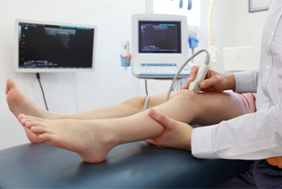
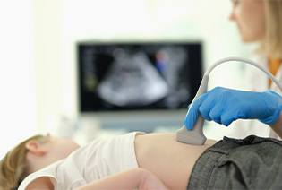
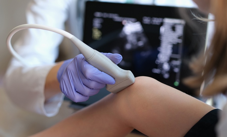
Ultrasound uses sound waves and does not involve ionising radiation. It is commonly used to assess abdominal, pelvic, kidney, liver, testicular, thyroid, and superficial soft-tissue masses. It may also guide a needle biopsy.
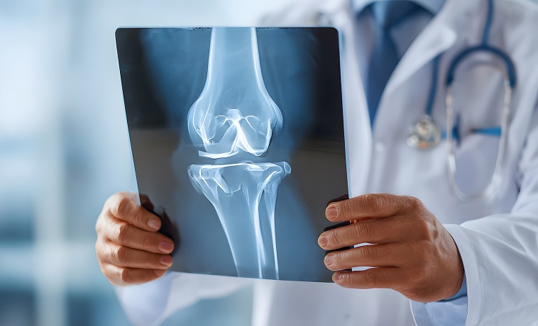
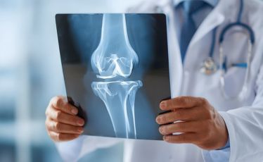
An X-ray may be the first test for persistent bone pain, swelling, breathing symptoms, or a suspected chest mass. It can show changes in bone, fractures, or abnormalities within the chest. Further imaging is usually required when a suspicious finding is identified.
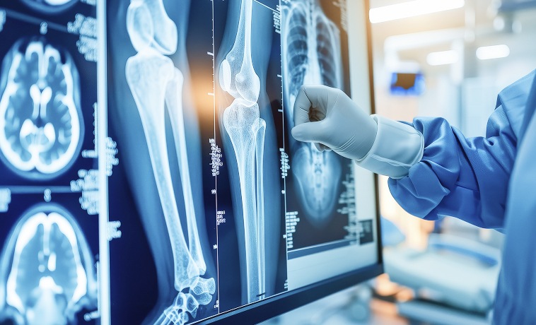
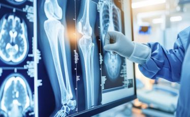
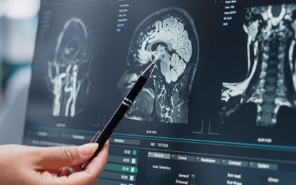
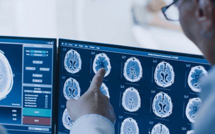
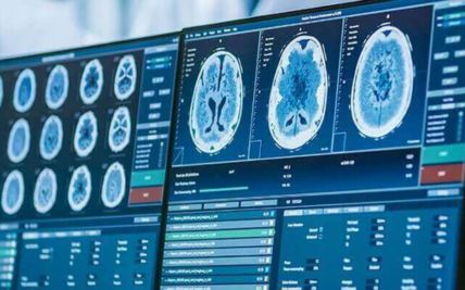
MRI uses magnetic fields and radio waves to produce detailed images without ionising radiation. It is commonly used for brain, spinal, bone, soft-tissue, abdominal, and pelvic tumours. MRI helps determine the tumour’s size and relationship to the brain, spinal cord, nerves, muscles, joints, and blood vessels. Sedation or anesthesia may be required when a young child cannot remain still.
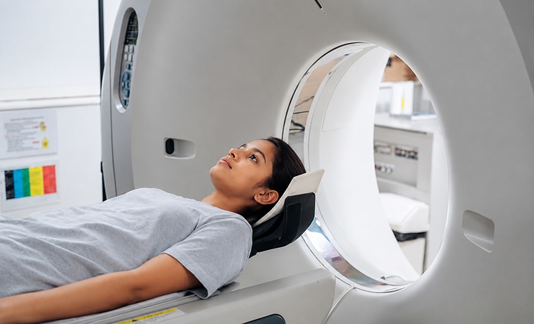
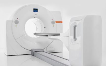
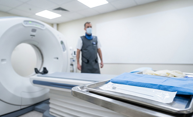
A CT scan provides rapid, detailed images of the chest, abdomen, pelvis, bones, and other structures. It may be used to assess tumour spread, lung involvement, bleeding, or an emergency complication. Pediatric CT protocols are designed to use the lowest radiation dose that can provide the required clinical information.


PET-CT combines metabolic and anatomical imaging. It may be used for staging and treatment-response assessment in selected lymphomas, sarcomas, and other cancers. It is not required for every pediatric cancer.
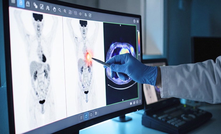
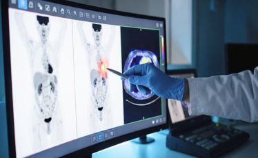
An MIBG scan uses a specialised radioactive tracer taken up by many neuroblastoma cells. It helps identify the primary tumour and disease in bones, bone marrow, or other areas. MIBG imaging may also help determine whether targeted radioactive MIBG treatment is an option in selected cases.
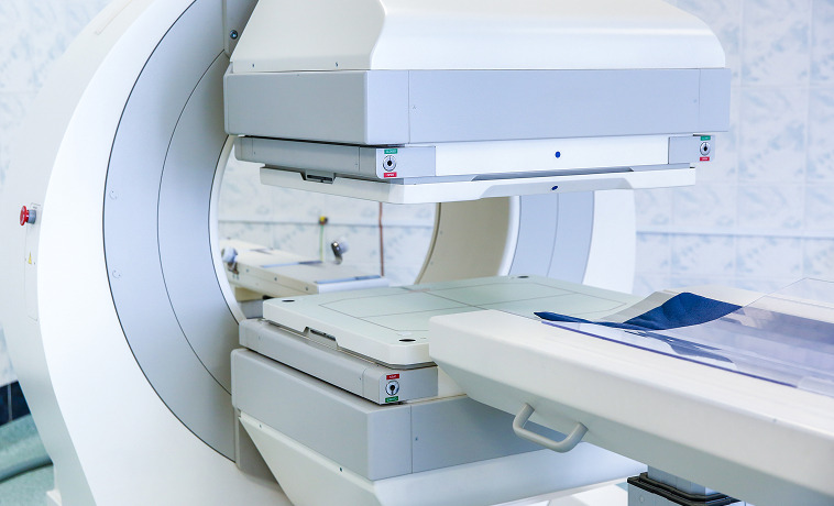
A bone scan may be used to assess whether a cancer has affected other bones. Its role depends on the cancer type, as PET-CT, MIBG, whole-body MRI, or other imaging may be preferred in some conditions.
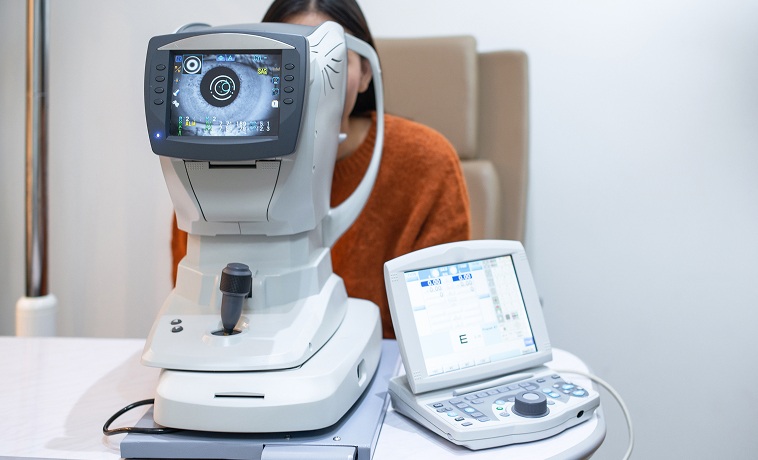
When retinoblastoma is suspected, a pediatric ophthalmologist examines the eyes, often under anesthesia. Retinal imaging, ocular ultrasound, and MRI may be used to assess the tumour and surrounding structures. A direct needle biopsy of suspected retinoblastoma is generally avoided.
A core needle collects small cylinders of tissue from a solid tumour, usually under ultrasound or CT guidance. It is commonly used for bone, soft-tissue, liver, kidney, lymph-node, and other masses when it can provide adequate tissue safely.
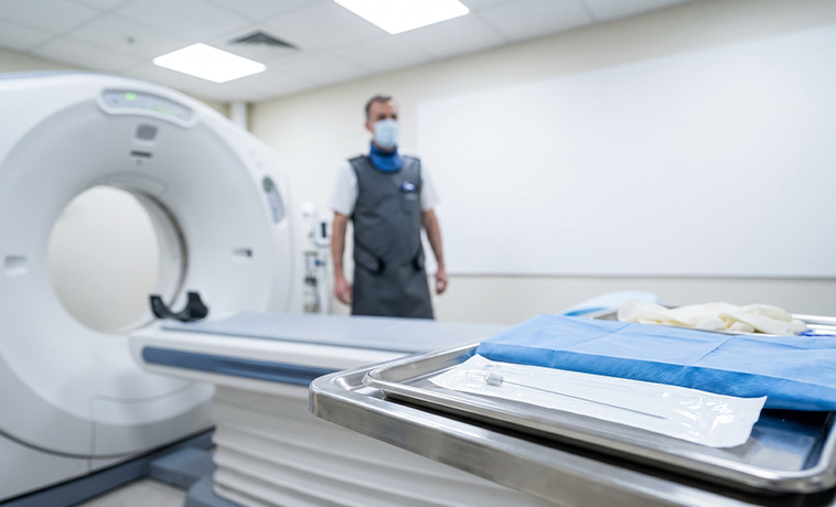
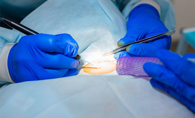
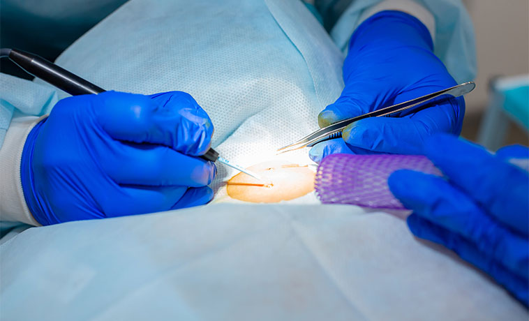
An incisional biopsy surgically removes part of a tumour. It may be performed when a needle biopsy is unsuitable or does not provide enough tissue. For suspected sarcoma, the biopsy path is carefully planned because it may need to be removed during later surgery.
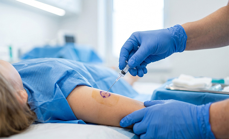
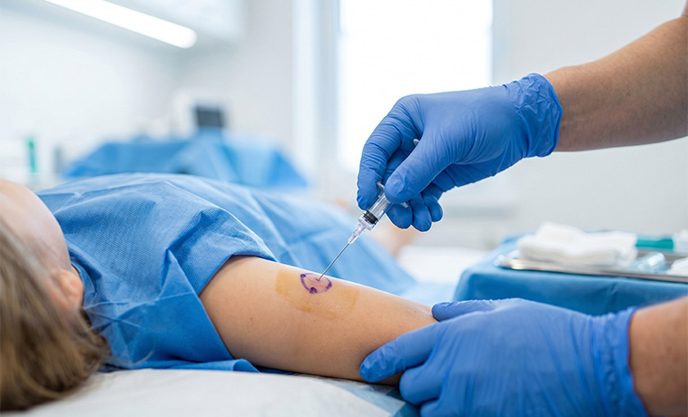
An excisional biopsy removes an entire small mass or lymph node. It may be preferred when lymphoma is suspected and the structure of the lymph node is required for accurate classification.
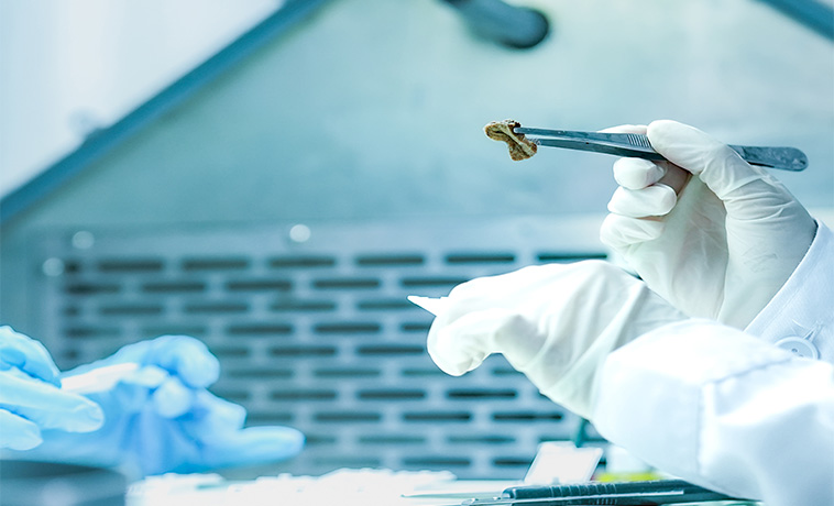
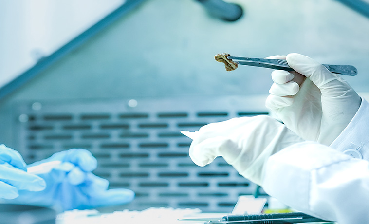
Bone marrow aspiration removes a small amount of liquid marrow, generally from the back of the pelvic bone. It is used to diagnose and monitor leukemia and to assess bone-marrow involvement in selected solid tumours and lymphomas.
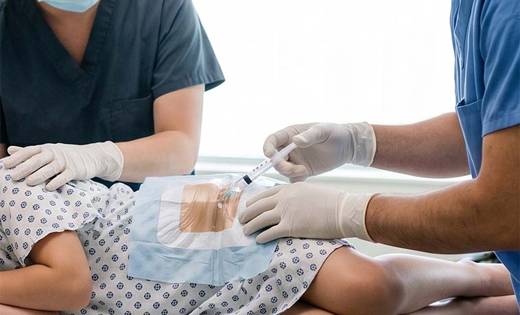
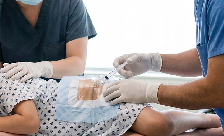
A lumbar puncture collects cerebrospinal fluid from the lower back. It may be used to check for leukemia, lymphoma, or a tumour that can spread through the fluid around the brain and spinal cord. Medicines may also be given through the same route in selected leukemias and lymphomas.
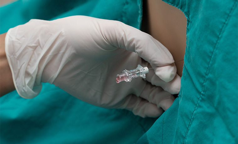
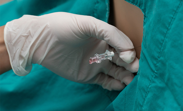
Some tumours are diagnosed during surgery intended to remove all or part of the mass. The surgeon may first obtain tissue for rapid assessment and then proceed according to the tumour’s location and the planned operation.
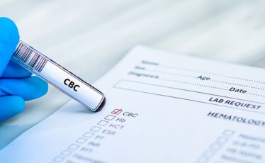
A complete blood count measures red blood cells, white blood cells, hemoglobin, and platelets. It can identify anemia, low platelets, abnormal white-cell levels, or other findings that require further investigation. A normal blood count does not exclude a solid tumour or every lymphoma.
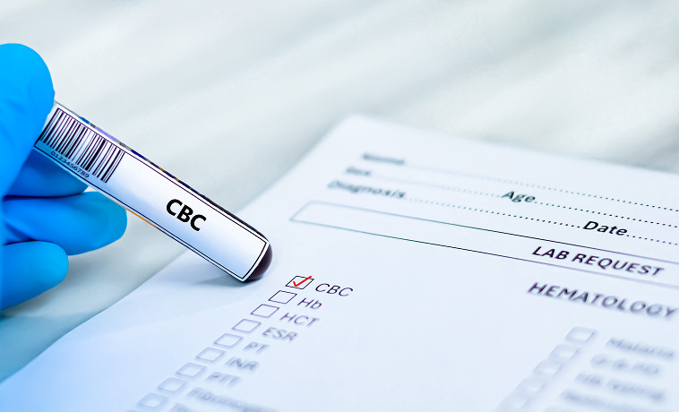
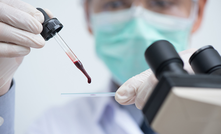
A blood smear allows cells to be examined under a microscope. It may show abnormal or immature cells suggesting leukemia or another bone-marrow disorder.
Flow cytometry identifies proteins on or inside cells. It is central to diagnosing and classifying many leukemias and lymphomas. It can also detect very small numbers of remaining leukemia cells after treatment.
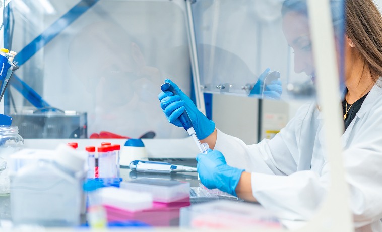
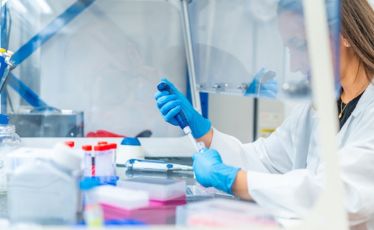
Blood tests assess kidney and liver function, electrolytes, calcium, uric acid, lactate dehydrogenase, and other markers. They help identify the effects of cancer and determine whether treatment can be given safely.


Selected pediatric cancers produce measurable substances in the blood or urine. These may include:
Alpha-fetoprotein in selected liver and germ-cell tumours
Beta-human chorionic gonadotropin in selected germ-cell tumours
Urinary catecholamine metabolites in neuroblastoma
Other tumour-specific markers where clinically relevant
Tumour markers support diagnosis and monitoring but do not confirm cancer on their own.


A pathologist examines biopsy or surgical tissue under a microscope to identify the tumour type, subtype, grade, and other important features. Specialist pediatric pathology review is important because childhood tumours can differ from adult cancers with a similar appearance.
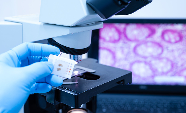
Immunohistochemistry uses specialised stains to identify proteins within cancer cells. It helps distinguish between leukemia, lymphoma, sarcoma, neuroblastoma, Wilms tumour, germ-cell tumours, and other pediatric cancers.
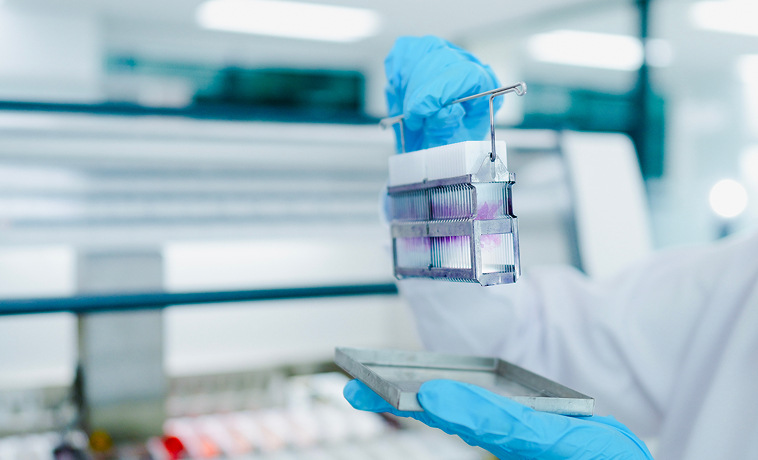

Testing for chromosomal and molecular changes helps confirm the diagnosis, define risk, select treatment, and monitor response. The relevant tests differ by cancer. Examples include molecular findings in ALL or AML, MYCN amplification in neuroblastoma, characteristic gene fusions in sarcoma, and molecular groups in pediatric brain tumours.

Genetic testing may be recommended when the child’s age, tumour type, physical features, or family history suggests an inherited cancer-predisposition syndrome. Genetic counselling helps families understand the results, treatment implications, and whether relatives may also require testing or surveillance.


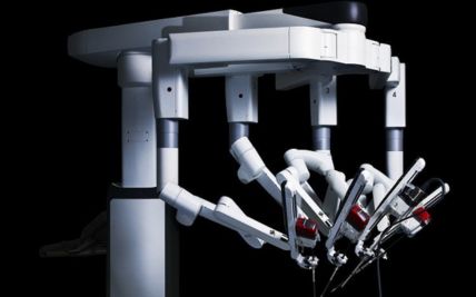
Surgery removes all or part of a solid tumour with an appropriate margin where required. It may be used for Wilms tumour, neuroblastoma, liver tumours, brain tumours, germ-cell tumours, and other solid cancers. The extent of surgery depends on the tumour’s location and relationship to nearby organs, nerves, and blood vessels.

Limb-sparing surgery removes a bone or soft-tissue sarcoma while preserving the affected arm or leg whenever this can provide adequate cancer clearance and a functional limb. Bone grafts, expandable implants, joint replacements, or soft-tissue reconstruction may be required as the child grows.
Neurosurgery may remove or reduce a brain or spinal tumour, relieve pressure, restore cerebrospinal fluid flow, and provide tissue for diagnosis. Image guidance, functional mapping, and neurophysiological monitoring may be used to protect important brain, spinal-cord, and nerve functions.
When clinically appropriate, part of the kidney may be preserved, particularly for selected children with tumours in both kidneys or an inherited predisposition. The decision balances cancer control with the need to maintain long-term kidney function.
Retinoblastoma may be treated using laser therapy, cryotherapy, chemotherapy delivered through an artery or into the eye, or other eye-preserving methods. Removal of the eye may be required when the tumour is extensive or the eye cannot be preserved safely.
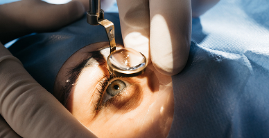
Surgery may remove selected metastatic deposits, such as lung nodules in osteosarcoma or other sarcomas, when this forms part of the cancer-specific treatment plan.

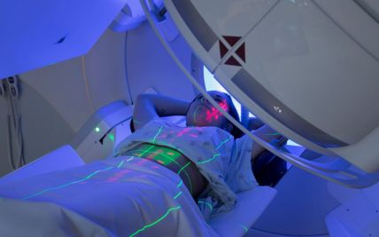
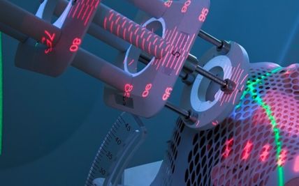
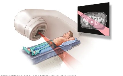
External beam radiation may be used after surgery, with chemotherapy, as the main local treatment, or to control recurrent or metastatic disease. Its use is carefully assessed because children’s growing tissues can be more sensitive to radiation and some effects may appear years later.
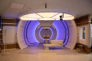
Three-dimensional conformal radiation shapes the treatment beams around the tumour using planning images.
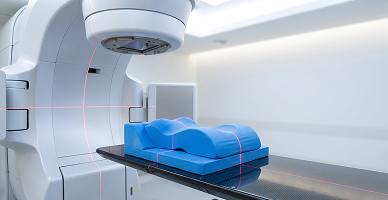
Intensity-modulated radiation therapy and volumetric-modulated arc therapy adjust the radiation dose across multiple beam angles. These techniques can help reduce exposure to selected nearby organs.
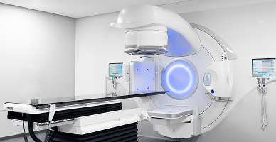
Image guidance confirms the child’s position and the target location during treatment. Accurate and reproducible positioning is particularly important when treating small children or tumours close to critical organs.
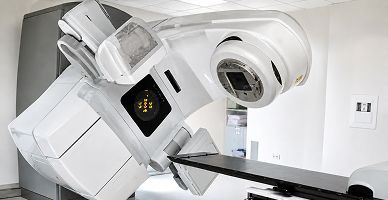
Stereotactic radiation delivers a focused dose to a small, well-defined target. It may be used in selected brain, spinal, bone, or metastatic tumours. It is not suitable for every childhood cancer or every child.
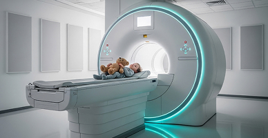
Craniospinal irradiation treats the brain and the length of the spinal fluid pathway. It is used for medulloblastoma and selected other tumours that can spread through cerebrospinal fluid. Treatment is adapted according to the child’s age, cancer type, molecular risk, and disease extent.
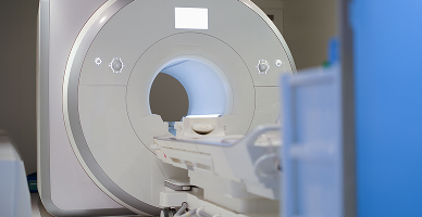
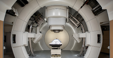
Proton therapy uses proton beams that release most of their radiation within the planned treatment area, with little radiation continuing beyond it. This can reduce the overall radiation dose received by some healthy tissues.
It may be considered for selected brain and spinal tumours, head and neck cancers, sarcomas, lymphomas, abdominal and pelvic tumours, and cancers requiring craniospinal irradiation. Its potential value is greatest when limiting radiation exposure to growing brain tissue, the heart, lungs, kidneys, bowel, endocrine organs, bones, or reproductive organs is clinically important.
Proton therapy does not eliminate radiation exposure or guarantee that long-term effects will not occur. It is not automatically the best option for every child. Suitability is determined through individual assessment and comparison with high-quality photon-radiation plans.
Chemotherapy is central to the treatment of many pediatric cancers, including leukemia, lymphoma, neuroblastoma, Wilms tumour, sarcoma, retinoblastoma, liver tumours, and selected brain tumours. It may be given before surgery, after surgery, with radiation, or as the main treatment. Doses and schedules are calculated according to the child’s diagnosis, body size, age, organ function, and treatment protocol.

Targeted medicines act on particular proteins or molecular changes within cancer cells. They may be used in selected leukemias, neuroblastomas, brain tumours, sarcomas, histiocytic disorders, and other cancers. The relevant target and evidence for treatment must be established before a targeted medicine is recommended.

Monoclonal antibodies recognise specific proteins on cancer cells. They may be used in selected leukemias, lymphomas, and neuroblastomas. Some work directly, while others recruit the immune system or deliver another treatment to the cancer cell.

Bispecific antibodies connect immune T cells with cancer cells, helping the immune system attack the cancer. They may be used for selected children with B-cell ALL according to the treatment setting and previous response.
CAR T-cell therapy collects the child’s T cells and modifies them to recognise a specific target on cancer cells. The cells are then returned after preparatory treatment. It may be used for selected relapsed or treatment-resistant B-cell ALL and other eligible cancers. Specialised monitoring is required for immune and neurological complications.

Immunotherapy may be used in selected pediatric leukemias, lymphomas, neuroblastomas, melanomas, and tumours with suitable biomarkers. Its role varies widely between cancers and continues to evolve.
Differentiation therapy encourages immature cancer cells to develop into more mature cells. It is central to the treatment of acute promyelocytic leukemia and may also be used in selected high-risk neuroblastomas.

High-dose chemotherapy followed by autologous stem-cell rescue may be used for selected high-risk neuroblastomas, lymphomas, brain tumours, or relapsed cancers. Allogeneic stem-cell transplantation may be recommended for selected high-risk or relapsed leukemias and other blood disorders. Suitability depends on disease risk, response, donor availability, organ function, and treatment history.
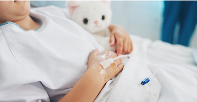
Pathology and molecular testing help define the cancer, estimate risk, select targeted treatment, and identify clinical-trial options. Precision oncology is used alongside established pediatric treatment protocols rather than as a replacement for them.
Prognosis for Pediatric Cancer
The outlook varies considerably between childhood cancers. Many can be treated successfully, but the likelihood of cure and long-term effects depends on the specific diagnosis rather than pediatric cancer as one group.
Factors influencing prognosis include:
Cancer type and subtype
Stage, grade, or risk group
Age at diagnosis
Tumour location and size
Molecular and genetic features
Spread to the bone marrow, brain, spinal fluid, or distant organs
Extent of safe surgical removal
Response to initial treatment
Minimal residual disease in leukemia
Whether the cancer has returned
Overall health and organ function
Ability to complete treatment and follow-up
ALL, Wilms tumour, retinoblastoma, Hodgkin lymphoma, and several other childhood cancers often have favourable outcomes with appropriate treatment. High-risk neuroblastoma, some brain tumours, metastatic sarcomas, and relapsed cancers may be more difficult to control.
Long-term follow-up is important because treatment can affect growth, learning, hormones, heart or lung health, fertility, hearing, vision, bones, kidneys, and the risk of later cancers.


Why Choose ACC for Pediatric Cancer Treatment
Dedicated pediatric oncology and hematology teams
Multidisciplinary care for leukemia, lymphoma, brain tumours, neuroblastoma, Wilms tumour, retinoblastoma, sarcomas, and other childhood cancers
Pediatric surgical, medical, radiation, and transplant expertise
Molecular pathology, flow cytometry, cytogenetics, and minimal residual disease testing
CAR T-cell therapy and stem-cell transplantation for eligible blood cancers
Organ-preserving and function-preserving surgery where oncologically appropriate
Specialist pediatric neuro-oncology, ocular oncology, sarcoma, and liver-tumour care
Advanced radiation techniques, including proton therapy for selected children
Radiation planning focused on reducing unnecessary exposure to growing tissues
Anesthesia and sedation support for imaging and radiation when required
Reconstruction planned around the child’s growth and future function
Genetic counselling for suspected inherited cancer risk
Age-appropriate, family-centred care supported by nutrition, psychological, educational, and child-development services
Long-term follow-up focused on growth, development, organ health, and survivorship
Health Blogs