
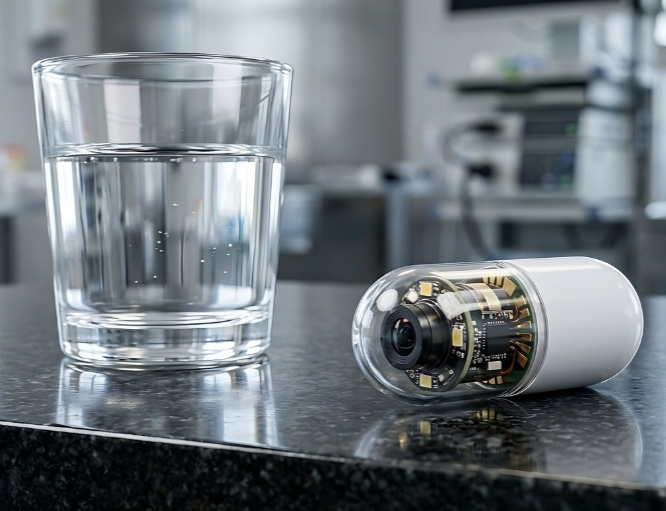
Capsule endoscopy is a non-invasive procedure that uses a swallowable, pill-sized camera to capture images from inside the digestive tract. It is primarily used to examine the small bowel, which can be difficult to assess completely with standard upper GI endoscopy or colonoscopy.
The capsule records images but cannot take a biopsy or provide treatment. If an abnormality is identified, another endoscopic, radiological or surgical procedure may be needed for tissue sampling or treatment.


Role in Cancer Care




What to Expect
Before the Test
You will receive instructions about diet, bowel preparation and when to stop eating and drinking. A laxative preparation may be prescribed to clear the bowel and improve the quality of the images.
Tell the clinical team about:
Difficulty swallowing or a history of choking
Previous bowel obstruction, narrowing or abdominal surgery
Crohn’s disease or other conditions that may narrow the bowel
All medicines and supplements you take, including iron tablets
Diabetes or medicines that may need adjustment while fasting
A pacemaker, defibrillator or another implanted electronic device
Pregnancy or the possibility of pregnancy
Do not stop or change prescribed medicines unless instructed. If narrowing or obstruction is suspected, imaging or a dissolvable patency capsule may be recommended first to check whether a capsule can pass safely through the bowel.

During the Test
Sensors will be attached to the abdomen or you will wear a sensor belt connected to a portable recorder. You will then swallow the capsule with water; sedation and anesthesia are not normally required.
You will receive instructions about when you may drink, eat and take medicines after swallowing the capsule. Avoid strenuous exercise, bending repeatedly or removing the equipment during the recording period. The examination usually records images for approximately 8–12 hours, depending on the capsule system and clinical protocol.
The capsule moves naturally through the digestive tract, and most people do not feel it. You do not usually need to remain at the healthcare facility throughout the recording period unless your clinical team advises otherwise.

After the Test
At the end of the recording period, the sensor belt or abdominal sensors and recorder are removed or returned as instructed. The disposable capsule normally passes naturally in a bowel movement, often without being noticed, and should not be retrieved unless specifically requested.
Do not undergo an MRI scan until the capsule has passed or your clinical team has confirmed that it is no longer inside your body. MRI exposure while a capsule remains in the digestive tract could cause injury.
Contact the clinical team promptly if the capsule has not been seen to pass within the period they specify. Seek urgent medical attention if you develop:
Increasing or severe abdominal pain
Persistent vomiting
Marked abdominal swelling
Inability to pass stool or gas
Fever or rapidly worsening symptoms
The recorded images require detailed specialist review, so results are not usually available immediately. Your clinical team will explain the findings and whether another investigation, biopsy or treatment is required.

Potential Benefits
- Cancer prevention: Precancerous polyps can be identified and removed.
- Early detection: Colorectal cancer may be detected before it causes symptoms.
- Direct visualisation: The lining of the rectum and colon can be examined in real time.
- Tissue diagnosis: Biopsies can be taken directly from suspicious areas.
- Polyp removal: Many polyps can be removed during the examination.
- Accurate localisation: The location of a tumour or lesion can be documented or marked for treatment.
- Diagnostic and therapeutic use: Bleeding, selected early lesions or obstruction may be treated endoscopically in suitable patients.
- Surveillance: The colon can be monitored after polyp removal or colorectal cancer treatment.