Eye Cancer
Eye cancer develops when abnormal cells grow in or around the eye. It may begin within the eyeball, on the surface of the eye, in the eyelid, or in tissues surrounding the eye. Cancer that begins in the eye is called primary eye cancer. Cancer that spreads to the eye from another part of the body is called secondary or metastatic eye cancer.
Eye cancer is uncommon and includes several distinct diseases. Uveal melanoma is the most common primary cancer inside the eye in adults, while retinoblastoma mainly affects young children. Other forms include conjunctival melanoma, intraocular lymphoma, and cancers of the eyelid or orbit.
Some eye cancers may not cause symptoms initially. Timely evaluation can help establish the diagnosis, guide treatment, preserve vision where possible, and reduce the risk of disease progression. Care often requires collaboration between ocular oncologists, medical oncologists, radiation oncologists, paediatric oncologists, pathologists, radiologists, and reconstructive specialists.
Types of Eye Cancer
Eye cancer is not a single disease. The site where the cancer begins and the type of cell involved influence its behaviour, treatment, and outlook.
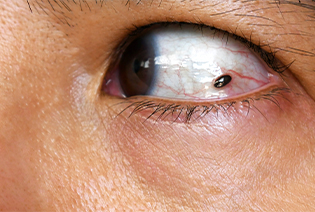
Uveal melanoma begins in the uvea, the middle layer of the eye. The uvea includes the iris, ciliary body, and choroid. Most uveal melanomas develop in the choroid.
Some tumours are found during a routine eye examination, while others cause blurred vision, flashes, floaters, or a dark area in the field of vision. Treatment depends on the tumour’s size and location, its effect on vision, and whether it has spread.
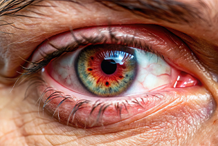
Conjunctival melanoma begins in the conjunctiva, the thin membrane covering the white part of the eye and the inner eyelids. It may appear as a new or changing pigmented patch on the eye, although some tumours contain little or no pigment.
Treatment commonly involves surgical removal with additional local treatment to reduce the risk of recurrence. Long-term follow-up is important because the cancer may return or spread.

Retinoblastoma develops in the retina and mainly affects children younger than five years. A white reflection in the pupil, particularly in photographs, is a common warning sign. Squinting or misalignment of the eyes may also occur.
The disease can affect one or both eyes and may be hereditary. Treatment is planned to cure the cancer while preserving the eye and useful vision whenever this can be achieved safely.
Intraocular lymphoma is a rare lymphoma involving structures inside the eye. It is often associated with lymphoma of the brain or central nervous system.
Symptoms may resemble inflammation and include blurred vision, floaters, or reduced vision. Diagnosis can require examination of fluid or tissue from the eye, along with imaging and assessment of the brain and other parts of the body.
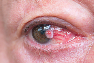
This cancer develops from the surface cells of the conjunctiva and may extend onto the cornea. It can appear as a raised, thickened, red, or irregular growth on the surface of the eye.
Treatment may include surgical removal, cryotherapy, or medicines applied directly to the eye. More extensive disease may require additional treatment.
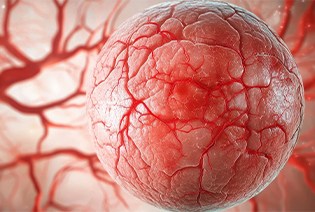
Cancer can spread to the eye from another part of the body. The breast and lung are among the more common primary sites, although other cancers can also spread to the eye.
Treatment depends on the original cancer, the extent of disease, eye symptoms, and the effect on vision. It is planned together with treatment for the primary cancer.
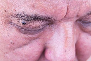
Cancers can arise in the eyelid or in the orbit, the bony cavity containing the eye and its surrounding tissues. These include skin cancers, lacrimal gland tumours, sarcomas, lymphomas, and cancers spreading from nearby areas.
Treatment may involve surgery, radiation therapy, medicines, or a combination of treatments. Reconstruction may be required to protect the eye and restore eyelid or facial function.
Most people with eye cancer do not have an identifiable preventable cause. Risk factors also differ considerably between the different types of eye cancer.
Modifiable Risk Factors
Ultraviolet exposure: Long-term exposure to ultraviolet radiation may increase the risk of some eyelid and conjunctival cancers. Its relationship with uveal melanoma is less certain.
Inadequate sun protection: Wearing UV-protective sunglasses and a broad-brimmed hat may help protect the eyelids and surface of the eyes.
Immunosuppression-related risk: People taking immune-suppressing medicines should follow the examination and cancer-surveillance plan recommended by their treating doctor.
Non-Modifiable Risk Factors
Increasing age, particularly for uveal melanoma and many eyelid cancers
Fair skin, light-coloured eyes, or a tendency to burn in the sun
Certain unusual or changing moles within or on the surface of the eye
Inherited variants involving genes such as RB1 or BAP1
A family history of retinoblastoma or BAP1-associated cancers
Certain congenital or genetic conditions
Previous cancer elsewhere in the body
Previous radiation exposure involving the eye or surrounding region
Reduced immune function due to an illness or medical treatment
Having a risk factor does not mean that eye cancer will develop. People with an inherited predisposition or a suspicious eye lesion may require an individualised surveillance plan.
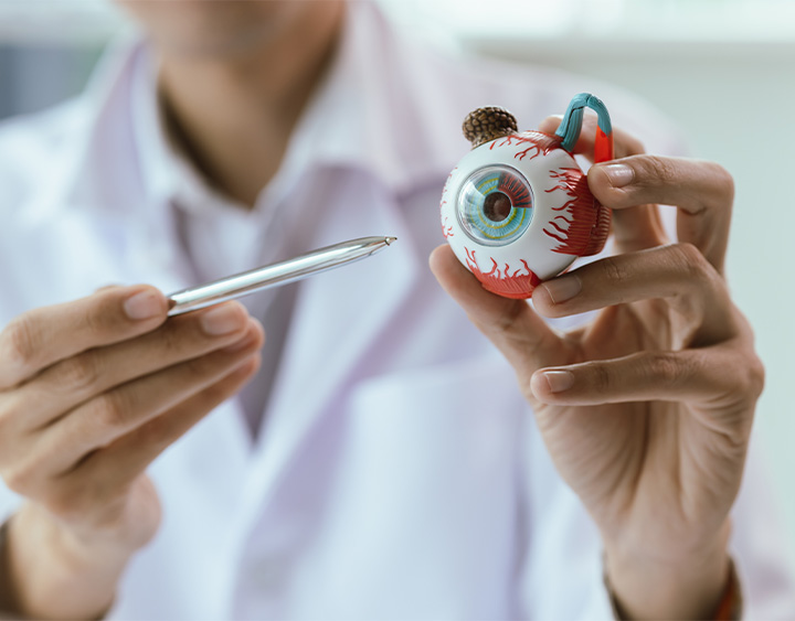
Signs and Symptoms
The symptoms of eye cancer depend on the tumour’s type, size, and location. Some tumours are discovered during an eye examination before symptoms develop.
Possible signs and symptoms include:
These symptoms can also result from non-cancerous eye conditions. However, a new or persistent change should be assessed by an eye specialist. A white pupil, sudden loss of vision, or a rapidly enlarging eye or orbital swelling requires prompt medical attention.
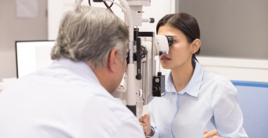
Diagnosis usually begins with a detailed eye examination. The ophthalmologist assesses vision, eye movements, visual fields, and the external and internal structures of the eye. Imaging is then used to determine the tumour’s location, size, and relationship to nearby structures. A biopsy may be needed to confirm the diagnosis, although some tumours can be diagnosed without one.
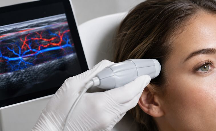
Ocular ultrasound uses sound waves to examine structures inside the eye. It can measure a tumour, assess its shape and internal characteristics, and identify involvement of nearby tissues.
A-scan ultrasound provides information about the tumour’s internal reflectivity, while B-scan ultrasound creates a two-dimensional image of the eye and tumour.
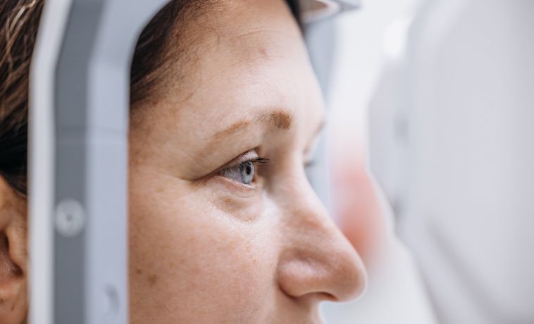
Optical coherence tomography, or OCT, uses light waves to produce detailed cross-sectional images of the retina and nearby structures. It can show retinal changes, fluid beneath or within the retina, and the effect of a tumour on the macula.
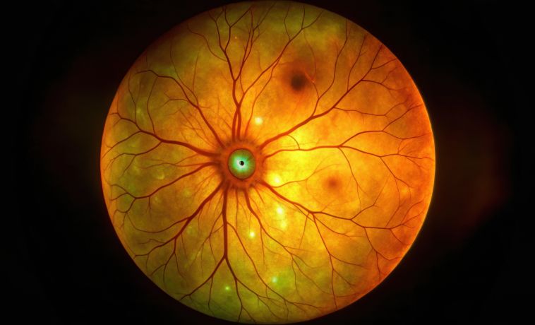
Colour fundus photography records the appearance and position of a tumour inside the eye. These images provide a baseline for comparing changes during observation, treatment, and follow-up. Fundus autofluorescence may provide additional information about the retina and the activity of certain lesions.
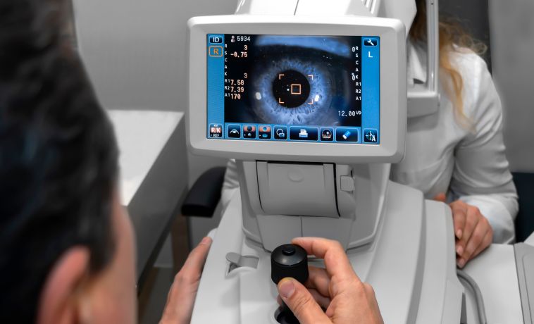
Fluorescein or indocyanine green angiography involves injecting a dye into a vein and photographing its circulation through blood vessels in the eye. It may help evaluate a tumour’s blood supply and distinguish it from other eye conditions.
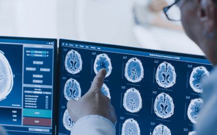
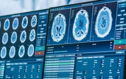
MRI uses magnetic fields and radio waves to produce detailed images of the eye, orbit, optic nerve, and brain. It can help determine the extent of the tumour and whether nearby structures are involved.
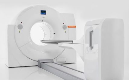
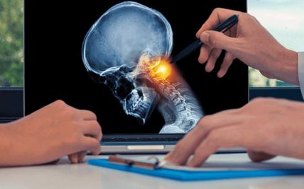
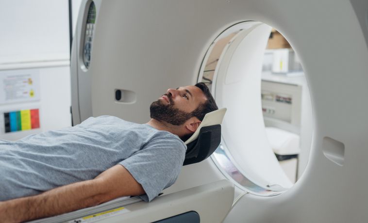
A CT scan uses X-rays to produce detailed images of the eye, orbit, and surrounding bones. It may be used to detect calcification in suspected retinoblastoma or assess cancers involving the orbit or bone.
A thin needle is passed into the tumour to collect cells. The procedure may be performed under ultrasound or direct visual guidance and is used for selected intraocular, orbital, or surface tumours.
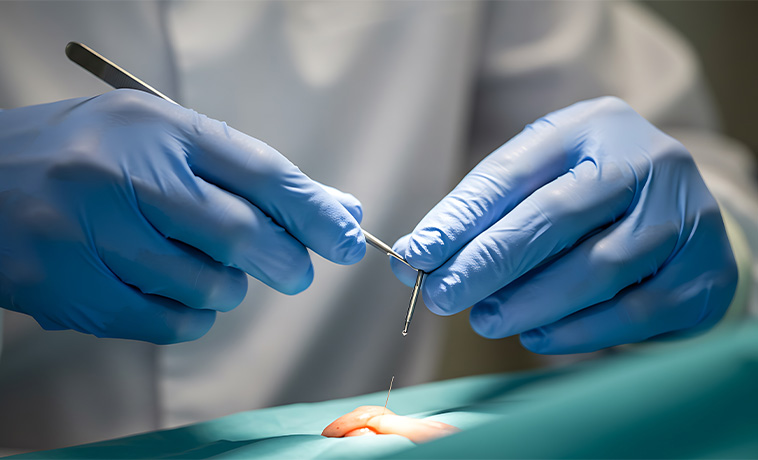
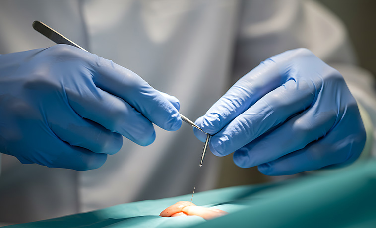
A small portion of the tumour is surgically removed for examination. It may be used for larger conjunctival, eyelid, or orbital tumours when removing the entire lesion during the initial procedure would not be appropriate.
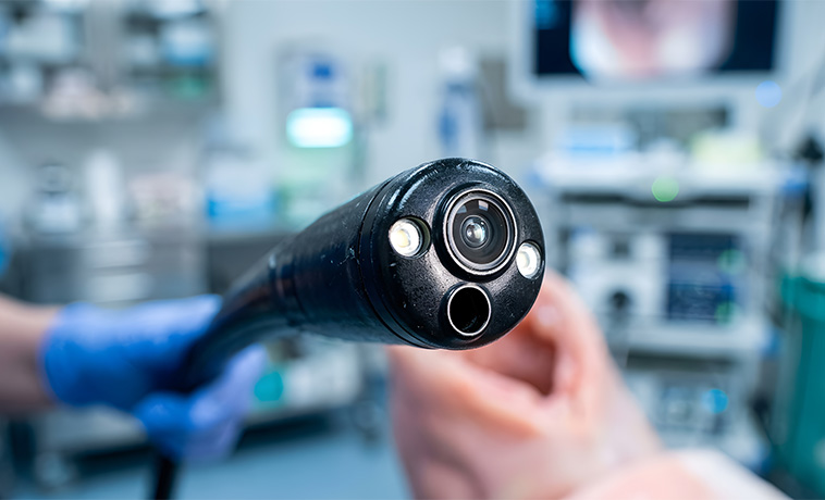
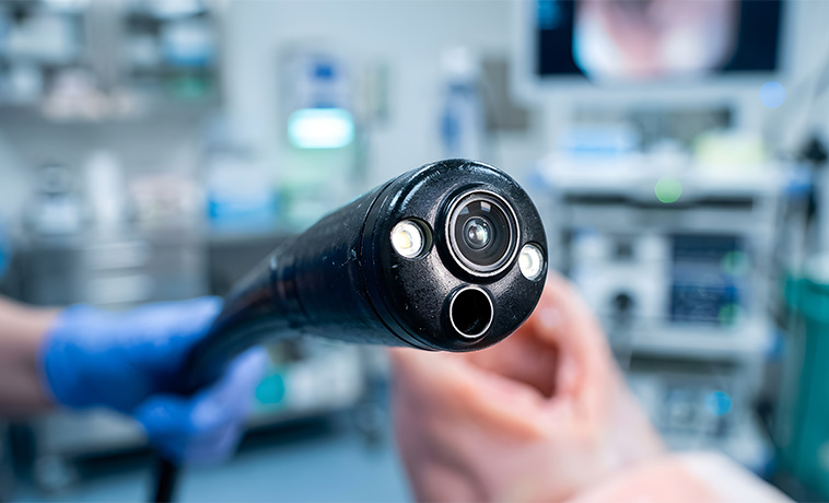
The entire lesion is removed and sent for examination. This may be appropriate for selected small tumours on the conjunctiva, eyelid, or another accessible area when complete removal can be performed safely.
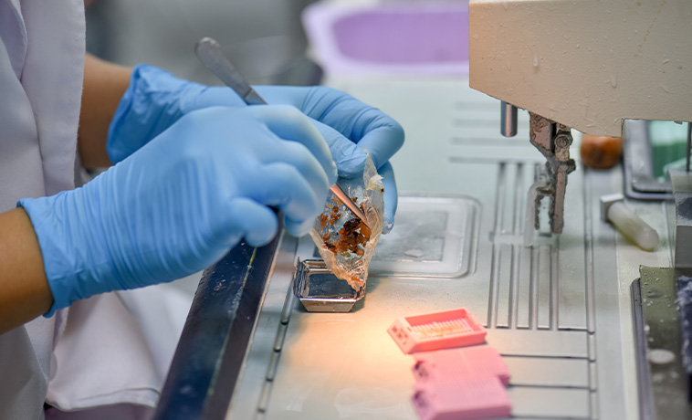
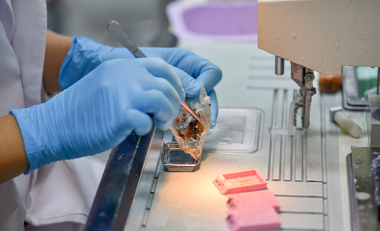
A small amount of fluid is collected from inside the eye. This is most commonly performed when intraocular lymphoma or another condition affecting the eye fluids is suspected.
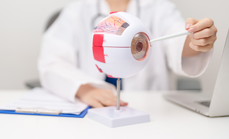
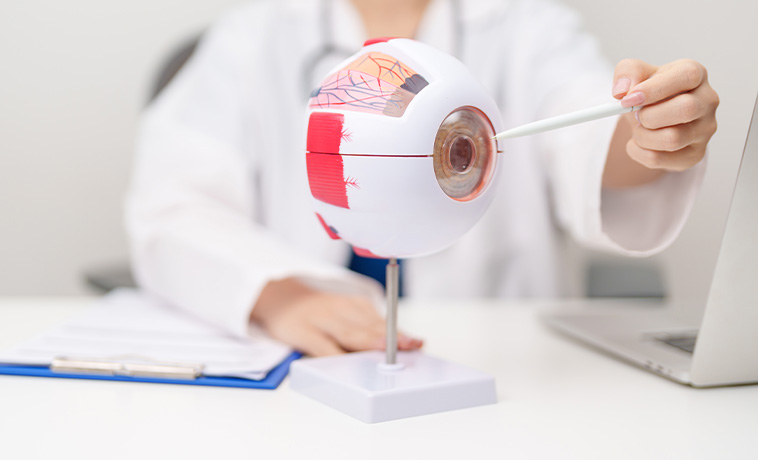
A specialist pathologist examines biopsy or surgical tissue to identify the cancer type. Immunohistochemistry and molecular tests may be used to clarify the diagnosis or identify treatment-related changes.
Genetic testing may be recommended for children with retinoblastoma and for selected people with suspected hereditary cancer syndromes. Molecular testing of uveal melanoma may provide information about the risk of the disease spreading, but it does not replace clinical staging.


Eye cancer stages describe how large a tumor is and how far it has spread using the TNM system (Tumor, Node, and Metastasis), ranging from Stage I (early) to Stage IV (advanced). Doctors group these factors to plan the best care.
Stage I: The tumor is small and stays in its original spot, such as the iris or choroid, without growing into other tissues or causing high pressure (glaucoma).
Stage II: The tumor is larger or has started to grow into nearby parts of the eye, like the ciliary body.
Stage III: The tumor is larger still, may have grown outside the eyeball, or causes issues like increased eye pressure.
Stage IV: The cancer has spread to distant parts of the body, most commonly the liver, or to far-away lymph nodes.
Small tumours on the conjunctiva, eyelid, iris, or other accessible areas may be removed with a margin of surrounding tissue. Cryotherapy or a topical medicine may be added to treat microscopic cells around the surgical area.
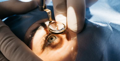
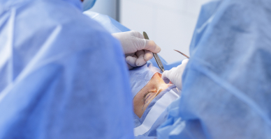
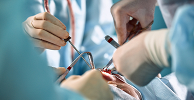
Selected iris, ciliary body, conjunctival, eyelid, or orbital tumours may be removed without removing the eye. The procedure is planned according to the tumour’s location and its relationship to structures needed for vision.
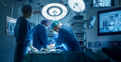
Enucleation removes the eyeball while preserving the eyelids and surrounding orbital tissues. It may be recommended for a large tumour, an eye with little visual potential, a painful eye, or disease that cannot be controlled safely with an eye-preserving treatment.
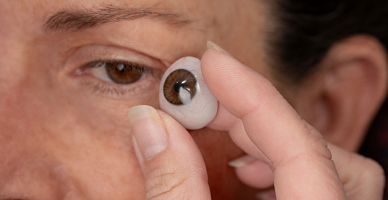
An orbital implant is generally placed during surgery. An artificial eye can later be fitted over the healed area to provide a natural appearance.
Cancers involving the eyelid, lacrimal gland, or orbit may require removal of the tumour and nearby affected tissue. Reconstructive surgery may be performed to protect the eye and restore eyelid closure, facial contour, and function.
More extensive removal of orbital contents is reserved for selected locally advanced cancers.
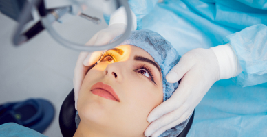
Laser treatment and cryotherapy may be used for small retinoblastomas and selected surface tumours. These treatments may be repeated and are sometimes combined with chemotherapy or radiation therapy.
Plaque brachytherapy is a common eye-preserving treatment for uveal melanoma. A small radioactive disc is temporarily attached to the outside wall of the eye over the tumour.
The plaque delivers radiation directly to the tumour over several days while limiting exposure to other areas. A second procedure is performed to remove it. Vision after treatment depends partly on the tumour’s size and its distance from the macula and optic nerve.
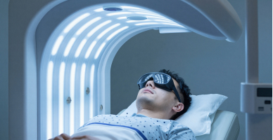
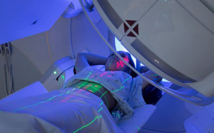
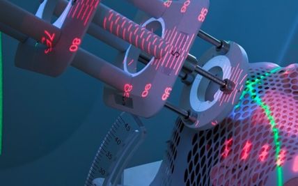
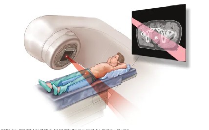
External beam radiation delivers radiation from a machine outside the body. It may be used for lymphoma, orbital or eyelid cancers, retinoblastoma in selected circumstances, and tumours that cannot be treated adequately with surgery or plaque therapy.
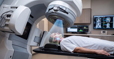
3D conformal radiation therapy, intensity-modulated radiation therapy, and image guidance help shape and position the radiation dose around the tumour. These techniques may reduce exposure to some nearby structures, although the benefit depends on the tumour’s location.
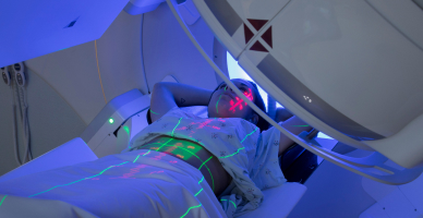
Proton therapy uses beams of protons that release most of their radiation within the planned treatment area, with little radiation continuing beyond it. This can help shape the dose around an eye or orbital tumour and limit radiation exposure to some nearby tissues.
It may be considered for selected uveal melanomas, tumours near sensitive structures, certain orbital cancers, or paediatric cases where reducing unnecessary radiation exposure is particularly important. Proton therapy cannot always protect the optic nerve or macula when these structures are very close to or involved by the tumour.
Suitability is determined after individual assessment and comparison with plaque therapy, photon radiation, surgery, and other appropriate treatments. Proton therapy is not required for every person with eye cancer.
Chemotherapy has an important role in retinoblastoma, intraocular lymphoma, and selected advanced or metastatic cancers.
For retinoblastoma, chemotherapy may be delivered:
Through a vein to treat one or both eyes
Through an artery leading directly to the eye
Directly into the vitreous cavity for selected tumour deposits
The route is chosen according to the extent of the disease, the child’s age, and the treatment plan.
Targeted medicines act on particular proteins or genetic changes in cancer cells. They may be considered for selected conjunctival melanomas, lymphomas, or advanced cancers when appropriate molecular changes are identified.
Their role in primary uveal melanoma is more limited and depends on the stage and individual clinical circumstances.
Immunotherapy helps the immune system recognise or attack cancer cells. It may be used for selected people with advanced melanoma or another responsive cancer type.
Uveal melanoma behaves differently from melanoma of the skin, and not all commonly used melanoma immunotherapies produce the same results. Treatment therefore requires disease-specific assessment.
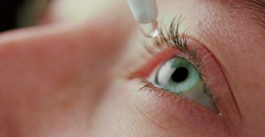
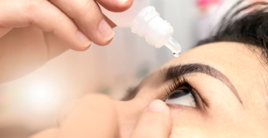
Medicines such as mitomycin C or 5-fluorouracil may be applied directly to the eye for selected precancerous or cancerous surface lesions. The medicine, concentration, and treatment schedule depend on the diagnosis and should be supervised by an experienced eye specialist.
Pathology and molecular testing can help confirm the diagnosis, assess inherited risk, estimate the likelihood of spread, and identify treatment options for selected patients. Not every eye cancer requires extensive genomic profiling.
Eyelid reconstruction may be required when cancer surgery removes part or all of an eyelid. Tissue from the remaining eyelid, nearby skin, or another part of the body may be used to restore eyelid closure, protect the eye’s surface, and improve appearance.
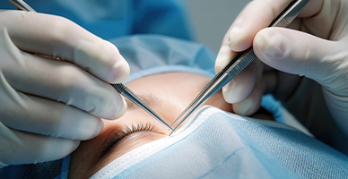
Orbital reconstruction may be performed after removing a tumour involving the tissues or bones around the eye. Implants, bone grafts, plates, or soft-tissue flaps may be used to restore the shape of the orbit, support the remaining structures, and maintain facial symmetry.
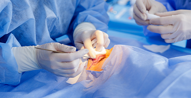
When the eye is removed through enucleation, an orbital implant is usually placed within the eye socket to replace lost volume. The eye muscles may be attached to the implant to help it move in coordination with the other eye.
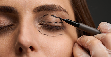
After the socket has healed, a customised artificial eye, or ocular prosthesis, can be fitted over the orbital implant. It is shaped and coloured to resemble the other eye and can be removed for cleaning and periodic adjustment.
More extensive cancers may require removal of tissue from the eyelid, orbit, or surrounding face. Reconstruction may involve skin grafts, local tissue flaps, or tissue transferred from another part of the body to close the surgical area, restore facial contour, and improve function and appearance.
Prognosis for Eye Cancer
The outlook for eye cancer varies significantly according to the cancer type. Many localised tumours can be controlled effectively, but the ability to preserve vision depends on the tumour’s size, location, and response to treatment.
Factors that influence prognosis include:
The cancer type and subtype
Tumour size and location
Stage at diagnosis
Involvement of the optic nerve, orbit, lymph nodes, or nearby structures
Spread to another part of the body
Molecular or genetic features
Response to treatment
Vision and eye function at diagnosis
Overall health and ability to complete treatment and follow-up
Uveal melanoma most commonly spreads through the bloodstream, particularly to the liver, so long-term systemic surveillance may be recommended. Retinoblastoma has a favourable outlook when treated while confined to the eye, although hereditary disease requires lifelong attention to the risk of additional cancers. Regular follow-up is important even after successful local treatment because some eye cancers can recur or spread several years later.


Screening for Eye Cancer
There is no routine population screening test specifically recommended for eye cancer. However, regular eye care and prompt evaluation of new symptoms can help identify suspicious changes.
Eye Examinations
An eye examination may detect a tumour that has not yet caused symptoms. The appropriate examination schedule depends on age, visual needs, general health, and individual risk rather than a single interval for everyone.
Childhood Eye Checks
Routine newborn and childhood examinations include assessment of the red reflex and the appearance and alignment of the eyes. A white pupil, persistent squint, abnormal red reflex, or unexplained visual difficulty should be evaluated promptly.
High-Risk Surveillance
More frequent specialist examinations may be recommended for:
Children with an inherited RB1 variant or a family history of retinoblastoma
People with a BAP1 tumour-predisposition syndrome
People with certain suspicious or changing ocular naevi
People previously treated for eye cancer
People with medical conditions or treatments that increase the risk of particular eye cancers
Why Choose ACC for Eye Cancer Treatment
Multidisciplinary care involving ocular oncology, medical oncology, radiation oncology, pediatric oncology, radiology, pathology, and reconstructive surgery
Specialised eye examination and ocular imaging for diagnosis and treatment planning
Eye-preserving treatment options for appropriately selected patients
Expertise in surgery, chemotherapy, targeted treatment, immunotherapy, and radiation therapy
Access to proton therapy for selected eye and orbital tumours
Genetic assessment and counselling for hereditary cancers such as retinoblastoma
Eyelid and orbital reconstruction, ocular prosthetic support, and visual rehabilitation
Coordinated monitoring for local recurrence and cancer spread
Age-appropriate care and family support for children with retinoblastoma
Individualised treatment planning focused on cancer control, vision, function, and quality of life

Health Blogs