Bone & Soft Tissue Cancer
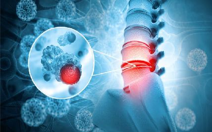
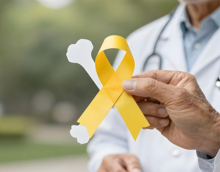
Bone and soft tissue cancers, known as sarcomas, are rare cancers that develop in the body’s connective and supporting tissues. Bone sarcomas begin in bone-forming or cartilage-forming cells, while soft tissue sarcomas may arise in muscle, fat, blood vessels, nerves, tendons, or other connective tissues.
Sarcomas can occur almost anywhere in the body and include many distinct subtypes that differ in their behaviour and response to treatment. Cancer that begins elsewhere and spreads to bone is called bone metastasis and is different from a primary bone cancer.
At Apollo Cancer Centres, sarcoma care is planned by a multidisciplinary team that may include surgical, medical and radiation oncologists, orthopedicians, radiologists, pathologists, reconstructive surgeons, and rehabilitation specialists. Accurate diagnosis and carefully planned biopsy are central to developing an appropriate, function-preserving treatment plan.
Types of Bone & Soft Tissue Cancer
Sarcomas are classified according to the tissue or cell in which they begin. Identifying the exact subtype and grade is essential because treatment can vary considerably between different sarcomas.
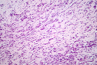
Osteosarcoma develops in bone-forming cells and is the most common primary bone cancer in children, adolescents, and young adults. It frequently affects the long bones around the knee or upper arm, although it can occur in any bone. Treatment commonly combines chemotherapy with surgery.
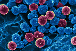
Ewing sarcoma is a fast-growing cancer that usually begins in bone but can also arise in soft tissue. It most often affects children, adolescents, and young adults and commonly develops in the pelvis, ribs, chest wall, or long bones. Treatment generally involves chemotherapy together with surgery, radiation therapy, or both.
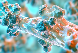
Chondrosarcoma develops in cartilage-forming cells and occurs mainly in adults. It commonly affects the pelvis, upper thigh, shoulder, or ribs. Its behaviour varies by subtype and grade; surgery is the main treatment for most localised cases because conventional chemotherapy and radiation are less effective for many common chondrosarcomas.
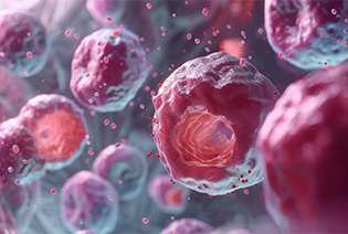
Liposarcoma develops from fat-forming cells, most often in the thigh or deep within the abdomen. It is not caused by body fat or obesity. Different subtypes can behave differently, ranging from slow-growing tumours to cancers with a greater risk of spreading.
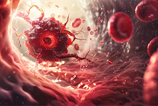
Leiomyosarcoma begins in smooth muscle, which is found in organs such as the uterus, stomach, intestines, bladder, and blood vessels. It is more common in adults and may develop in the abdomen, uterus, limbs, or major blood vessels. Treatment depends on its location, size, and whether it has spread.
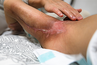
Rhabdomyosarcoma develops from cells that normally form skeletal muscle. It occurs mainly in children and adolescents but can also affect adults. Common sites include the head and neck, urinary and reproductive organs, and limbs. Treatment usually combines chemotherapy with surgery and/or radiation therapy.
Most people who develop sarcoma have no identifiable risk factor, and these cancers are not generally linked to everyday injuries or lifestyle habits.
Known risk factors may include:
Previous radiation therapy, particularly to the area where the sarcoma develops
Certain inherited conditions, including Li-Fraumeni syndrome, hereditary retinoblastoma, and neurofibromatosis type 1
Paget disease of bone and selected inherited bone conditions
Certain previous cancer treatments, depending on the treatment received
Long-standing lymphoedema, which is associated with a rare type of soft tissue sarcoma
Increasing age for some sarcomas, while osteosarcoma, Ewing sarcoma, and rhabdomyosarcoma occur more often in younger people
Having a risk factor does not mean that sarcoma will develop. In most cases, there is no known way to prevent it.
Signs and Symptoms
Sarcomas may not cause obvious symptoms when they are small. Symptoms depend on where the tumour develops and whether it affects nearby bones, muscles, nerves, blood vessels, or organs.
Possible signs and symptoms include:
Most lumps and musculoskeletal pain are not caused by cancer. However, a growing lump, unexplained swelling, persistent bone pain, or an unexpected fracture should be assessed promptly.
Diagnosis begins with a review of symptoms and medical history, followed by an examination of the affected area. Imaging helps define the tumour, but a biopsy is required to confirm the diagnosis and identify the exact sarcoma subtype.
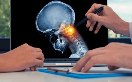
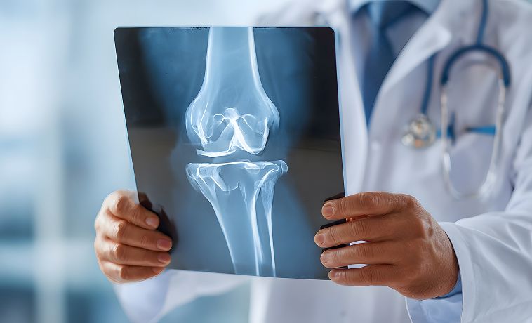
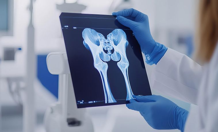
An X-ray is usually the first test when a primary bone tumour is suspected. It can show whether an area of bone has been damaged, weakened, thickened, or replaced by abnormal tissue.
Certain patterns may suggest that a bone lesion requires further investigation, but an X-ray cannot confirm whether it is cancerous. If the findings are concerning or symptoms persist, MRI and other tests are usually recommended.
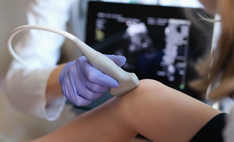
Ultrasound uses sound waves to examine a lump within the soft tissues. It can help determine whether the lump is solid or fluid-filled and assess its size, depth, and blood flow.
Ultrasound is often used as an initial test for a superficial soft tissue lump. It can also guide a needle accurately into the tumour during biopsy. Deeper or suspicious abnormalities generally require MRI for further assessment.
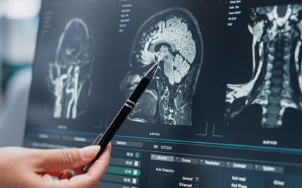
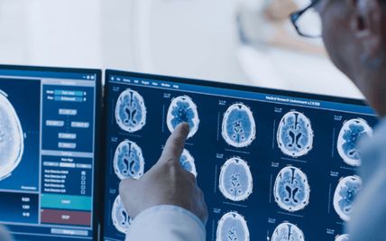
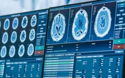
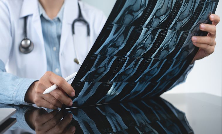
MRI provides detailed images of bone marrow and soft tissues without using X-rays. It is usually the most important test for defining the local extent of a suspected bone or soft tissue sarcoma.
MRI helps determine the tumour’s size, depth, and relationship to nearby muscles, joints, nerves, blood vessels, and other structures. This information is essential for planning the biopsy, surgery, and possible reconstruction. The scan usually examines the entire affected area so that the treatment team can assess the tumour fully.
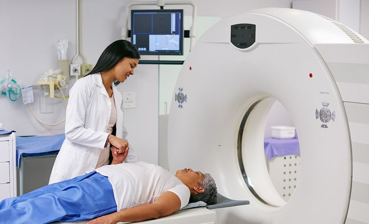
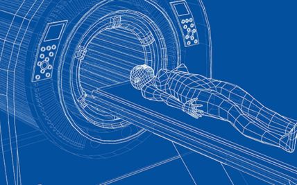
A CT scan uses X-rays and computer processing to create detailed cross-sectional images. It may provide clearer information about bone destruction, calcification within a tumour, and sarcomas arising in deep areas such as the chest, abdomen, or pelvis.
A CT scan of the chest is commonly performed after sarcoma is suspected or confirmed because the lungs are a frequent site of spread. CT may also be used to guide a biopsy when a tumour is deep or cannot be seen clearly with ultrasound.
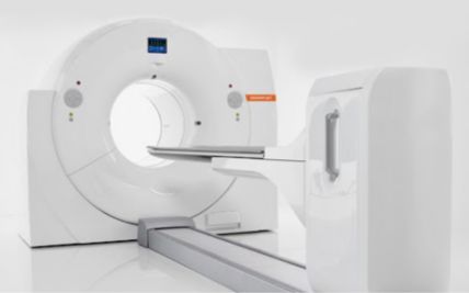
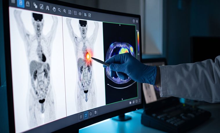
PET-CT combines metabolic information from a PET scan with the anatomical detail of CT. A small amount of radioactive tracer is given through a vein, and areas using more of the tracer may appear more active on the scan.
PET-CT may be recommended for selected sarcoma subtypes to assess disease activity, look for spread, or evaluate the response to treatment. It is not required for every patient, and the decision depends on the type of sarcoma and the clinical situation.
A bone scan uses a small amount of radioactive material to identify areas of increased activity within the skeleton. It may help determine whether a primary bone cancer has affected other bones.
Areas of increased activity are not always cancerous and may also result from arthritis, fractures, or infection. Any abnormal finding may therefore require comparison with X-rays, MRI, CT, or other tests.
Core needle biopsy is commonly preferred for suspected bone and soft tissue sarcomas. After the area is numbed with local anaesthesia, a hollow needle is used to collect several small cores of tissue.
Ultrasound, CT, or other imaging may be used to guide the needle into the most appropriate part of the tumour while avoiding nearby structures. Collecting multiple tissue samples helps the pathologist identify the tumour and perform additional laboratory tests. Occasionally, another biopsy may be needed if the sample is insufficient or does not represent the tumour adequately.
An incisional biopsy is a surgical procedure in which a small portion of the tumour is removed for examination. It may be recommended when a core needle biopsy is not possible, has not provided a clear diagnosis, or when more tissue is needed for specialised testing.
The incision is carefully planned in line with the expected definitive surgery so that the biopsy scar and tract can later be removed with the tumour. An incisional biopsy should therefore be performed by, or in close consultation with, an experienced sarcoma surgical team.
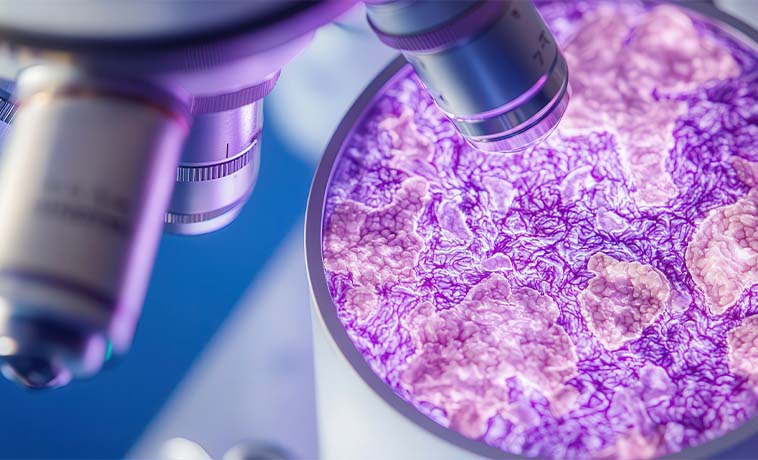
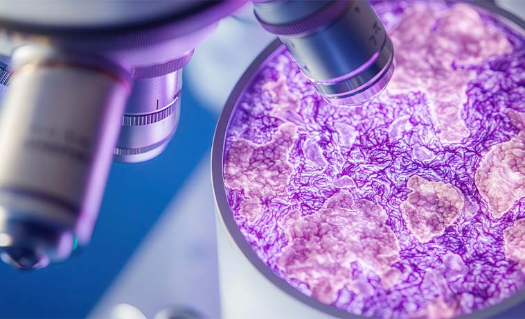
The biopsy sample may undergo:
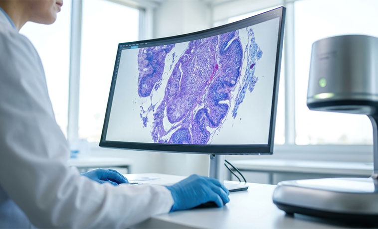
Histopathology to identify the sarcoma subtype
Tumour grading to assess how abnormal and active the cells appear
Immunohistochemistry to identify proteins within the tumour
Molecular or genetic testing to confirm selected subtypes
Biomarker testing when it may help guide targeted treatment
Staging describes the extent of the cancer and helps guide treatment. Staging generally considers tumour size and location, whether the tumour is superficial or deep, tumour grade, involvement of nearby structures or lymph nodes, and spread to distant organs, particularly the lungs.
Stage I: The cancer is generally low-grade and remains confined to the site where it began.
Stage II: The cancer remains localised but is usually higher-grade. There is no spread to distant organs.
Stage III: The cancer is generally high-grade, larger, or more locally extensive.
Stage IV: The cancer has spread to a distant part of the body. The lungs are the most common site of distant spread, it may also spread to other bones, the liver, or other organs.
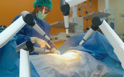
Wide local excision removes the tumour along with a planned margin of surrounding healthy tissue. The amount of tissue removed depends on the tumour’s size, location, grade, and proximity to important structures.
After surgery, a pathologist examines the edges of the removed tissue. If no cancer cells are present at the edges, the tumour is said to have a clear or negative margin. If the margin contains cancer cells, further surgery or radiation therapy may be considered to reduce the risk of local recurrence.
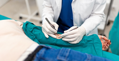
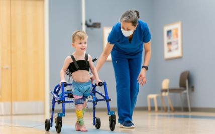
Limb-sparing surgery removes the cancer while retaining the affected arm or leg. It may involve removing part of a bone, muscle, or joint together with the tumour, followed by reconstruction to restore stability and movement.
Reconstruction may use a custom joint replacement, metal implant, bone graft, or a combination of techniques. Blood vessels, nerves, muscles, and skin may also require reconstruction. The decision depends not only on whether the limb can be preserved, but also on whether it is likely to remain functional after treatment.
Chemotherapy may be given before surgery for osteosarcoma or Ewing sarcoma. This can treat cancer cells elsewhere in the body and may help the surgical team assess how the tumour responds to treatment.
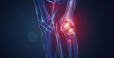
Sarcomas may develop in anatomically difficult areas such as the pelvis, spine, chest wall, head and neck, abdomen, or retroperitoneum. Removing these tumours may require close coordination between orthopaedic oncologists, surgical oncologists, neurosurgeons, vascular surgeons, thoracic surgeons, urologists, gastrointestinal surgeons, and reconstructive surgeons.
The operation may involve removing and reconstructing bone, soft tissue, blood vessels, nerves, or parts of nearby organs. Detailed imaging and multidisciplinary planning are essential to balance complete tumour removal with the preservation of neurological function, organ function, mobility, and quality of life.
Amputation is no longer required for most sarcomas of the arms or legs. It may, however, be the safest option when the tumour extensively involves major nerves or blood vessels, cannot be removed with clear margins, has caused severe infection or tissue damage, or when limb preservation would leave a painful or non-functional limb.
When amputation is required, the level of surgery is planned to remove the cancer while preserving as much useful limb length as possible. Prosthetic planning, physiotherapy, pain management, and psychological support begin early to help the patient regain mobility and independence.

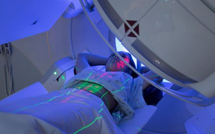
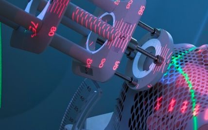
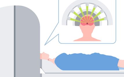
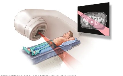
External beam radiation therapy delivers radiation from a machine outside the body. A planning CT scan is used to map the tumour, surgical area, and nearby healthy structures.
Treatment is usually divided into several sessions, called fractions, delivered over a planned period. The total dose and number of sessions depend on the sarcoma subtype, treatment goal, location, and whether radiation is given before or after surgery.
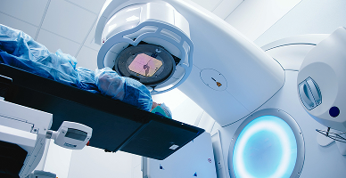
3D conformal radiation therapy uses CT-based planning to shape radiation beams around the tumour. Beams are delivered from several directions so that the prescribed dose covers the treatment area while reducing unnecessary exposure to surrounding tissues.
It may be used when the tumour and nearby structures can be adequately treated with a conformal beam arrangement.
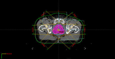
IMRT is an advanced form of external beam radiation in which the intensity of individual radiation beams is adjusted across the treatment area. This allows the dose to conform more closely to irregularly shaped tumours.
IMRT can be particularly useful for sarcomas near sensitive structures such as the spinal cord, bowel, kidneys, bladder, nerves, or major joints. It may help reduce radiation exposure to these structures, although some low-dose radiation may be spread across a wider area.
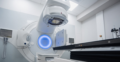
Image-guided radiation therapy uses imaging before or during each treatment session to confirm the tumour’s position and the patient’s alignment. This is important because small differences in positioning or changes in the tumour can affect where radiation is delivered.
The treatment team can make adjustments before delivering the dose, allowing smaller safety margins where appropriate. IGRT is a method of improving treatment accuracy and is commonly combined with 3D conformal radiation, IMRT, or proton therapy rather than being a separate treatment.
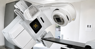
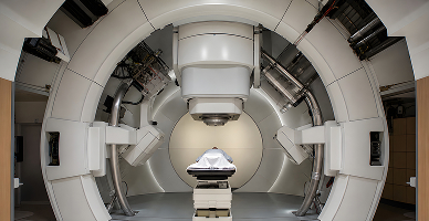
Proton therapy is an advanced form of external beam radiation that uses protons instead of X-rays. Protons release most of their radiation within the planned treatment area and deliver little radiation beyond it. This may reduce exposure to healthy tissues located behind the tumour.
Proton therapy may be considered for selected sarcomas:
Near the spine, brain, head and neck, pelvis, or other critical structures
In children and adolescents, where reducing radiation to growing tissues may be particularly important
When a large treatment area is required
When conventional radiation plans expose nearby organs to clinically significant doses
In selected patients who previously received radiation near the same area
For appropriate patients, Apollo Proton Cancer Centre offers proton therapy after individual evaluation. A proton plan may be compared with advanced photon-radiation plans to determine whether it provides a meaningful clinical advantage. Proton therapy is not required for every sarcoma and does not necessarily provide better tumour control than other techniques when both can deliver the required dose safely.
Chemotherapy uses anti-cancer medicines to destroy cancer cells or slow their growth. It may be given before surgery to treat microscopic disease and assess response, after surgery to reduce recurrence risk, or for recurrent or metastatic cancer.
Chemotherapy is an established part of treatment for:
Osteosarcoma, usually before and after surgery
Ewing sarcoma, as part of treatment combining chemotherapy with surgery and/or radiation
Rhabdomyosarcoma, usually alongside surgery and radiation therapy
Chemotherapy may also be used for selected high-grade or advanced soft tissue sarcomas, including some leiomyosarcomas and liposarcomas. However, it is not routinely beneficial for every sarcoma. Conventional chondrosarcoma, for example, generally responds poorly to standard chemotherapy, although treatment may differ for rarer aggressive subtypes.
The medicines, number of cycles, and sequence of treatment are selected according to the sarcoma subtype, age, stage, overall health, and response to therapy.
Targeted therapies act on specific proteins or molecular pathways that help cancer cells grow and survive. Unlike chemotherapy, which affects rapidly dividing cells more broadly, targeted treatment is chosen according to particular features of the tumour.
These medicines may be considered for selected advanced or recurrent sarcomas when testing identifies a relevant molecular target or when a treatment has demonstrated benefit in that subtype. Some targeted medicines inhibit signals involved in tumour growth or blood-vessel formation, while others act on specific gene alterations.
Targeted therapy is not available for every sarcoma and does not automatically replace surgery, chemotherapy, or radiation. Its use depends on the tumour subtype, previous treatment, biomarker findings, and current evidence.

Immunotherapy helps the immune system recognise or attack cancer cells. Its role in sarcoma is more limited than in cancers such as melanoma or lung cancer because most sarcomas do not respond consistently to currently available immunotherapies.
It may be considered for selected sarcoma subtypes, tumours with particular biomarkers, advanced disease after other treatments, or as part of a clinical trial. Biomarker findings such as high microsatellite instability or other uncommon molecular features may influence treatment in selected cases.
The potential benefit must be weighed against immune-related side effects, which can affect organs such as the skin, bowel, lungs, liver, or hormone-producing glands.
Precision oncology uses detailed information about the tumour to support diagnosis and treatment selection. In sarcoma care, its first role is often to confirm the exact subtype because several sarcomas are associated with characteristic gene rearrangements, mutations, or other molecular changes.
Testing may include immunohistochemistry, fluorescence in situ hybridisation, polymerase chain reaction, or next-generation sequencing. In selected cases, the results may identify a targeted treatment, clarify prognosis, or determine eligibility for a clinical trial.
Not every patient requires broad molecular profiling. Testing is selected when it can help confirm the diagnosis or meaningfully influence treatment.
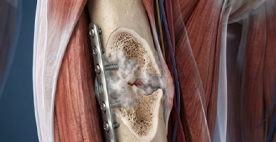
When a tumour involves part of a major bone or joint, the removed section may be replaced with a metal implant or custom endoprosthesis. This is commonly considered for tumours near the knee, hip, shoulder, or other major joints.
The implant restores structural support and may allow movement and weight-bearing after recovery. Physiotherapy is essential to rebuild strength and help the patient adapt to the reconstructed joint.

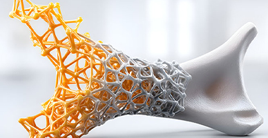
Selected bone defects may be reconstructed using the patient’s own bone, donated bone, or a combination of biological material and implants. In some cases, a treated section of the patient’s bone may be reused after the tumour has been removed.
Biological reconstruction may be particularly useful in younger patients because it can provide living bone that may integrate with the body. Healing generally takes longer than with a metal replacement and may require temporary restrictions on weight-bearing.
Removing a soft tissue sarcoma can leave a defect involving skin, muscle, or other tissues. Plastic and reconstructive surgeons may move healthy skin, fat, or muscle from another part of the body to cover the area.
A flap can protect exposed bone, implants, nerves, and blood vessels while supporting wound healing. Microsurgery may be used to reconnect small blood vessels when tissue is transferred from a distant part of the body.
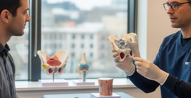
When a tumour closely involves a major nerve or blood vessel, specialist surgeons may be required during removal and reconstruction. A damaged or removed blood vessel may be replaced with a graft to maintain circulation to the limb.
Selected nerves may be repaired or reconstructed, although the degree of functional recovery varies. Sometimes a nerve must be removed to achieve safe cancer clearance, and rehabilitation then focuses on adapting movement and function.
Prognosis for Bone & Soft Tissue Cancer
The outlook for bone and soft tissue cancer varies considerably because sarcomas include many different diseases. Many localised sarcomas can be treated successfully, particularly when the tumour can be completely removed and, where indicated, combined with chemotherapy or radiation therapy.
Factors that influence prognosis include:
Sarcoma subtype and tumour grade
Tumour size, depth, and location
Stage at diagnosis and whether the cancer has spread
Ability to remove the tumour with clear margins
Response to chemotherapy or radiation therapy
Molecular features, where relevant
Age, overall health, and ability to complete treatment
Whether the cancer is newly diagnosed or recurrent
Regular follow-up is important because recurrence may occur at the original site or in another part of the body, particularly the lungs.


Why Choose ACC for Bone & Soft Tissue Cancer Treatment
Dedicated multidisciplinary expertise in bone and soft tissue cancers
Specialist musculoskeletal imaging and pathology review
Biopsies planned in coordination with the definitive surgical team
Limb-sparing surgery and complex bone and soft tissue reconstruction
Subtype-specific chemotherapy, targeted therapy, and precision oncology
Advanced radiation techniques, including proton therapy for selected patients
Integrated physiotherapy, rehabilitation, pain management, and functional recovery support
Structured long-term surveillance following treatment
Health Blogs