




Cancer imaging plays an important role throughout the cancer care journey. It may be used to screen eligible individuals, investigate symptoms, detect and characterise abnormalities, determine the extent of disease, support treatment planning, assess response, and monitor for recurrence.
At Apollo Cancer Centres, radiologists, nuclear medicine specialists, gastroenterologists, pulmonologists, oncologists, and other specialists work together to interpret imaging findings in the context of the patient’s clinical history, pathology, laboratory results, and previous examinations. Complex cases may be reviewed by a multidisciplinary tumour board to support coordinated treatment decisions.
Read MoreApollo Cancer Centres provides access to a range of imaging technologies. The appropriate examination or combination of examinations is selected according to the individual clinical need.
CT uses X-rays and computer processing to create detailed cross-sectional images of the body. In cancer care, it may be used to identify and characterise tumours, assess lymph nodes and distant spread, determine the relationship of a tumour to nearby organs and blood vessels, plan treatment, evaluate complications, and monitor response or recurrence. Depending on the area being examined and the clinical question, oral or intravenous contrast material may be used to improve visualisation of organs, blood vessels, and abnormal tissue.
Read More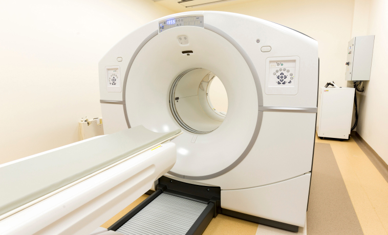
Low-dose CT is a specialised CT protocol that produces detailed images of the lungs while using a lower radiation dose than a conventional diagnostic chest CT. Its principal role in cancer care is lung cancer screening for eligible individuals who have an increased risk of developing the disease. LDCT does not usually require contrast material. It is not a substitute for a diagnostic chest CT when symptoms or suspicious findings require more detailed evaluation. Eligibility for LDCT screening is determined after clinical assessment.
Read More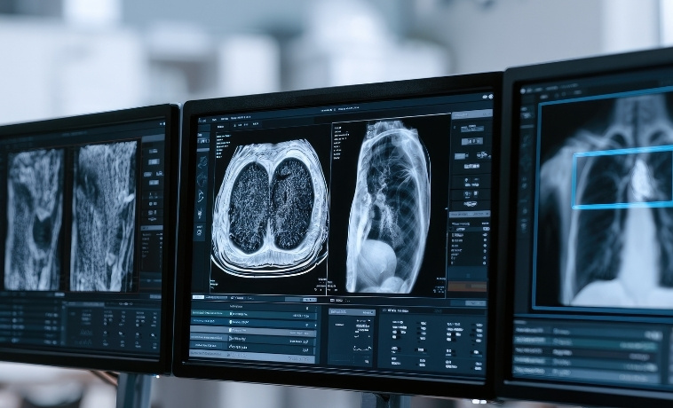
PET-CT combines metabolic or molecular information from positron emission tomography with anatomical detail from CT. A small amount of radiotracer is administered before imaging. The radiotracer selected depends on the cancer type and the clinical question. PET-CT may help stage certain cancers, assess lymph nodes or distant spread, detect recurrence, evaluate treatment response, support radiation-treatment planning, or determine suitability for selected molecularly targeted radionuclide treatments.Not every cancer is equally well assessed with PET-CT. Small lesions or cancers with low tracer uptake may be difficult to detect, while inflammation and infection can sometimes also show increased uptake. Findings are therefore interpreted alongside other imaging, pathology, and clinical information.
Read More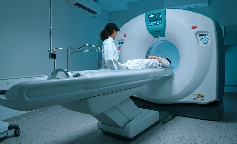
Single-photon emission computed tomography combined with CT, or SPECT-CT, uses a small amount of radiotracer to provide functional information together with cross-sectional anatomical localisation. In cancer care, SPECT-CT may be used for bone imaging, sentinel lymph-node mapping, thyroid imaging, renal-function assessment, or other specialised nuclear medicine examinations. The type of radiotracer, preparation, and imaging protocol depend on the clinical indication.
Read More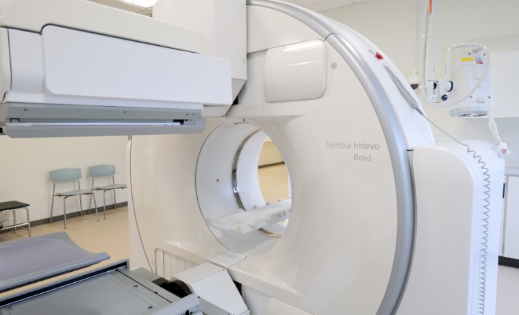
Magnetic resonance imaging uses a strong magnetic field and radiofrequency waves rather than ionising radiation to produce detailed images. Both 1.5T and 3T MRI systems can provide high-quality cancer imaging. The appropriate system and imaging protocol are selected according to the body part, clinical question, patient factors, and type of detail required. MRI is particularly useful for cancers involving the brain, spine, head and neck, breast, liver, rectum, pelvis, prostate, bones, soft tissues, and bone marrow. Specialised techniques such as diffusion-weighted imaging, dynamic contrast-enhanced imaging, multiparametric MRI, or magnetic resonance cholangiopancreatography may be used when clinically appropriate.
Read More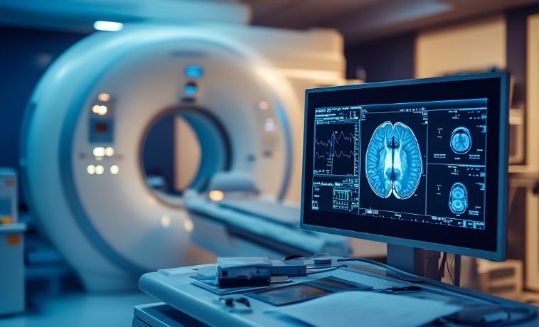
X-ray imaging uses a small amount of ionising radiation to produce two-dimensional images. Although it provides less detail than CT or MRI, it can offer a rapid initial assessment of the chest, lungs, bones, treatment devices, and certain complications associated with cancer or its treatment. Additional imaging may be required if an X-ray identifies a suspicious finding or does not adequately answer the clinical question.
Read More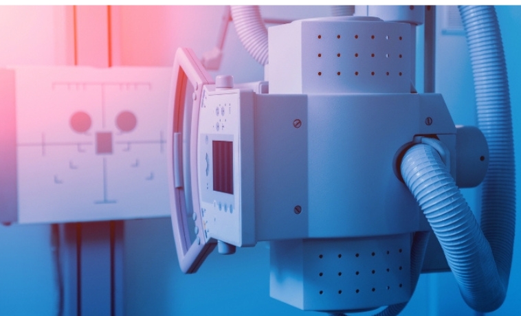
Ultrasound uses high-frequency sound waves to create real-time images without ionising radiation. It may be used to assess abnormalities involving the breast, thyroid, liver, abdomen, pelvis, testes, lymph nodes, and superficial soft tissues.Doppler ultrasound can evaluate blood flow within or around an abnormality. Ultrasound may also provide real-time visual guidance during biopsies, drainage procedures, and selected interventional oncology treatments.
Read More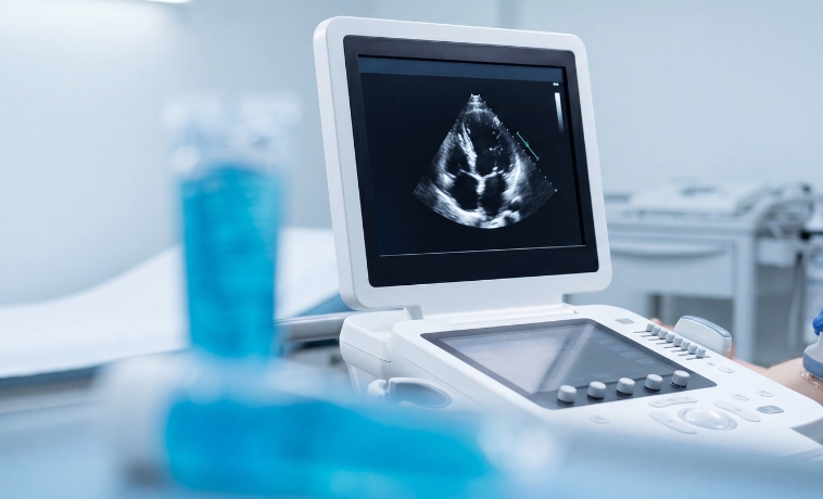
Mammography uses low-dose X-rays to examine breast tissue. It may be used for routine breast cancer screening in eligible individuals or as a diagnostic examination for symptoms such as a breast lump, nipple changes, or an abnormality identified on another test. Diagnostic mammography may include additional or magnified views of a specific area. Depending on the findings, breast ultrasound, tomosynthesis, contrast-enhanced mammography, MRI, or biopsy may be recommended.
Read More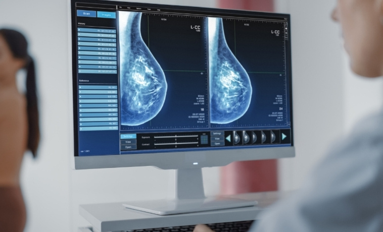
Digital breast tomosynthesis, sometimes called 3D mammography, obtains multiple low-dose X-ray images of the breast from different angles. These images are reconstructed into thin sections, reducing the effect of overlapping breast tissue. Tomosynthesis may improve the detection and characterisation of certain breast abnormalities, particularly in patients with dense breast tissue. It may be used for screening or diagnostic evaluation, depending on the individual’s needs and local clinical protocols.
Read More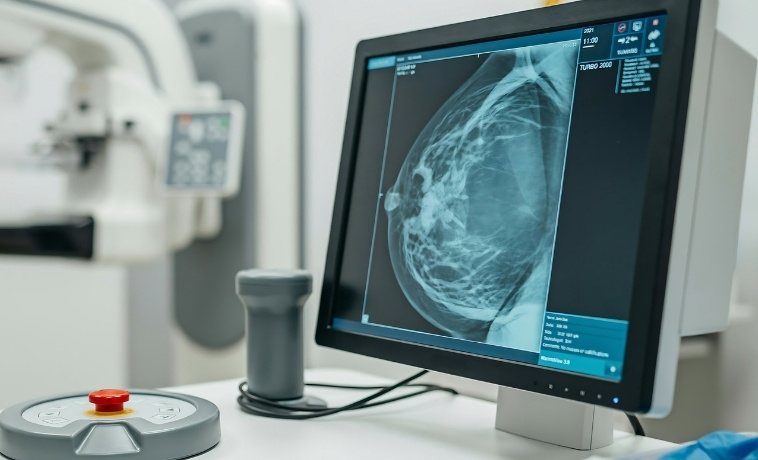
Contrast-enhanced mammography combines mammographic imaging with an intravenous iodine-based contrast agent. It produces images that show both the structure of the breast and areas of increased contrast enhancement. It may be used to evaluate an abnormality, assess the extent of a known breast cancer, investigate selected inconclusive findings, or monitor response to treatment in appropriate patients. Kidney function, previous contrast reactions, pregnancy status, and the clinical indication are reviewed before the examination.
Read More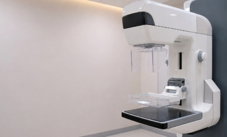
Upper gastrointestinal endoscopy uses a thin, flexible camera to directly examine the oesophagus, stomach, and first part of the small intestine. In cancer care, it may help identify and characterise suspicious mucosal abnormalities, determine their location and visible extent, and evaluate symptoms such as persistent swallowing difficulty, unexplained upper gastrointestinal bleeding, or other concerning findings. High-definition imaging and image-enhancement technologies may improve the visibility of subtle abnormalities. Tissue samples may be collected during the examination when clinically indicated, but the biopsy and any therapeutic intervention form part of the procedure rather than the imaging technology itself.
Read More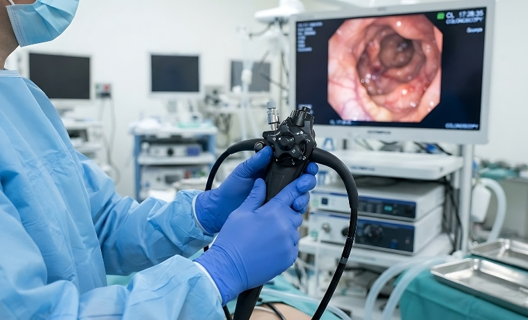
Colonoscopy uses a thin, flexible camera to examine the rectum and colon. It may be used for colorectal cancer screening in eligible individuals or to investigate symptoms and abnormal findings. High-definition imaging, magnification, chromoendoscopy, or electronic image-enhancement technologies may help detect and characterise polyps and other mucosal abnormalities. Tissue sampling or removal of certain polyps may be performed when clinically appropriate.
Read More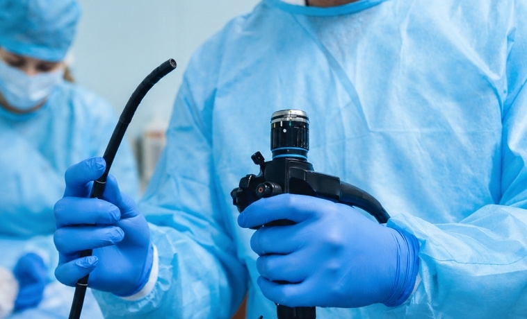
Bronchoscopy uses a flexible or rigid camera system to visualise the trachea and airways. In cancer care, it may help assess centrally located lung tumours, airway narrowing, bleeding, or abnormalities identified on CT or PET-CT. High-definition and enhanced bronchoscopic imaging may improve visualisation of selected airway abnormalities. Bronchoscopy may also provide access for tissue sampling or other procedures when required.
Read More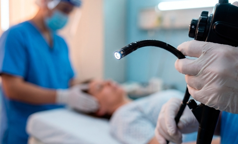
Capsule endoscopy uses a swallowable capsule containing a miniature camera. As the capsule passes through the digestive tract, it captures images that are transmitted to a recording device worn by the patient. It is particularly useful for examining areas of the small intestine that may be difficult to reach with conventional upper GI endoscopy or colonoscopy. Its role in cancer evaluation is selective and depends on the symptoms, suspected site, previous investigations, and risk of the capsule becoming retained in a narrowed area of the bowel.
Read More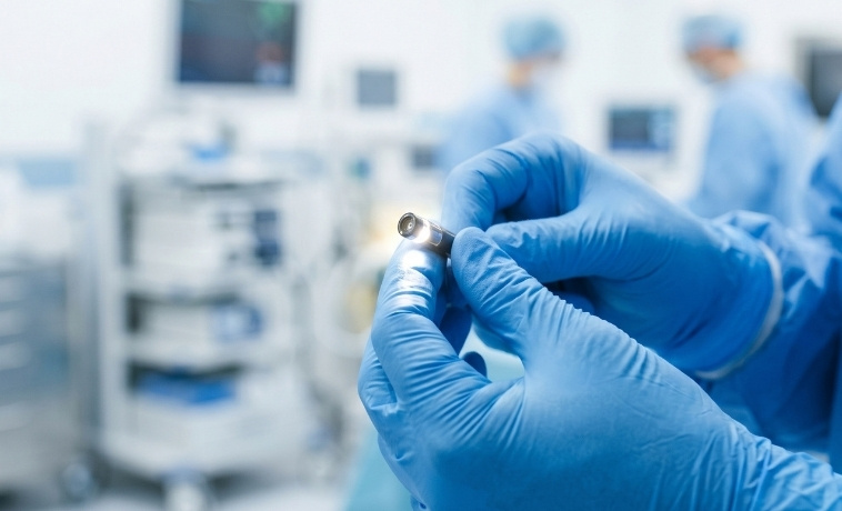
Endoscopic ultrasound (EUS) combines endoscopy with high-frequency ultrasound. An ultrasound transducer positioned at the tip of the endoscope produces detailed images of the wall of the digestive tract and nearby organs, lymph nodes, blood vessels, and tissues. EUS may be used to assess cancers involving the esophagus, stomach, pancreas, bile ducts, rectum, and other nearby structures. It can help determine the depth of tumour involvement, assess local spread, and evaluate nearby lymph nodes. Tissue sampling may be performed during EUS when clinically indicated.
Read More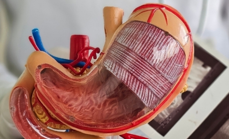
Endobronchial ultrasound, or EBUS, combines bronchoscopy with ultrasound imaging from within the airways. It allows doctors to visualise structures beyond the airway wall, including lymph nodes and lesions within the chest. EBUS is commonly used in the evaluation and staging of suspected or confirmed lung cancer. It can help assess mediastinal and hilar lymph nodes and other abnormalities that may not be directly visible during standard bronchoscopy. Ultrasound-guided tissue sampling may be performed during the examination when clinically indicated.
Read More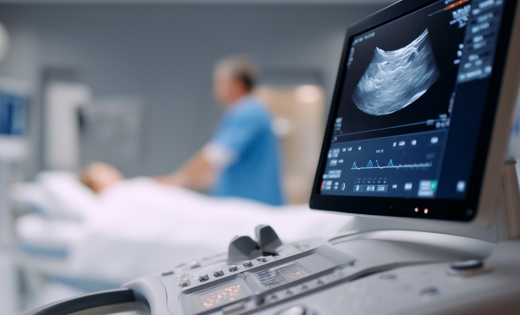

How Imaging Fits into Your Overall Care Plan
Imaging may be used at several points in the cancer care journey. Screening examinations such as mammography, low-dose CT (LDCT), or colonoscopy may be recommended for eligible individuals according to their age, risk factors, clinical history, and applicable screening guidance.
When cancer is suspected, imaging can identify an abnormality, define its location and extent, and help determine whether further evaluation or tissue sampling is needed. After diagnosis, imaging may be used to establish the stage of the cancer and help the multidisciplinary team plan surgery, radiation therapy, systemic treatment, interventional treatment, or a combination of approaches.
During treatment, imaging may help assess whether the disease is responding and identify complications. After treatment, follow-up imaging may be recommended to evaluate symptoms, monitor previously identified findings, or look for recurrence when clinically indicated.
The same examination is not repeated automatically for every patient. The type and timing of imaging are selected according to the cancer, treatment received, previous results, current symptoms, and whether the findings are likely to influence care.



