Neurological Cancer
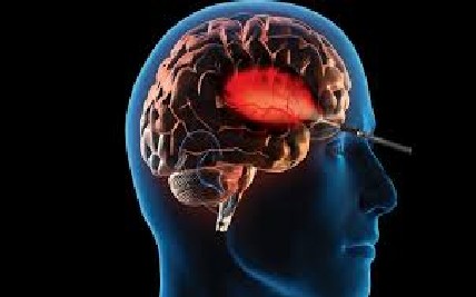
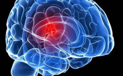
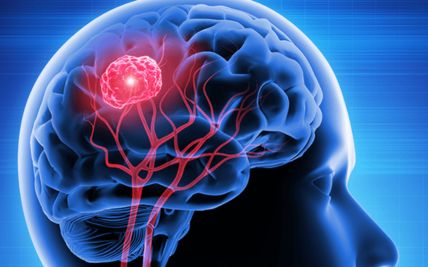
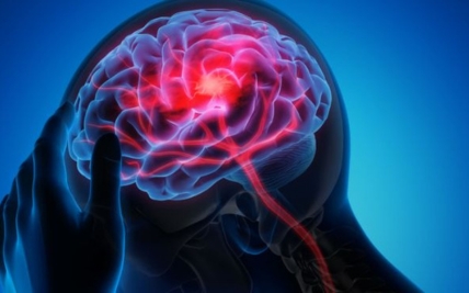
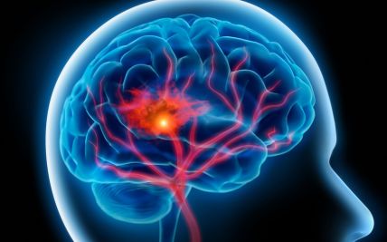
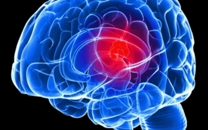
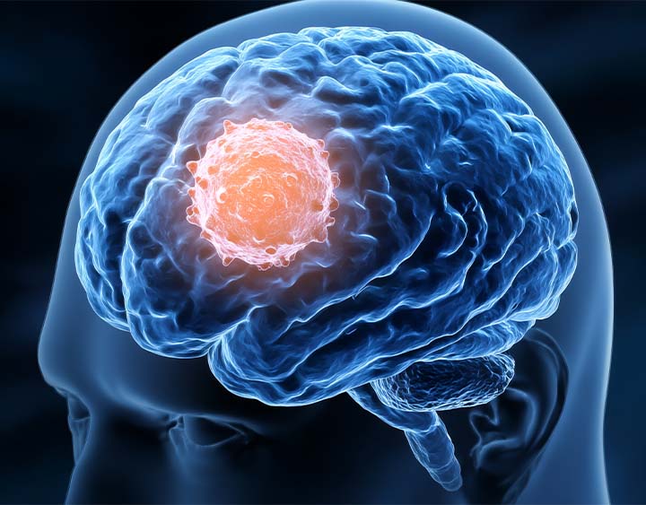
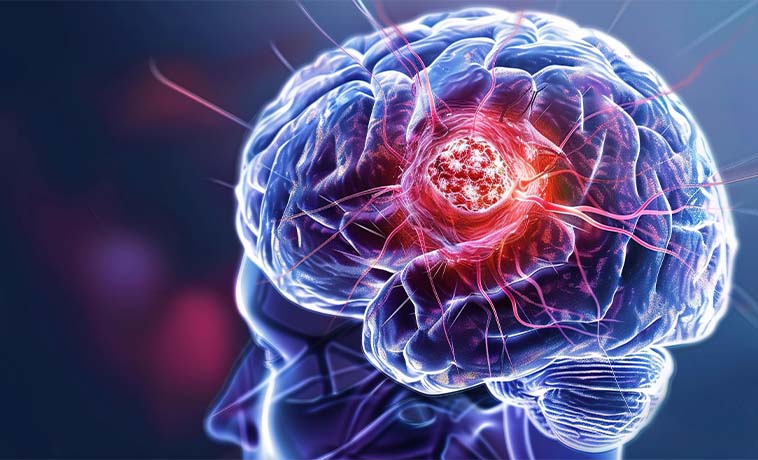
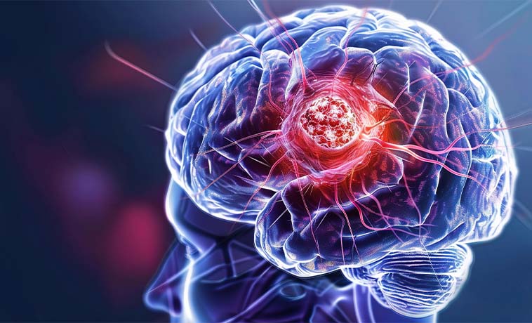
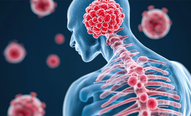
Neurological cancers are malignant tumours that develop in the brain, spinal cord, or other parts of the central nervous system. Cancer from another organ can also spread to the brain or spine, forming a secondary or metastatic tumour.
Not every brain or spinal tumour is cancerous. Some are non-cancerous or slow-growing, but they may still require treatment if they press on areas responsible for movement, speech, vision, memory, hormones, breathing, or other important functions. The seriousness of a tumour therefore depends not only on whether it is cancerous but also on its type, location, grade, and effect on nearby structures.
Treatment requires close coordination between neurosurgeons, neuro-oncologists, radiation oncologists, neuroradiologists, neuropathologists, medical oncologists, and rehabilitation specialists. The aim is to control the tumour while preserving neurological function and quality of life wherever possible.
Types of Neurological Cancer
Brain and spinal tumours are classified according to the cells in which they begin, their location, molecular features, and expected behaviour.
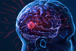
Gliomas arise from glial cells, which support and protect nerve cells within the brain and spinal cord. Their behaviour ranges from relatively slow-growing to highly aggressive. Modern classification combines microscopic appearance with molecular features. Important groups include astrocytomas, oligodendrogliomas, glioblastomas, and ependymomas.
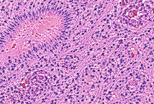
Astrocytomas develop from cells related to astrocytes. They may occur in the brain or spinal cord and can affect both children and adults. Some are slow-growing, while others are infiltrative and more aggressive. Molecular changes, including IDH status in adult-type diffuse gliomas, are central to classification and treatment planning.
Glioblastoma is an aggressive, fast-growing primary brain tumour. It usually infiltrates surrounding brain tissue, which means complete microscopic removal is generally not possible through surgery alone. Treatment commonly combines maximal safe surgery, radiation therapy, and chemotherapy. Additional treatment depends on the tumour’s molecular features, response, and whether it returns.
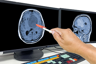
Oligodendroglioma is a diffuse glioma defined by an IDH mutation and the combined loss of chromosome arms 1p and 19q. These tumours often grow more slowly than glioblastoma but can still infiltrate surrounding brain tissue and recur. Treatment depends on grade, symptoms, tumour location, and the amount that can be safely removed.

Ependymomas develop from cells lining the fluid-filled spaces of the brain or the central canal of the spinal cord. They can occur at different ages and in different parts of the central nervous system. Treatment commonly involves surgery, with radiation recommended in selected cases according to tumour location, grade, molecular type, and completeness of removal.
Meningiomas arise from the membranes covering the brain and spinal cord. Most are non-cancerous and slow-growing, although some are atypical or malignant. Small, asymptomatic meningiomas may be monitored. Surgery or radiation may be required if the tumour grows, causes symptoms, or is located near critical structures.
Medulloblastoma is a high-grade tumour that usually develops in the cerebellum at the back of the brain. It is more common in children but can also occur in adults. It may spread through the cerebrospinal fluid to other parts of the brain or spinal cord. Treatment generally involves surgery followed by risk-adapted craniospinal radiation and chemotherapy.
Schwannomas develop from Schwann cells, which form the covering around certain nerves. Most are non-cancerous. Vestibular schwannomas affect the nerve responsible for hearing and balance. Treatment may involve observation, surgery, or focused radiation depending on the tumour’s size, growth, symptoms, hearing, and individual priorities.
Pituitary tumours arise in the gland at the base of the brain that regulates hormones. Most are non-cancerous, but they can cause problems by producing excess hormones or pressing on the optic nerves and surrounding structures. Treatment may include medicines, endoscopic surgery through the nose, radiation, or a combination, depending on the hormone produced and the tumour’s size and behaviour.
Craniopharyngiomas develop near the pituitary gland and optic pathways. They are generally non-cancerous but can affect vision, hormone production, growth, memory, and other brain functions. Treatment may involve surgery, cyst drainage, radiation, or combined approaches planned to reduce damage to nearby critical structures.
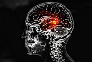
Primary central nervous system lymphoma is a lymphoma arising in the brain, spinal cord, eyes, or cerebrospinal fluid. It behaves differently from most other brain tumours. Treatment commonly involves high-dose systemic medicines that can enter the central nervous system. Surgery is generally used to obtain a diagnosis rather than remove the entire tumour.
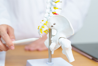
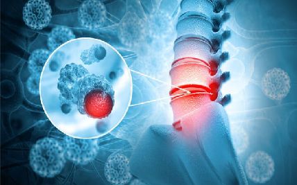
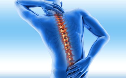
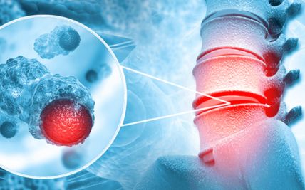
Tumours may develop within the spinal cord, its coverings, spinal nerves, or the bones surrounding it. They may be primary tumours or metastases from cancer elsewhere. Symptoms can include persistent back pain, weakness, numbness, walking difficulty, or loss of bladder or bowel control. Treatment depends on the tumour type, stability of the spine, and degree of pressure on the spinal cord or nerves.
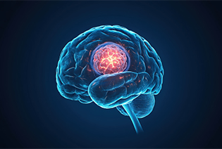
Metastatic tumours spread to the brain or spine from cancer elsewhere in the body. Common primary sites include the lung, breast, kidney, skin melanoma, and gastrointestinal tract. Treatment is planned according to the original cancer, number and location of metastases, neurological symptoms, molecular features, and the extent of cancer elsewhere.
Skull-base tumours arise in or extend into the bones and tissues beneath the brain. They include chordomas, chondrosarcomas, meningiomas, nerve-sheath tumours, and cancers spreading from the sinuses or other nearby structures. Their location near major nerves and blood vessels often requires coordinated skull-base surgery and specialised radiation planning.
The cause of most primary brain and spinal tumours is unknown. There are few clearly established modifiable risk factors.
Modifiable Risk Factors
No lifestyle change has been proven to prevent most primary central nervous system tumours. Avoiding unnecessary exposure to ionising radiation and following appropriate workplace safety measures may reduce general health risks, but most brain and spinal tumours cannot be linked to a preventable exposure.
Tobacco, mobile-phone use, and everyday electromagnetic-field exposure have not been established as causes of primary brain cancer.
Non-Modifiable Risk Factors
Previous therapeutic radiation to the head, particularly during childhood
Inherited conditions such as neurofibromatosis type 1 or 2
Li–Fraumeni syndrome
Tuberous sclerosis
Von Hippel–Lindau syndrome
Lynch syndrome and other selected inherited conditions
A weakened immune system, which increases the risk of primary CNS lymphoma
Increasing age for several adult brain tumours
Childhood or adolescence for certain embryonal and paediatric tumours
Sex-related differences in the frequency of selected tumour types
A personal history of cancer elsewhere in the body, which increases the risk of brain or spinal metastases
Most people diagnosed with a brain or spinal tumour do not have an inherited syndrome or recognised exposure.
Signs and Symptoms
Symptoms depend on the tumour’s location, growth rate, surrounding swelling, and effect on the brain, spinal cord, nerves, or cerebrospinal fluid.
Possible signs and symptoms include:
These symptoms can also result from migraine, epilepsy, stroke, infection, spinal disease, hormonal disorders, or other conditions. A first seizure, sudden neurological deficit, rapidly worsening confusion, or symptoms of spinal-cord compression require urgent medical assessment.
Diagnosis begins with a review of symptoms, previous cancers and treatments, medical and family history, and current medicines. A neurological examination assesses strength, sensation, reflexes, coordination, balance, vision, hearing, speech, memory, and other cognitive functions. Imaging identifies the tumour and its relationship to critical structures, while biopsy and laboratory testing establish its type, grade, and molecular features.
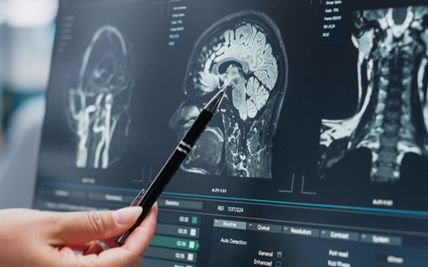
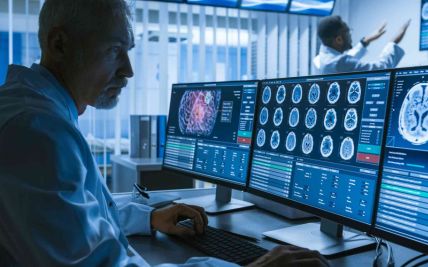
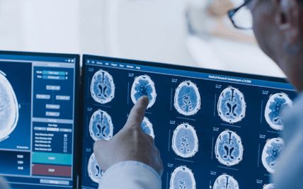
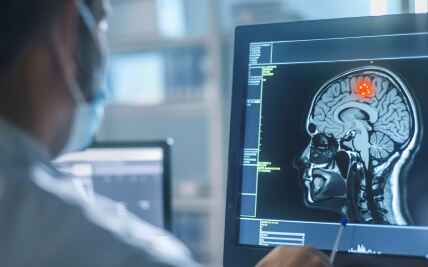
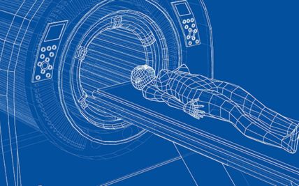
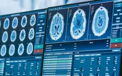
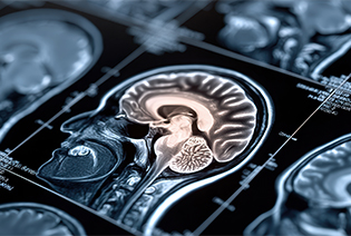
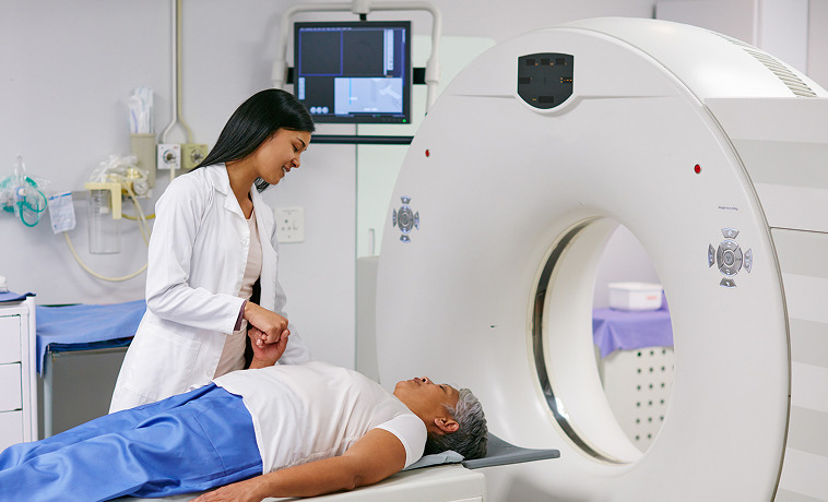
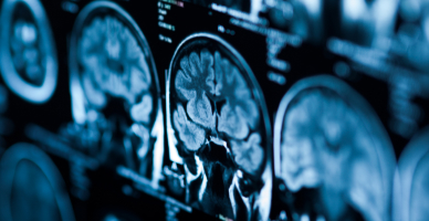
MRI uses magnetic fields and radio waves to produce detailed images of the brain and spinal cord. Contrast material may be administered to show the tumour and its blood supply more clearly. MRI is the main imaging test for most brain and spinal tumours. It can also show swelling, bleeding, fluid obstruction, nerve compression, and changes after treatment.
Functional MRI identifies areas of the brain involved in movement, language, and other functions. It may be used before surgery when a tumour lies close to an important functional region. The information helps the surgical team plan an approach that reduces the risk of neurological injury.
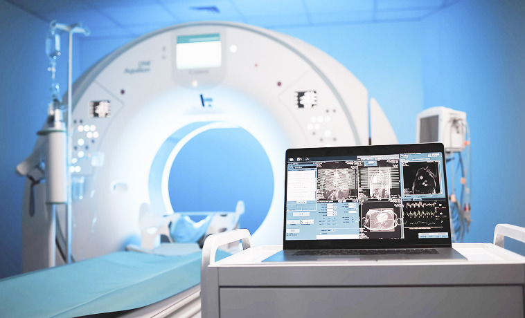
Diffusion tensor imaging maps major white-matter pathways carrying signals through the brain. Tractography creates visual representations of these pathways. These techniques may help plan surgery around pathways involved in movement, language, vision, or other functions.
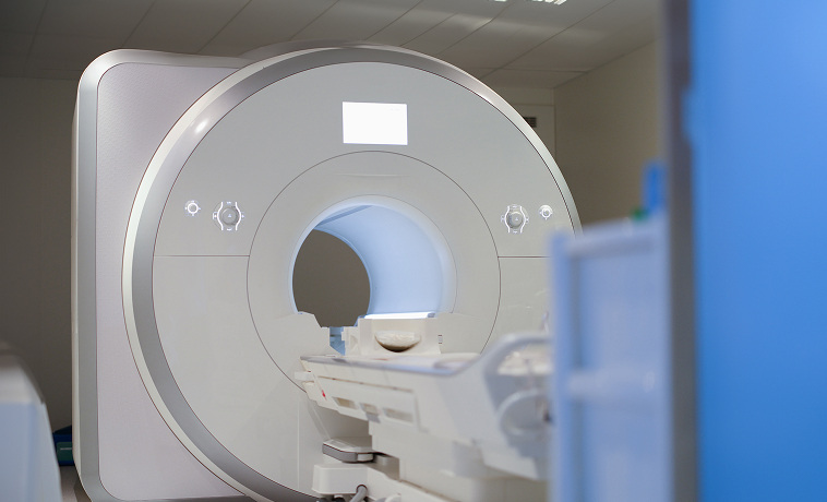
Perfusion MRI assesses blood flow within and around a tumour. It may help distinguish active tumour from treatment-related change and provide information about tumour behaviour.
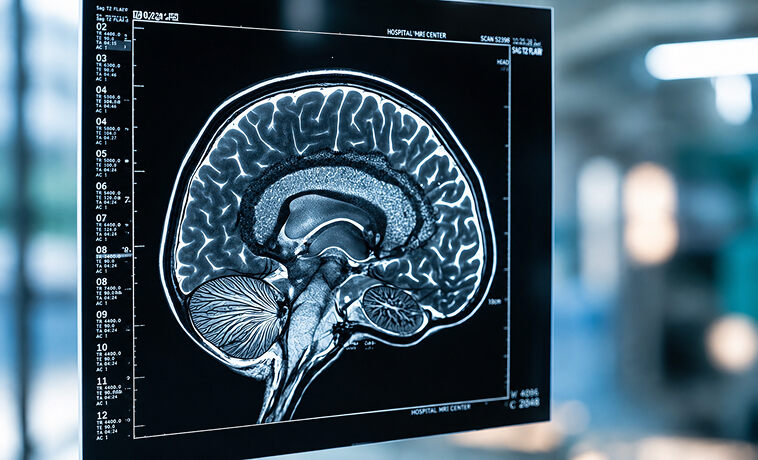
MR spectroscopy measures selected chemicals within tissue. It may support tumour characterisation or assessment of treatment-related changes but does not replace biopsy when tissue diagnosis is required.


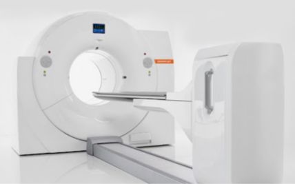
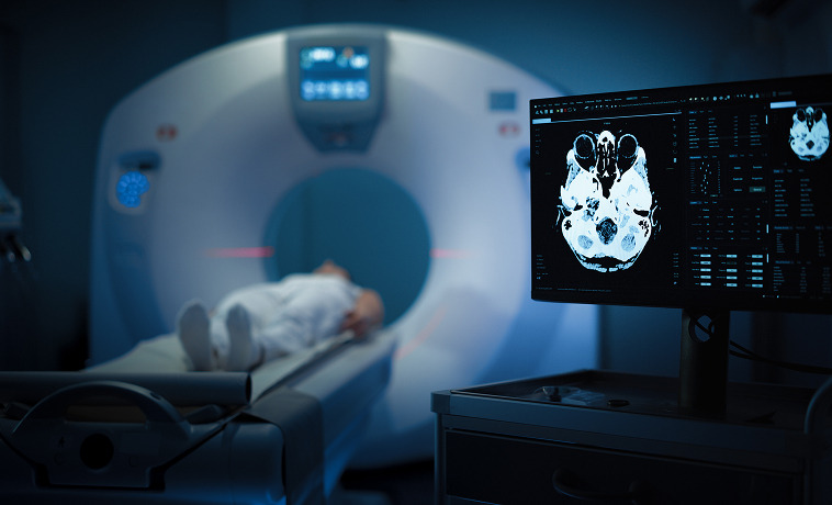
A CT scan uses X-rays to produce rapid images of the brain, skull, spine, and surrounding structures. It is particularly useful in emergencies and for detecting bleeding, calcification, bone involvement, or hydrocephalus. MRI usually provides greater soft-tissue detail for treatment planning.
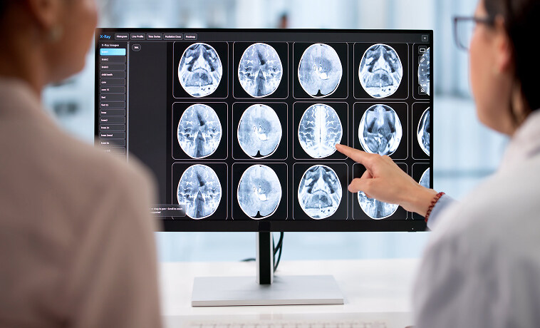
PET imaging evaluates metabolic or molecular activity. Selected amino-acid or other PET tracers may help assess tumour activity, guide biopsy, plan radiation, or distinguish recurrence from treatment-related change. Standard FDG PET has limitations in the brain because normal brain tissue naturally uses large amounts of glucose.
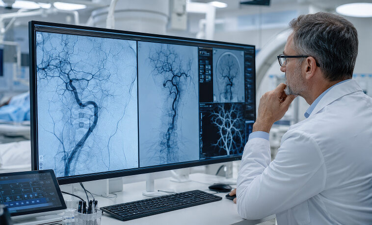
Cerebral angiography examines blood vessels supplying the brain or tumour. It may be used for selected highly vascular tumours or skull-base lesions. In some cases, embolisation may be performed before surgery to reduce the tumour’s blood supply.
Contrast-enhanced MRI of the spine evaluates spinal-cord, nerve, meningeal, and vertebral tumours. Whole-spine imaging may be required for tumours that can spread through cerebrospinal fluid.
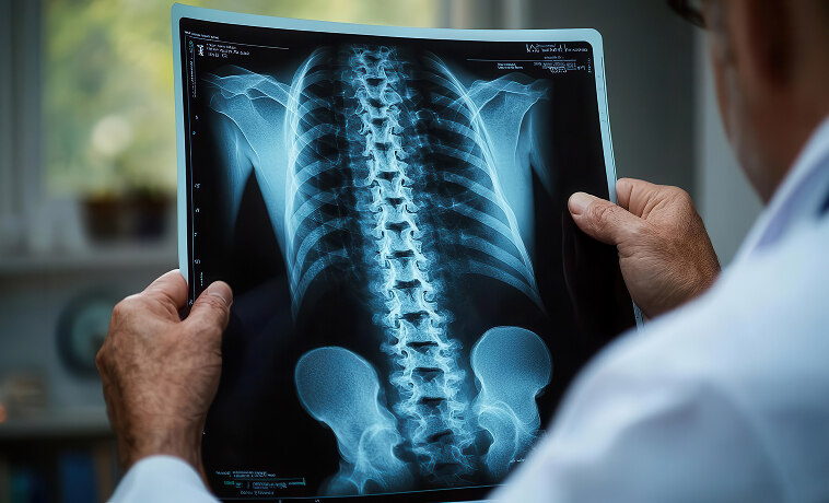
Stereotactic biopsy uses three-dimensional image guidance to direct a needle into the tumour through a small opening in the skull. Several tissue samples are collected from carefully selected areas. It may be used for deep, diffuse, or surgically inaccessible tumours and when treatment depends on obtaining a molecular diagnosis.
An open biopsy is performed through a craniotomy or spinal operation. It may be used when a larger tissue sample is required or when biopsy and tumour removal can be performed during the same procedure.
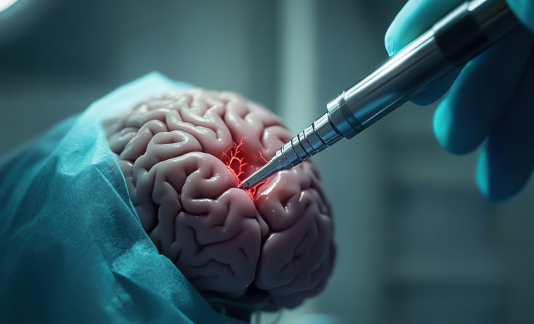
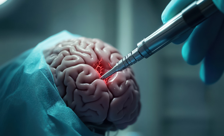
An endoscope may be passed through a small opening in the skull or through the nose to reach selected ventricular, pituitary, or skull-base tumours. The same procedure may sometimes be used to treat fluid obstruction or remove part of the tumour.
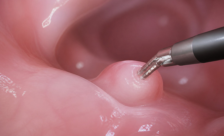
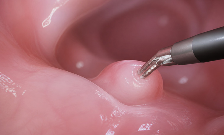
A spinal tumour may be sampled through image-guided needle biopsy or during surgery. The approach depends on whether the tumour involves the spinal cord, nerve coverings, vertebrae, or surrounding tissues.
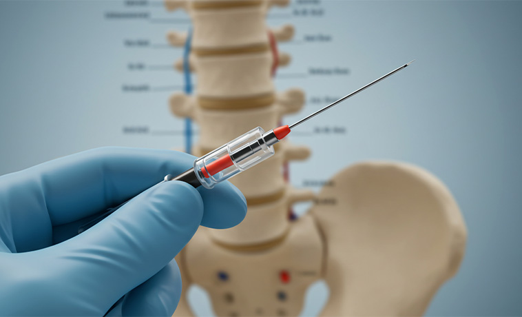
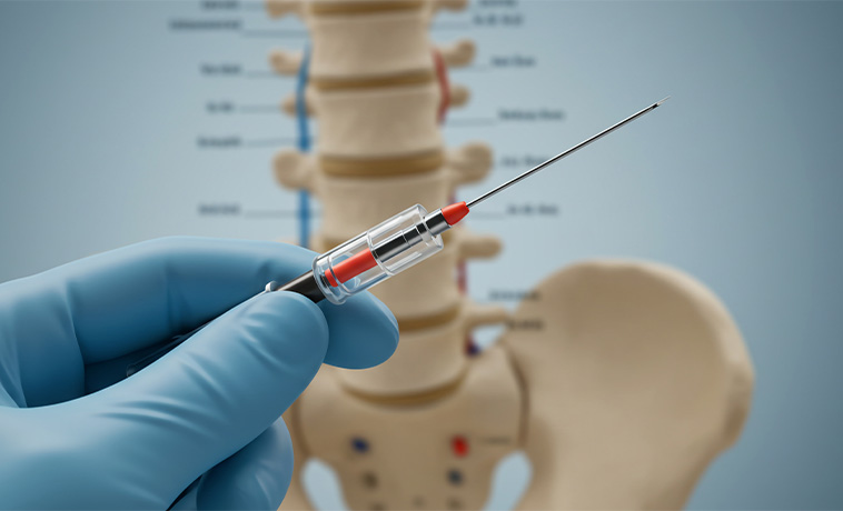
A lumbar puncture may collect cerebrospinal fluid to look for cancer cells, proteins, or tumour markers. It is used in selected lymphomas, germ-cell tumours, medulloblastomas, and cancers involving the brain or spinal coverings. Lumbar puncture may be unsafe when pressure inside the skull is raised and is performed only after appropriate assessment.
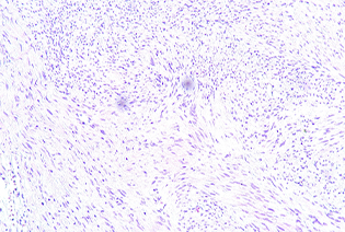
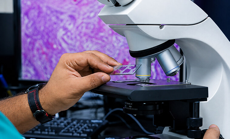
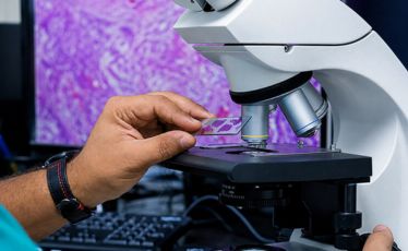
A neuropathologist examines tumour tissue under a microscope to identify the cell type, growth pattern, mitotic activity, and other features. The microscopic findings are combined with molecular results because tumours that appear similar can behave differently.
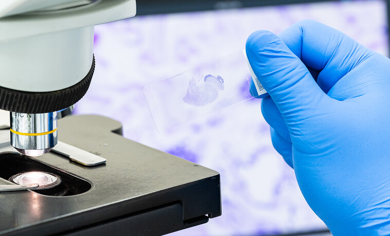
Immunohistochemistry uses specialised stains to identify proteins within tumour cells. It helps classify gliomas, meningiomas, lymphomas, metastases, and other central nervous system tumours.
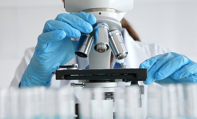

Molecular testing identifies genetic or epigenetic changes that define the tumour and may influence prognosis or treatment. Depending on the diagnosis, testing may include:
IDH1 and IDH2
1p/19q codeletion
ATRX and TP53
MGMT promoter methylation
H3 K27 or H3 G34 alterations
BRAF alterations
TERT promoter changes
EGFR amplification
Other tumour-specific markers
Not every tumour requires every test. Testing is selected according to its location, appearance, and suspected type.


Cerebrospinal fluid is examined for abnormal cells when lymphoma, leukaemia, medulloblastoma, germ-cell tumour, or leptomeningeal spread is suspected. A negative sample does not always exclude disease, and repeat testing may sometimes be required.
Genetic testing may be recommended when the tumour type, age at diagnosis, family history, or presence of multiple tumours suggests an inherited condition. Genetic counselling helps explain the results and whether relatives may also require testing or surveillance.
Most primary brain and spinal tumours are graded rather than staged from I to IV. The grade reflects the tumour’s biological features and expected behaviour. Modern classification also uses molecular findings, and the meaning of each grade varies between tumour types.
Grade 1: Grade 1 tumours are generally slow-growing and have relatively well-defined boundaries. Some can be treated effectively with surgery alone if complete removal is safe.
Grade 2: Grade 2 tumours are usually slower-growing than high-grade tumours but may infiltrate nearby tissue. They can recur and, in some tumour types, may progress to a higher grade.
Grade 3: Grade 3 tumours show more aggressive biological features and tend to grow more rapidly.
Grade 4: Grade 4 tumours are high-grade and biologically aggressive. They may grow rapidly, infiltrate surrounding tissue, and require intensive combined treatment.
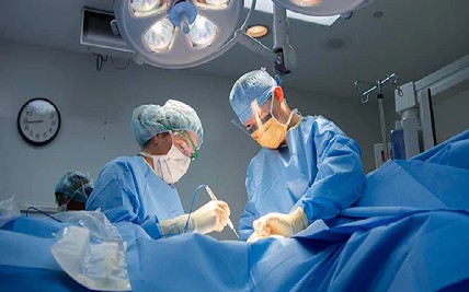
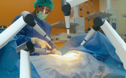
Maximal safe resection removes as much of the tumour as possible without causing unacceptable neurological injury. It may provide tissue for diagnosis, reduce pressure, relieve symptoms, and improve the effect of other treatments. Complete removal is not always possible when a tumour infiltrates normal brain or lies near areas responsible for language, movement, vision, or other critical functions.
Neuronavigation combines preoperative imaging with real-time surgical tracking. It helps the surgeon locate the tumour and plan a precise route through the brain or spine. The system improves anatomical orientation but does not replace surgical judgement or functional monitoring.
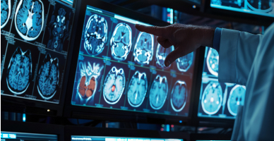
During an awake craniotomy, the patient is kept awake for selected parts of the operation so that language, movement, or other functions can be tested. It may be used when a tumour lies close to important functional areas and the patient is medically and psychologically suitable.
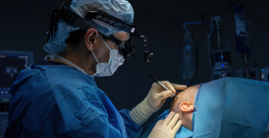
Electrical stimulation and neurophysiological monitoring assess brain, spinal-cord, and nerve function during surgery. These techniques help identify functional tissue and provide warning of changes that could lead to injury.
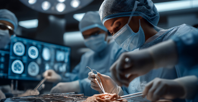
Stereotactic techniques use three-dimensional image guidance to reach a precisely defined target. They may be used for biopsy, drainage, electrode placement, or selected tumour procedures.
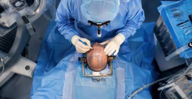
Neuroendoscopy uses a small camera and instruments passed through a limited opening. It may be used for selected ventricular, pituitary, skull-base, or cystic tumours. Endoscopy can also create a pathway for cerebrospinal fluid when a tumour causes hydrocephalus.
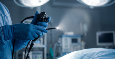
Endoscopic endonasal surgery reaches selected pituitary and skull-base tumours through the nose. It avoids a traditional open cranial incision. Suitability depends on the tumour’s location, extent, relationship to blood vessels and nerves, and the ability to reconstruct the skull base safely.
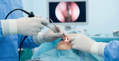
Complex skull-base surgery may combine endoscopic and open microsurgical approaches. Neurosurgeons may work with head and neck, vascular, ophthalmic, or reconstructive specialists. The approach is selected according to the tumour’s origin and involvement of major nerves and blood vessels.
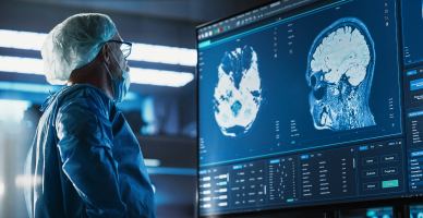
Spinal surgery may remove or reduce a tumour, relieve pressure on the spinal cord, and obtain tissue for diagnosis. Neuronavigation and neurophysiological monitoring may be used. Spinal stabilisation may be required if tumour removal or bone involvement makes the spine unstable.

A tumour can block normal cerebrospinal fluid circulation and cause hydrocephalus. A temporary drain, shunt, or endoscopic third ventriculostomy may be used to relieve pressure.
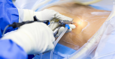
Laser interstitial thermal therapy uses a thin laser probe placed into the tumour under image guidance. Heat is used to destroy selected tumour tissue. It may be considered for carefully selected deep, recurrent, or difficult-to-access lesions and is not suitable for every brain tumour.
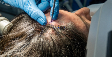
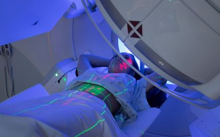
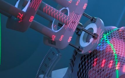
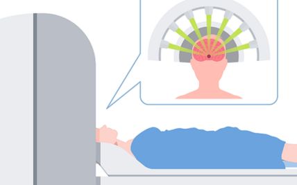
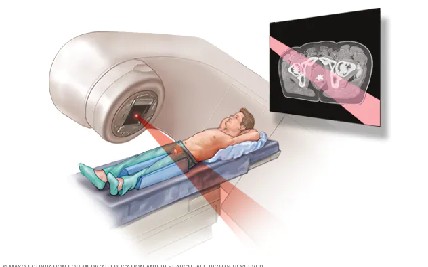
External beam radiation may be used after surgery, as the main treatment when surgery is not possible, or to control recurrent or metastatic disease. The treatment area and dose depend on the tumour type, grade, location, age, and proximity to critical structures.
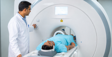
Three-dimensional conformal radiation shapes the treatment beams around the tumour using planning scans. It may be used when the target can be treated safely with a conformal approach.
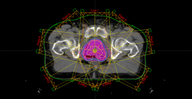
Intensity-modulated radiation therapy and volumetric-modulated arc therapy adjust the radiation dose across multiple beam angles. These techniques can help reduce exposure to the optic nerves, brainstem, spinal cord, cochlea, pituitary gland, and other nearby structures.
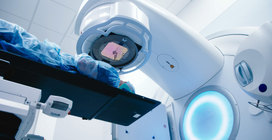
Proton therapy uses proton beams that release most of their radiation within the planned treatment area, with little radiation continuing beyond it. This can reduce the total radiation dose received by some surrounding tissues.
It may be considered for selected paediatric brain tumours, medulloblastoma requiring craniospinal irradiation, ependymoma, low-grade glioma, skull-base chordoma or chondrosarcoma, spinal tumours, and selected recurrent cancers. Its potential value is greatest when reducing radiation exposure to developing brain tissue, the heart, lungs, bowel, endocrine organs, or other distant structures is clinically important.
Proton therapy cannot guarantee preservation of neurological, cognitive, hormonal, visual, or hearing function when the tumour involves or lies close to the structures responsible. Suitability is determined through individual assessment and comparison with established photon-radiation techniques.
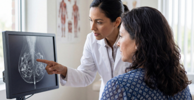
Image guidance confirms positioning before or during treatment. This is important when small differences in position could affect the tumour or a nearby critical structure.
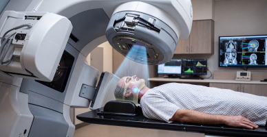
Stereotactic radiosurgery delivers a highly focused radiation dose to a small target, usually in one session. Despite its name, it does not involve an incision. It may be used for selected brain metastases, vestibular schwannomas, meningiomas, pituitary tumours, arteriovenous malformations, and other suitable lesions.
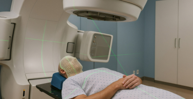
Stereotactic radiotherapy delivers focused radiation over several sessions. Dividing the dose may be appropriate when a lesion is larger or close to a sensitive structure.
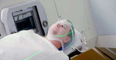
Craniospinal irradiation treats the brain and the length of the spinal fluid pathway. It is used for tumours such as medulloblastoma that can spread through cerebrospinal fluid. The dose and treatment fields are adapted according to age, tumour type, molecular risk, and disease spread.
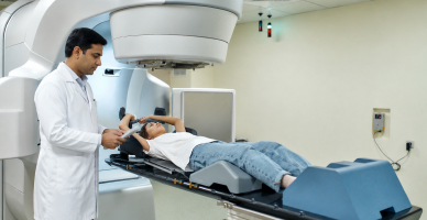
Chemotherapy may be used for selected gliomas, medulloblastomas, ependymomas, lymphomas, germ-cell tumours, and paediatric brain cancers. Its effectiveness depends partly on the tumour type and the ability of the medicine to reach the central nervous system.
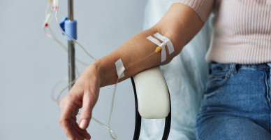
Targeted medicines act on particular proteins or molecular changes within tumour cells. They may be used for selected tumours with BRAF, NTRK, or other actionable changes. A molecular alteration does not automatically mean that a targeted medicine will be appropriate. The tumour type, evidence, stage, and previous treatment must also be considered.
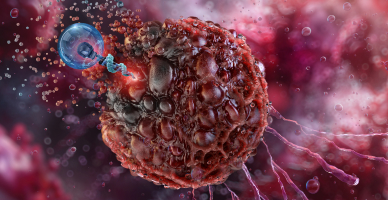
Immunotherapy has an established role in treating selected brain metastases from cancers such as melanoma or lung cancer. Its role in most primary brain tumours remains limited and continues to be studied.
Medicines are the first treatment for many hormone-producing pituitary tumours, particularly prolactinomas. Other pituitary tumours may require surgery, radiation, hormone replacement, or combined treatment.
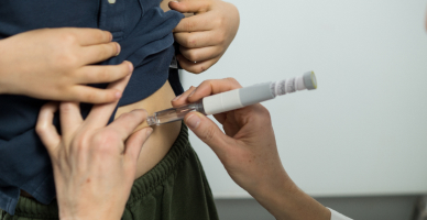
High-dose chemotherapy followed by return of the patient’s stored stem cells may be used in selected primary CNS lymphomas, recurrent germ-cell tumours, or paediatric brain tumours. This is a specialised treatment and is not routinely used for most neurological cancers.
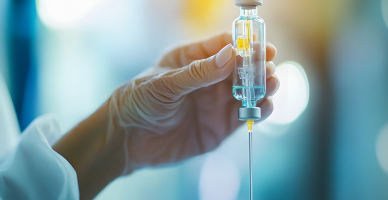
Integrated pathology and molecular testing can refine the diagnosis, estimate prognosis, and identify treatment options or suitable clinical trials. Precision oncology does not replace established treatments such as surgery, radiation, and tumour-specific chemotherapy.
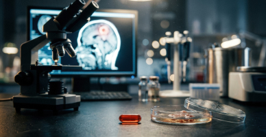
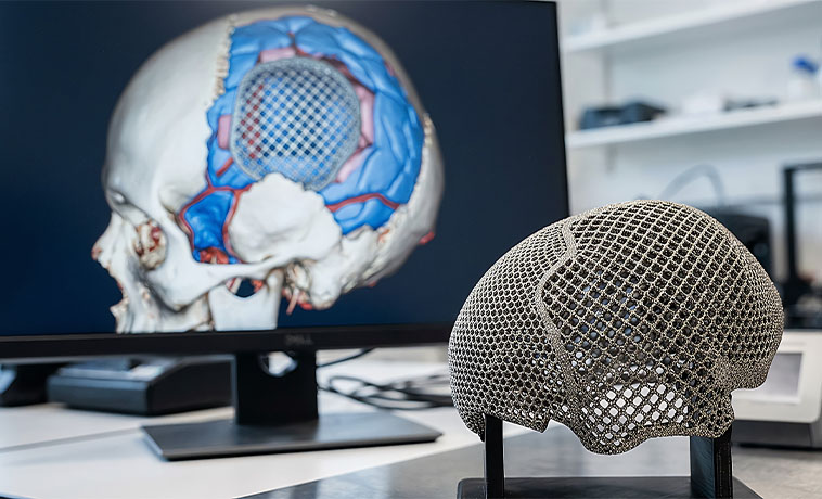
Cranioplasty repairs an opening or defect in the skull using the patient’s own bone or a customised implant. It may be performed during tumour surgery or after swelling or infection has resolved. The procedure protects the brain and restores the contour of the skull.
The dura is the protective membrane surrounding the brain and spinal cord. If it is opened or removed during surgery, a graft may be used to create a watertight closure. This helps protect the nervous system and reduce the risk of cerebrospinal fluid leakage.
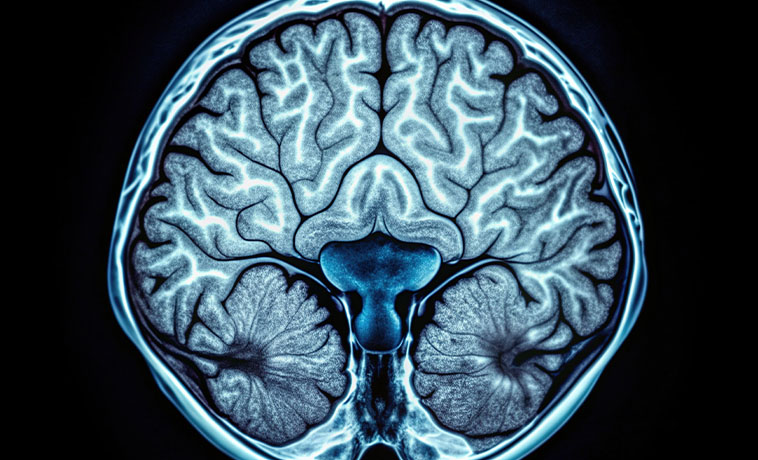
After endoscopic or open skull-base surgery, tissue from within the nose, nearby areas, or another part of the body may be used to separate the brain from the nasal passages or sinuses. The reconstruction provides a barrier against cerebrospinal fluid leakage and infection.
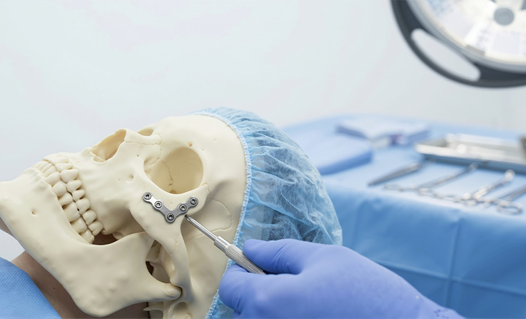
Large or previously treated surgical areas may require skin grafts, local flaps, or free-tissue transfer to cover the skull, implants, or deeper structures.
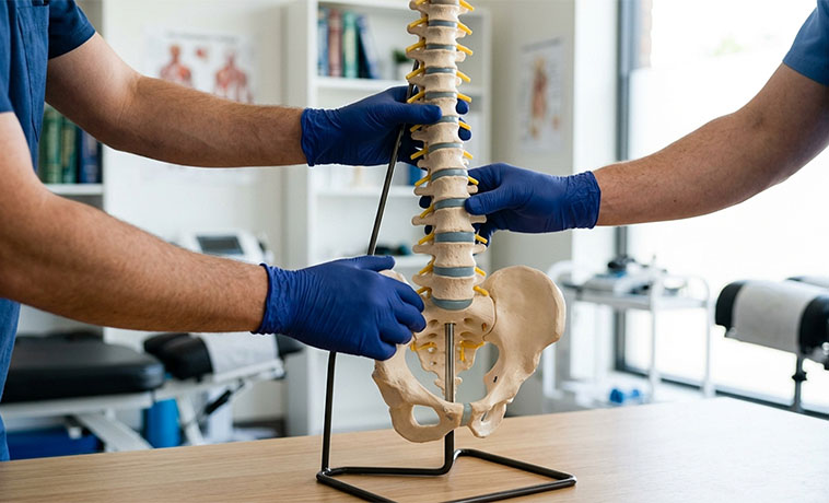
Tumours involving the vertebrae or surgery that removes supporting bone can make the spine unstable. Screws, rods, cages, bone grafts, or other implants may be used to restore alignment and protect the spinal cord.
Prognosis for Neurological Cancer
The outlook varies widely between brain and spinal tumours. Some can be controlled or treated successfully, while others are infiltrative, recurrent, or biologically aggressive.
Factors influencing prognosis include:
Tumour type and molecular subtype
Grade
Tumour location and size
Spread through the brain, spine, or cerebrospinal fluid
Whether the tumour is primary or metastatic
Extent of safe surgical removal
Age and overall health
Neurological function at diagnosis
Molecular features such as IDH status or MGMT promoter methylation
Response to radiation or systemic treatment
Whether the tumour has returned
Ability to complete treatment and follow-up
Some meningiomas, schwannomas, pituitary tumours, low-grade gliomas, and paediatric tumours can have favourable long-term outcomes. Glioblastoma and certain other high-grade tumours are more difficult to control.
Prognosis should be discussed according to the integrated diagnosis rather than using one survival figure for all neurological cancers.


Why Choose ACC for Neurological Cancer Treatment
Multidisciplinary neuro-oncology teams
Specialist care for primary, metastatic, adult, paediatric, brain, spinal, and skull-base tumours
Advanced MRI, functional imaging, tractography, and molecular pathology
Maximal safe resection supported by neuronavigation, functional mapping, and neurophysiological monitoring
Awake craniotomy and minimally invasive neuroendoscopic approaches for selected tumours
Complex skull-base and spinal tumour surgery
Stereotactic radiosurgery and fractionated stereotactic radiotherapy
Advanced radiation techniques, including IMRT, IGRT, craniospinal irradiation, and proton therapy for selected patients
Tumour-specific chemotherapy, targeted therapy, and precision-oncology services
Specialist paediatric neuro-oncology and anaesthesia support
Cranioplasty, skull-base, scalp, dural, and spinal reconstruction
Integrated neurological, cognitive, speech, swallowing, and physical rehabilitation
Multidisciplinary tumour-board review focused on tumour control and preservation of neurological function
Health Blogs