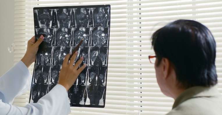
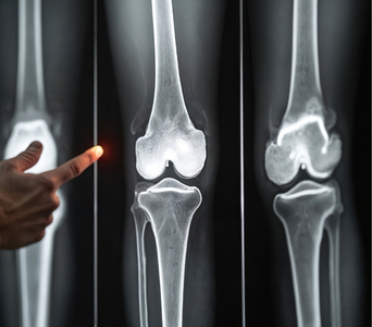
An X-ray, also called radiography, is a quick and non-invasive imaging test that uses a small amount of ionising radiation to create pictures of structures inside the body. It is particularly useful for examining the lungs, chest and bones.
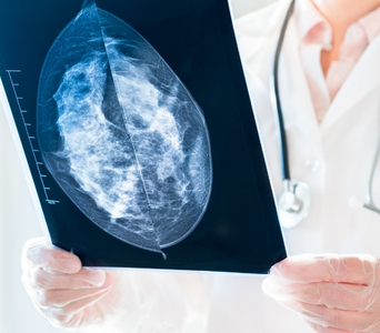
In cancer care, X-rays may help investigate symptoms, identify certain chest or bone abnormalities and assess complications related to cancer or its treatment. However, X-rays provide less detail than CT or MRI and usually cannot confirm or rule out cancer on their own.
If an X-ray shows a suspicious or unclear finding, additional assessment with CT, MRI, ultrasound, PET-CT, endoscopy or biopsy may be required.



Role in Cancer Care
What to Expect
Before the Test
Most X-ray examinations require little or no preparation. You can usually eat, drink and take your regular medicines unless given different instructions.
Before the examination, inform the team if you:
Are pregnant or may be pregnant
Have recently undergone imaging of the same area
Have difficulty standing, changing position or holding your breath
Have pain that may make positioning uncomfortable
Have any tubes, drains or medical devices in the area being examined
You may be asked to remove jewellery, eyeglasses, removable dental work or clothing containing metal. Depending on the area being examined, you may need to change into a hospital gown.
Bring previous images and reports whenever possible. Comparison with earlier examinations can help determine whether a finding is new, stable or changing.

During the Test
The technologist will position you standing, sitting or lying on an examination table. The position depends on the body area and the views required.
You may be asked to remain still and hold your breath for a few seconds while the image is taken. This helps prevent movement from blurring the image. The technologist may reposition you to obtain additional views from different angles.
The X-ray exposure itself lasts only a fraction of a second. The entire examination is usually completed within a few minutes, although it may take longer if several views are required or movement is difficult.
The examination is painless, but positioning may cause temporary discomfort if the area is injured or painful. Tell the technologist if you need assistance.

After the Test
No recovery period is required, and you can usually return to your normal activities immediately. No radiation remains in your body after the examination.
You may be asked to wait briefly while the images are checked for quality. If additional views are required, they may be taken before you leave.
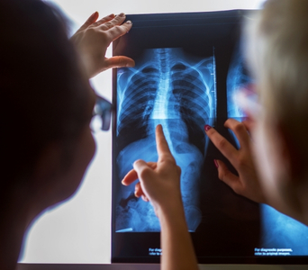
A radiologist will examine the images and prepare a report for the doctor who requested the test. Further imaging or other investigations may be advised if a finding is suspicious, unclear or not fully evaluated by X-ray.

Potential Benefits
- Quick examination: Most X-rays can be completed within a few minutes.
- Non-invasive imaging: The test does not usually require injections, instruments or sedation.
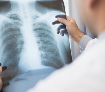
- Chest assessment: Chest X-rays can identify some lung abnormalities, fluid collections and treatment-related complications.
- Bone evaluation: X-rays can show certain bone lesions, areas of bone destruction and fractures.
- Complication assessment: Imaging may help investigate acute symptoms or complications during cancer treatment.
- Device-position confirmation: X-rays may be used to check the position of selected tubes, drains, lines or ports.
- Widely available: X-ray equipment is available in many outpatient, emergency and inpatient settings.
- Low radiation exposure: A conventional X-ray generally uses a relatively small amount of ionising radiation, although the dose varies by examination.