icon
icon
Overview

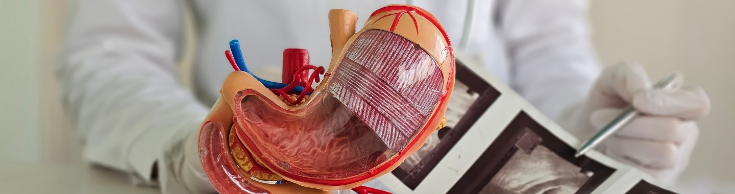
Endoscopic ultrasound (EUS) combines endoscopy with high-frequency ultrasound to produce detailed images of the digestive tract and nearby organs. It can examine the layers of the gastrointestinal wall and structures such as the pancreas, bile ducts, gallbladder, liver and surrounding lymph nodes.

EUS may be performed for imaging alone or combined with fine-needle aspiration or fine-needle biopsy to collect cells or tissue. Upper EUS is performed through the mouth, while lower EUS is introduced through the anus to examine the rectum and nearby structures.
endoscopic
How the Technology Works
EUS places an ultrasound probe close to the area being examined, allowing detailed imaging from inside the digestive tract.
work-icon
Endoscope Placement
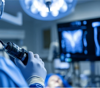
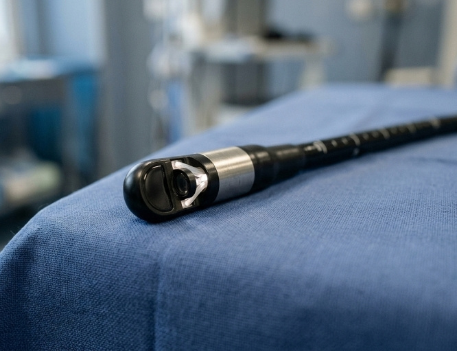
A thin, flexible endoscope with a miniature ultrasound probe at its tip is guided through the mouth into the oesophagus, stomach and duodenum, or through the anus into the rectum. Its camera allows the doctor to examine the inner lining while positioning the ultrasound probe near the area of interest.
work-icon
Ultrasound Imaging
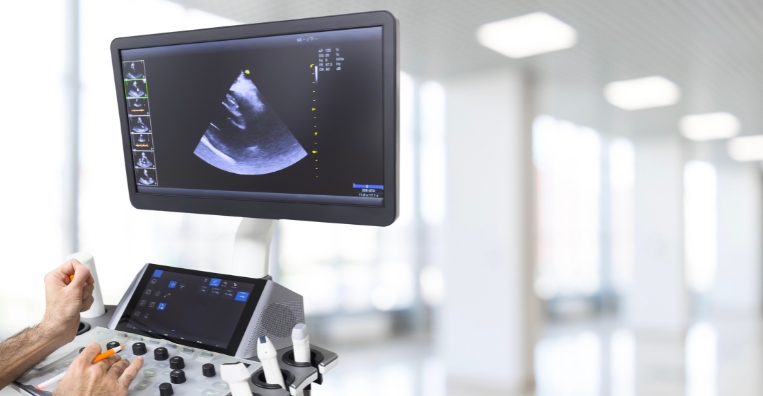
The probe emits high-frequency sound waves that reflect from the gastrointestinal wall, nearby organs, blood vessels and lymph nodes. A computer converts these returning echoes into detailed images that can show the depth, structure and relationship of an abnormality to surrounding tissues.
work-icon
Needle Guidance
When tissue sampling is required, real-time ultrasound helps guide a thin needle through the gastrointestinal wall and into a mass, lymph node, cyst or other abnormality. The collected cells or tissue are sent to a laboratory for cytological, histopathological and, where appropriate, biomarker or molecular analysis.

Role in Cancer Care

Evaluating Pancreatic Abnormalities
EUS provides detailed images of the pancreas and can help assess solid masses, cystic lesions and changes that may be difficult to characterise on other tests. It can show a lesion’s size, location and relationship to the pancreatic duct and nearby blood vessels. EUS-guided sampling may help determine whether an abnormality is cancerous and identify its type.
Evaluating Pancreatic Abnormalities
Assessing Biliary Cancers
EUS can examine the bile ducts, gallbladder and nearby structures when cancer or another cause of obstruction is suspected. It may help evaluate a mass, unexplained bile-duct narrowing or enlarged lymph nodes identified on imaging. Tissue sampling may be performed when the abnormality can be reached safely.
Assessing Biliary Cancers
Examining Gastrointestinal Tumours
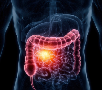
EUS can assess cancers and other tumours affecting the oesophagus, stomach, duodenum and rectum. It can show which layers of the gastrointestinal wall are involved and whether nearby structures appear affected. It is also useful for evaluating subepithelial lesions that develop beneath an apparently normal surface lining.
Examining Gastrointestinal Tumours
Assessing Local Spread
EUS can help determine how deeply a tumour has grown into or through the gastrointestinal wall. It can also examine nearby lymph nodes, blood vessels and organs for possible involvement. These findings contribute to local staging but do not replace CT, MRI, PET-CT or other tests used to assess disease elsewhere in the body.
Assessing Local Spread
Obtaining Tissue Samples
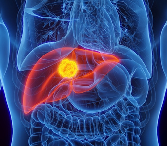
EUS-guided fine-needle aspiration or fine-needle biopsy can collect samples from abnormalities that may be difficult to reach through the skin or standard endoscopy. These can include pancreatic masses, gastrointestinal-wall lesions, lymph nodes and selected liver or other nearby lesions. Occasionally, the sample may be insufficient or inconclusive, and repeat sampling or another biopsy method may be required.
Obtaining Tissue Samples
Planning Cancer Treatment
Information from EUS can help determine whether surgery, systemic therapy, radiotherapy or another approach may be appropriate. It may clarify a tumour’s relationship to important blood vessels and help identify lymph nodes or lesions that require sampling before treatment. EUS findings are interpreted alongside pathology, other imaging and multidisciplinary clinical assessment.
Planning Cancer Treatment

What to Expect

Before the Test 

Preparation depends on whether upper or lower EUS is planned. For upper EUS, you will be asked to stop eating and drinking for a specified period; lower or rectal EUS may require a clear-liquid diet, laxative preparation or an enema.

Tell the clinical team about:

  • All medicines and supplements you take, particularly anticoagulants and antiplatelet medicines

  • Diabetes and any medicines that may require adjustment while fasting

  • Bleeding disorders or significant heart or lung conditions

  • Allergies or previous reactions to sedatives, anaesthetics, latex or medicines

  • Pregnancy or the possibility of pregnancy

  • Dentures, removable dental work or loose teeth before upper EUS

Do not stop prescribed medicines unless instructed by your clinical team. Blood tests may be required before needle sampling, and antibiotics may be advised for selected procedures. If sedation or general anaesthesia is planned, arrange for a responsible adult to take you home and follow the instructions about supervision afterwards.

Before the Test

During the Test 

You will usually lie on your side while your heart rate, blood pressure and oxygen level are monitored. Upper EUS is commonly performed with throat-numbing medicine and intravenous sedation; sedation requirements for lower EUS depend on the examination planned.

For upper EUS, the endoscope is passed through the mouth into the oesophagus, stomach and duodenum. For lower EUS, it is passed through the anus into the rectum or lower bowel. The scope does not interfere with breathing.

The doctor positions the ultrasound probe near the area being assessed and examines the resulting images. If required, a needle is guided into the abnormality to collect cells or tissue. The examination commonly takes approximately 30–60 minutes, although tissue sampling or additional procedures may take longer.

During the Test

After the Test

You will be monitored until the effects of sedation have reduced. Following upper EUS, do not eat or drink until the throat numbness has worn off and swallowing is safe. A temporary sore throat, bloating, mild abdominal discomfort or increased wind may occur.

If you received sedation, do not drive, operate machinery, drink alcohol, make important decisions or sign legal documents for the period specified by the clinical team. Arrange for someone to accompany you home and remain with you if advised.

The doctor may explain the initial imaging findings shortly after the procedure. Cytology or biopsy results take longer and will be discussed once laboratory analysis is complete.

Seek urgent medical advice if you develop:

  • Severe or increasing chest or abdominal pain

  • Persistent vomiting

  • Fever or chills

  • Breathing or swallowing difficulty

  • Vomiting blood or passing black or bloody stools

  • Fainting, marked weakness or rapidly worsening symptoms

After the Test

Potential Benefits

EUS may provide several benefits when investigating or managing suspected or confirmed cancer:
  • Detailed Imaging: It provides high-resolution images of the gastrointestinal wall and nearby organs.
  • Close Evaluation: It examines abnormalities from inside the digestive tract, placing the ultrasound probe close to the area of interest.
  • Layer Assessment: It can show how deeply a tumour has grown through the layers of the gastrointestinal wall.
  • Nodal Assessment: It can examine nearby lymph nodes and guide sampling when required.
  • Targeted Sampling: It enables real-time needle sampling of selected masses, cysts, lymph nodes and other abnormalities.
  • Treatment Planning: Its findings can support decisions about surgery, systemic therapy, radiotherapy and other treatments.
  • Minimal Incisions: It can obtain tissue from some difficult-to-reach areas without requiring an external surgical incision.
  • Radiation-Free Imaging: The ultrasound component creates images using sound waves rather than ionising radiation.
Endoscopic Ultrasound (EUS)
Frequently Asked Questions
01 How is EUS different from standard endoscopy?
icon icon
Standard endoscopy mainly allows the doctor to inspect the inner lining of the digestive tract. EUS adds an ultrasound probe that can show the deeper layers of the gastrointestinal wall and organs, lymph nodes and blood vessels beyond it. Both visual endoscopy and ultrasound imaging can be performed during the same examination.
02 Will EUS be painful?
icon icon
Most people receive sedation for upper EUS and do not find the procedure painful, although temporary pressure, gagging or bloating may occur. Lower EUS may be performed with or without sedation, depending on the extent of the examination. The team can adjust comfort measures when necessary.
03 Is a biopsy always performed during EUS?
icon icon
No. Some examinations are performed for imaging alone. Fine-needle aspiration or fine-needle biopsy is added only when tissue or fluid is needed and the abnormality can be sampled safely.
04 Can EUS confirm cancer?
icon icon
EUS images may show features that raise or lower suspicion for cancer, but imaging alone usually cannot provide a definitive diagnosis. When a sample is collected, a pathologist examines it to determine whether cancer is present and identify its type. An inconclusive sample may need to be repeated or followed by another biopsy method.
05 Can EUS determine the complete stage of cancer?
icon icon
EUS is particularly useful for assessing the depth of selected gastrointestinal tumours and nearby lymph nodes or structures. It cannot reliably evaluate every part of the body or exclude distant spread. Complete staging may also require CT, MRI, PET-CT, surgery or other investigations.
06 What complications are possible?
icon icon
Most EUS examinations are completed without a serious complication. Possible problems include a reaction to sedation, aspiration, bleeding, infection or, rarely, a tear in the digestive tract; needle sampling can also carry a small risk of pancreatitis when the pancreas is sampled. The individual risk depends on the area examined and whether a biopsy or another intervention is performed.
image image
Book an Appointment
Please Enter Your Name
Please Enter Your Mobile Number
Please Enter OTP