




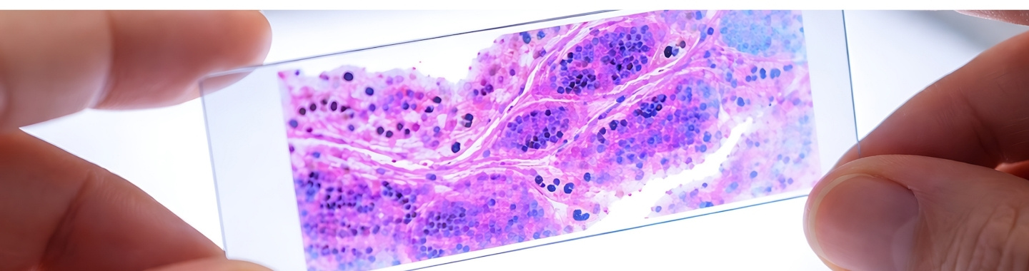
Histopathology is the examination of tissue under a microscope to identify disease and study its characteristics. In cancer care, it plays a central role in confirming whether a tissue sample is cancerous, identifying the type and subtype of tumour, and assessing features that may influence prognosis and treatment.
Tissue may be obtained through a needle biopsy, endoscopic biopsy, surgical biopsy or cancer surgery. Depending on the specimen, histopathology may also provide information about tumour grade, local extent, lymph-node involvement, surgical margins and response to treatment given before surgery. The findings are interpreted alongside the patient’s clinical history, imaging and, when required, immunohistochemistry or molecular test results.
Samples and Methods


How It Is Performed
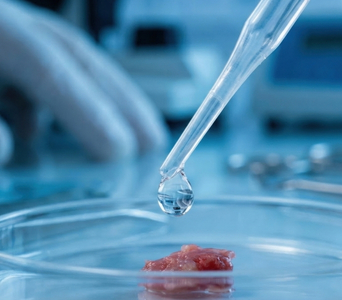
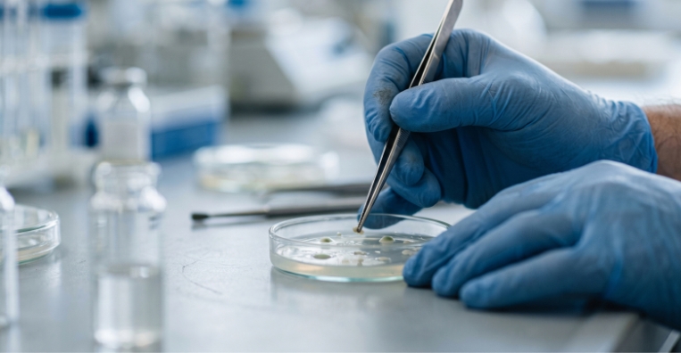
- Sample Receipt: The tissue is received in the laboratory with identifying details, the specimen site and relevant clinical information. Correct labelling and appropriate preservation are essential for reliable evaluation.
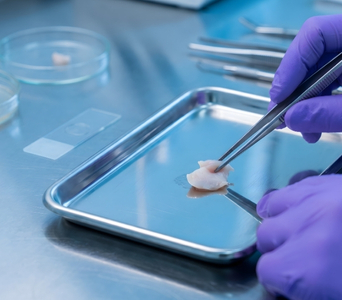
- Gross Examination: The specimen is inspected and described, including its size, appearance and visible abnormalities. Representative areas are selected for microscopic examination.
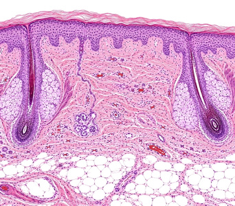
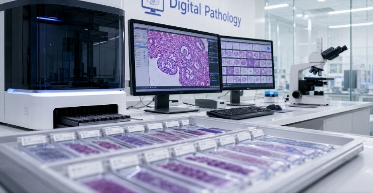
- Tissue Processing: The selected tissue is usually preserved in formalin, processed and embedded in paraffin wax. Very thin sections are then cut, placed on glass slides and stained.
- Microscopic Examination: A pathologist examines the prepared slides and correlates the findings with the available clinical and imaging information. Additional stains or molecular tests may be requested before the diagnosis is finalised.
- Pathology Reporting: The final report summarises the diagnosis and other clinically relevant findings. For surgical specimens, this may include tumour type, grade, margins, lymph-node status and pathological stage, where applicable.
Advantages
- Confirms many cancers and distinguishes malignant tumours from non-cancerous conditions.
- Identifies the tumour’s type, subtype and, where applicable, grade.
- Helps assess tumour extent, lymph-node involvement and pathological stage.
- Determines whether cancer cells are present at the examined surgical margins.
- Supports biomarker testing that may help guide treatment decisions.
- Allows stored tissue blocks and slides to be reviewed or tested again when required.



