




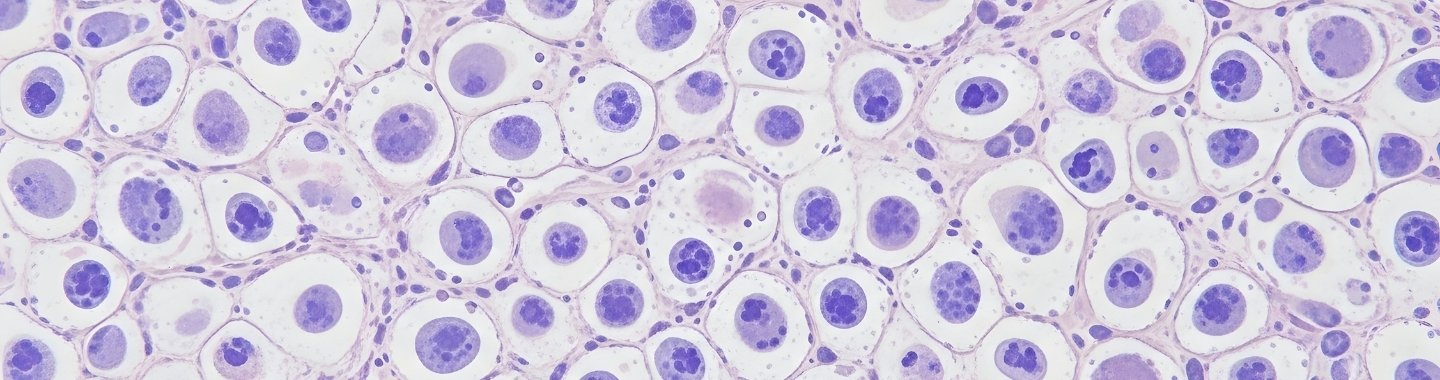
Overview
Hematopathology is the specialised examination of diseases affecting the blood, bone marrow, lymph nodes and other lymphatic tissues. It plays a central role in diagnosing and classifying leukemias, lymphomas, plasma-cell neoplasms, myelodysplastic neoplasms and myeloproliferative neoplasms.
Diagnosis often requires an integrated assessment of blood counts, microscopic appearance, flow cytometry, tissue histopathology, immunohistochemistry, cytogenetics and molecular findings. These results can also provide prognostic information, help guide treatment and support disease monitoring.
Samples and Methods
Peripheral Blood
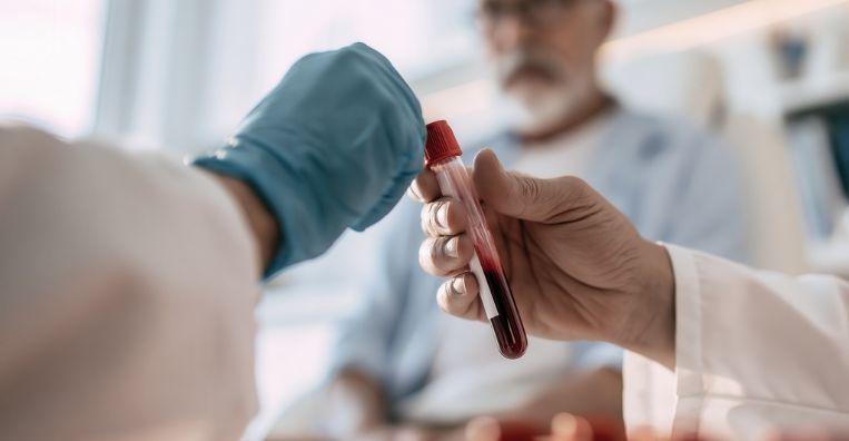
A complete blood count and blood-film examination can identify abnormal cell numbers, immature cells and changes in cell appearance.
Bone Marrow
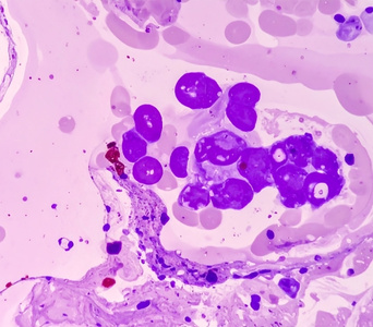
Bone marrow aspiration provides individual cells for detailed assessment, while a bone marrow biopsy shows the organisation and cellularity of the marrow. The two methods often provide complementary information.

Lymphatic Tissue
Lymph nodes or other involved tissues may be sampled through core or excision biopsy. Preserving tissue architecture is particularly important when investigating lymphoma.
Flow Cytometry
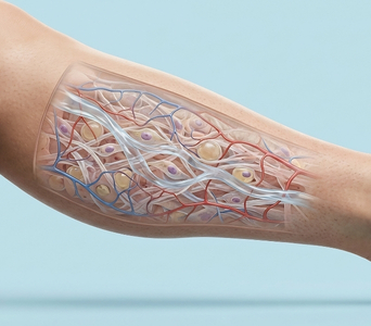
Flow cytometry examines patterns of markers on or within individual cells. It can identify abnormal cell populations and help classify selected leukemias, lymphomas and plasma-cell disorders.
Cytogenetic Analysis
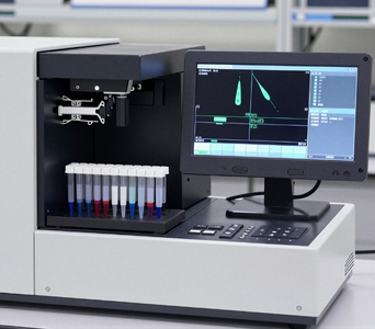
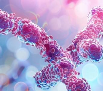
Karyotyping and fluorescence in-situ hybridisation can detect selected chromosomal changes associated with particular hematological malignancies.
Molecular Analysis
PCR and next-generation sequencing may identify mutations, gene rearrangements or other changes relevant to diagnosis, risk assessment, treatment selection or disease monitoring.

How It Is Performed
- Clinical Review: The laboratory reviews the clinical history, blood counts, imaging findings and previous treatment information supplied with the sample.
- Morphological Assessment: Blood films, bone marrow preparations or tissue sections are examined under a microscope to assess the appearance, number and distribution of cells.
- Ancillary Testing: Flow cytometry, immunohistochemistry, cytogenetic analysis and molecular testing are selected according to the suspected condition and initial findings.
- Integrated Interpretation: The results are considered together using recognised diagnostic criteria. A diagnosis is not generally based on a single isolated laboratory finding.
- Final Reporting: An integrated report summarises the diagnosis and relevant prognostic or predictive features. Some findings may be issued later as an addendum because specialised tests have different processing times.

Advantages
- Combines multiple laboratory findings to classify complex blood and lymphatic cancers accurately.
- Distinguishes between conditions that may appear similar on routine blood tests or microscopy.
- Helps place selected diseases into recognised prognostic or risk groups.
- Identifies biomarkers that may help guide treatment selection.
- Assesses treatment response and detects persistent or recurrent disease in selected conditions.
- May detect measurable residual disease that is not identified through routine examination.
Frequently Asked Questions
01
Why are several tests needed to diagnose blood cancer?


Many blood cancers can appear similar on a single test. Combining morphology, immunophenotyping, cytogenetics and molecular findings allows more accurate classification.
02
Are bone marrow aspiration and biopsy the same?
No. Aspiration collects liquid marrow and individual cells, while a biopsy removes a small core of tissue so that marrow structure can be examined. They are often performed together.
03
What is flow cytometry?
Flow cytometry assesses markers on or within individual cells. It can detect abnormal cell populations and help classify certain leukemias, lymphomas and plasma-cell disorders.
04
What is measurable residual disease?
Measurable residual disease refers to small numbers of cancer cells that remain during or after treatment but may not be detected through routine examination. It is assessed only in selected diseases using validated tests.
05
Can the diagnosis change after molecular results are received?
The initial diagnosis may be refined when genetic or molecular findings become available because the classification of many hematological malignancies incorporates these features.


Book an Appointment