




Overview
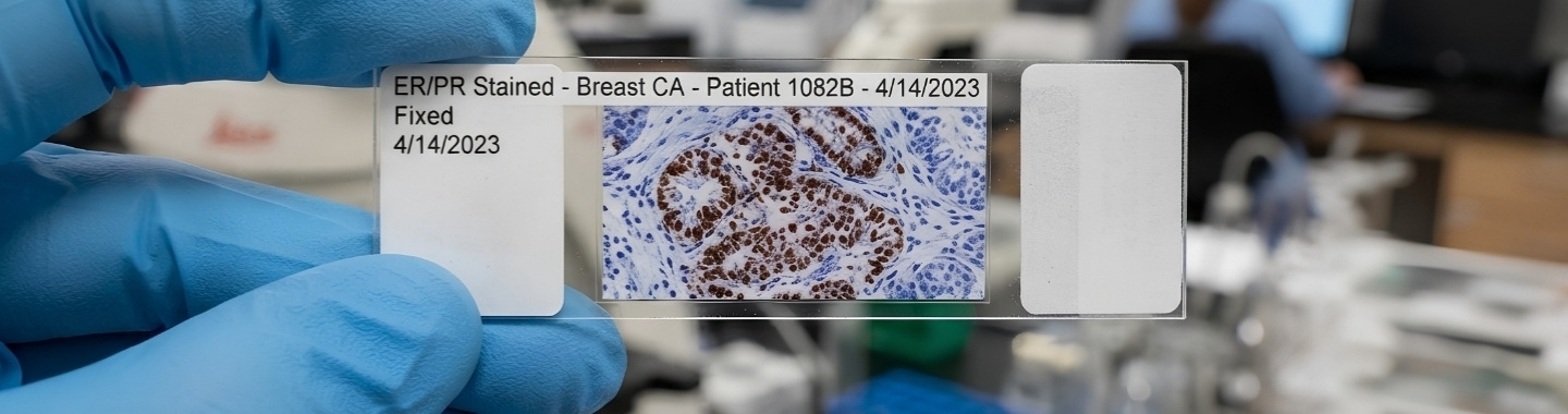
Immunohistochemistry, or IHC, is a laboratory method that uses antibodies to detect specific proteins in tissue. The resulting staining pattern is examined under a microscope and interpreted alongside the tissue’s appearance and the patient’s clinical information.
In cancer care, IHC can help determine a tumour’s cell lineage or likely site of origin, distinguish between tumours with similar appearances and assess selected biomarkers that may influence prognosis or treatment. It complements routine histopathology rather than replacing it.
Samples and Methods
Tissue Biopsies
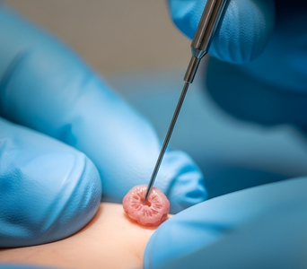
IHC can be performed on tissue obtained through needle, endoscopic or surgical biopsy. The tissue must contain sufficient preserved cells for reliable assessment.

Surgical Specimens
Tissue removed during surgery may be tested to refine tumour classification or assess biomarkers relevant to further treatment.
Cell Blocks
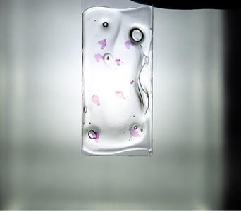
Cellular material collected through fine-needle aspiration or from body fluids may be processed into a cell block suitable for selected IHC tests.
Marker Panels
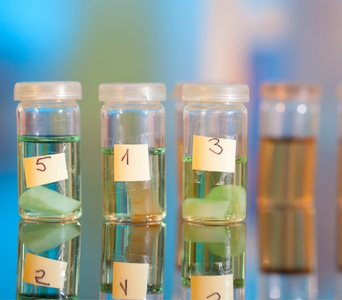
Pathologists usually select a panel of antibodies based on the tumour’s microscopic appearance and the possible diagnoses. A panel is often more informative than a single marker.
Biomarker Scoring
Some biomarkers are assessed using validated cancer-specific scoring systems based on staining intensity, distribution or the proportion of cells stained.
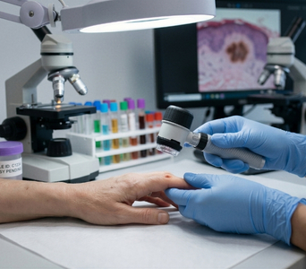
How It Is Performed
- Tissue Selection: The pathologist reviews the specimen and selects a representative tissue block containing sufficient viable tumour.
- Slide Preparation: Thin sections are cut from the tissue block and placed on specially prepared glass slides.
- Antibody Staining: The tissue sections are treated with antibodies designed to bind to specific proteins. A detection system makes the resulting staining visible.
- Quality Control: Appropriate controls are examined to confirm that the staining process has worked as expected and that the result can be interpreted.
- Result Interpretation: The pathologist assesses the location, intensity and distribution of staining in the context of the tumour’s microscopic features. The results are then incorporated into the pathology report or issued as an addendum.

Advantages
- Helps determine the cell lineage and type of a tumour when routine microscopy is inconclusive.
- May help identify the likely primary site of a metastatic tumour.
- Distinguishes between tumours with similar microscopic appearances.
- Provides selected prognostic or predictive information relevant to treatment planning.
- Allows multiple markers to be assessed using tissue already collected during biopsy or surgery.
- Complements histopathology and molecular testing to provide a more complete tumour profile.
Frequently Asked Questions
01
Why is IHC performed after a biopsy?


Routine microscopic examination may not fully identify a tumour or determine its origin. IHC provides additional information about the proteins expressed by the tumour cells.
02
Can one IHC marker confirm the type of cancer?
Usually not. Most diagnoses require interpretation of a panel of markers alongside the tumour’s microscopic appearance, clinical history and imaging findings.
03
Can IHC determine where a metastatic cancer began?
IHC may help narrow down the likely primary site, but it is not always conclusive. Imaging, endoscopy or molecular testing may still be required.
04
Does a positive biomarker guarantee that treatment will work?
No. A positive biomarker may indicate that a treatment can be considered, but it cannot guarantee a response. Treatment decisions also depend on the cancer type, validated test criteria and the patient’s clinical condition.
05
Why might IHC need to be repeated?
Repeat testing may be needed if the sample is inadequate, the controls are unsatisfactory, the result is borderline or a more recent tumour sample is clinically preferable.


Book an Appointment