Hematological Cancer
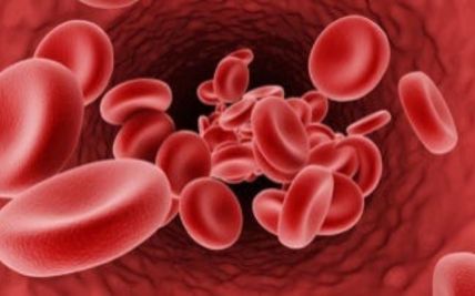
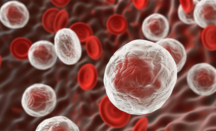
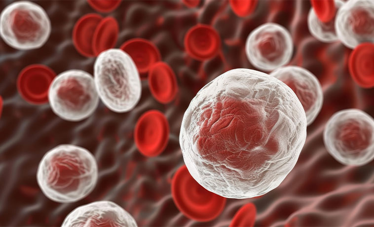
Hematological cancers, more commonly known as blood cancers, are cancers that begin in the bone marrow (the spongy tissue inside our bones where blood cells are produced) or in the lymphatic system (the network of tissues and organs that helps the body fight infection). They occur when abnormal blood cells begin growing out of control, crowding out and interrupting the function of healthy blood cells that are needed to fight infection, carry oxygen and help blood clot.
Blood cancers broadly fall into three major groups: leukemia, which involves abnormal white blood cells crowding out healthy cells in the marrow and bloodstream; lymphoma, which begins in the lymph nodes or lymphatic tissue; and myeloma, which arises from plasma cells in the bone marrow. Because these cancers affect blood formation and immunity throughout the entire body rather than forming a single, localised tumour, treatment calls for deep hematology expertise combined with access to advanced, hospital-wide supportive care.
At Apollo Cancer Centres, hematological cancer care is led by dedicated hemato-oncologists working alongside transplant physicians, radiation and medical oncologists, pathologists and transfusion medicine specialists — supported by one of the country's most experienced bone marrow transplant (BMT) programmes. Together, this team designs a treatment plan built around each patient's specific cancer type, genetic profile and overall health, with the goal of achieving remission and, wherever possible, long-term cure.
Types of Hematological Cancer
Blood cancer is not a single disease. Each type affects different blood or immune cells and requires its own diagnostic, risk-classification, and treatment approach.
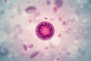
Acute lymphoblastic leukemia, or ALL, develops from immature lymphoid cells in the bone marrow. It progresses quickly and can affect the blood, lymph nodes, spleen, liver, brain, spinal fluid, or other organs. ALL is the most common childhood cancer but also occurs in adults. It is classified as B-cell or T-cell ALL, with further molecular subtypes that influence treatment and prognosis.
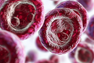
Acute myeloid leukemia, or AML, develops from immature myeloid cells and can rapidly disrupt the production of normal blood cells. It occurs at all ages but is more common in older adults. AML includes several molecular and genetic subtypes. Acute promyelocytic leukemia is a distinct subtype requiring urgent treatment with medicines that help the abnormal cells mature.

Chronic lymphocytic leukemia, or CLL, develops from mature B lymphocytes. It usually progresses more slowly than acute leukemia and may be discovered during a routine blood test before symptoms develop. Some people can be monitored for years without treatment. Others develop enlarging lymph nodes or spleen, falling blood counts, infections, or other signs that require therapy.
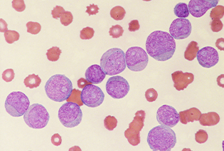
Chronic myeloid leukemia, or CML, develops from myeloid cells in the bone marrow. Most cases contain an abnormal gene called BCR::ABL1, formed by the Philadelphia chromosome. Medicines that specifically block the BCR::ABL1 protein have transformed the management of CML. Many patients can achieve long-term disease control with tablets, although regular molecular monitoring remains essential.
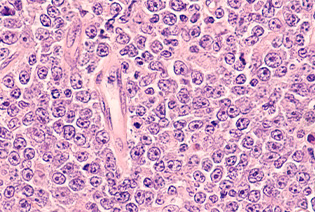
Hodgkin lymphoma begins in lymphocytes and is characterised by abnormal cells known as Reed–Sternberg cells in classical Hodgkin lymphoma. It commonly causes painless lymph-node enlargement and may also produce fever, night sweats, or weight loss. Treatment depends on the stage, disease bulk, symptoms, and response shown on PET-CT.
Non-Hodgkin lymphoma includes a large and diverse group of B-cell, T-cell, and natural killer-cell cancers. Some are slow-growing, while others progress rapidly and require prompt treatment. Common subtypes include diffuse large B-cell lymphoma, follicular lymphoma, mantle-cell lymphoma, Burkitt lymphoma, peripheral T-cell lymphoma, and several others. Accurate tissue diagnosis is essential because treatment differs substantially between subtypes.
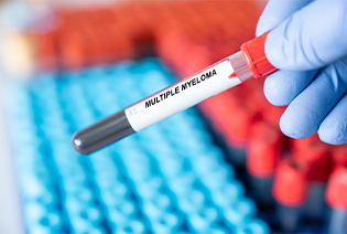
Multiple myeloma develops from plasma cells, which normally produce antibodies. The abnormal plasma cells accumulate in the bone marrow and produce an abnormal protein. The disease can cause bone damage, anemia, kidney impairment, high calcium levels, infections, or other organ problems. Related conditions include monoclonal gammopathy of undetermined significance, smouldering myeloma, solitary plasmacytoma, and AL amyloidosis.
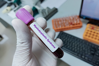
Myelodysplastic neoplasms, or MDS, develop when the bone marrow produces blood cells that are abnormal or do not mature properly. This may lead to anemia, infection, bleeding, or a combination of these problems. MDS ranges from lower-risk disease requiring monitoring or supportive treatment to higher-risk disease with a greater likelihood of progressing to AML.
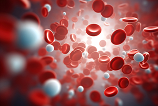
Myeloproliferative neoplasms, or MPNs, cause the bone marrow to produce too many blood cells. They include polycythemia vera, essential thrombocythemia, and primary myelofibrosis. These conditions may increase the risk of blood clots, bleeding, an enlarged spleen, bone-marrow scarring, or transformation to acute leukemia. Treatment is based on the MPN subtype and individual risk.
The cause of most blood cancers cannot be identified. Risk factors also differ significantly between leukemia, lymphoma, myeloma, MDS, and MPNs.
Modifiable Risk Factors
Few blood cancers have clearly preventable risk factors. Possible modifiable risks include:
Tobacco use: Associated with an increased risk of AML and several other cancers.
Benzene exposure: Long-term occupational exposure can increase the risk of AML and MDS.
Certain pesticide or chemical exposures: These have been associated with selected blood cancers, although individual risk is difficult to determine.
Some infections: Preventing or treating infections such as HIV, hepatitis C, or Helicobacter pylori may reduce the risk of particular lymphoma subtypes.
Unnecessary radiation or chemical exposure: Appropriate workplace protection can help limit exposure to recognised carcinogens.
Non-Modifiable Risk Factors
Increasing age
Previous chemotherapy or radiation therapy
A weakened immune system or long-term immunosuppressive treatment
HIV infection or previous organ transplantation
Certain viral infections, including Epstein–Barr virus and human T-cell lymphotropic virus type 1
Genetic conditions such as Down syndrome
Inherited bone-marrow failure or leukemia-predisposition syndromes
A family history of selected leukemias, lymphomas, myeloma, or related disorders
Monoclonal gammopathy of undetermined significance
Monoclonal B-cell lymphocytosis
Previous MDS or another bone-marrow disorder
Autoimmune or chronic inflammatory conditions associated with particular lymphoma subtypes
Increasing age and male sex for several blood cancers
Having a risk factor does not mean that blood cancer will develop. Most people with common symptoms such as tiredness or fever do not have blood cancer.
Signs and Symptoms
Symptoms depend on the cancer type, the blood cells affected, and the organs involved. Some slow-growing blood cancers are found before symptoms develop.
Possible signs and symptoms include:
These symptoms can also result from infection, nutritional deficiencies, autoimmune disease, or other non-cancerous conditions. Persistent symptoms, unexplained lymph-node enlargement, unusual bleeding, or significantly abnormal blood counts should be medically assessed.
Diagnosis begins with a detailed review of symptoms, previous illnesses and treatments, family history, medicines, infections, and possible occupational exposures. The doctor may examine the lymph nodes, skin, abdomen, liver, spleen, and bones. Imaging can assess disease in lymph nodes, organs, and bones, while biopsy and laboratory tests establish the exact subtype and its molecular features.
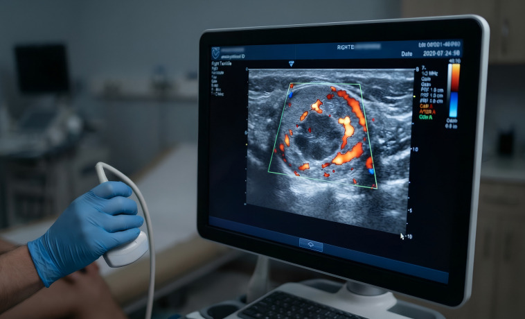
Ultrasound may be used to examine an enlarged lymph node, liver, spleen, or another superficial or abdominal abnormality. It can also guide a needle biopsy. Ultrasound alone cannot reliably determine whether a lymph node is cancerous.
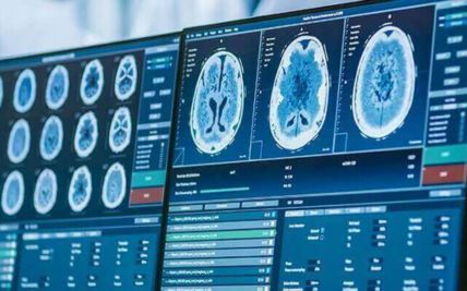
A CT scan produces detailed images of the neck, chest, abdomen, pelvis, and other areas. It can show enlarged lymph nodes, organ involvement, infection, bleeding, or other complications. CT is commonly used for lymphoma staging and treatment planning. It may also be used when PET-CT is unsuitable or not required.
Whole-body low-dose CT can identify areas of bone destruction caused by myeloma. It is more sensitive than conventional skeletal X-rays for many bone lesions. It may be combined with MRI or PET-CT according to symptoms and the clinical question.
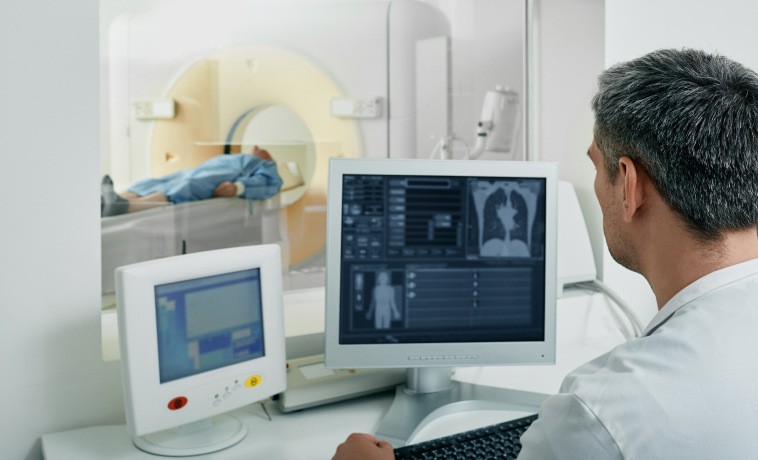
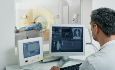
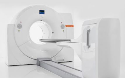
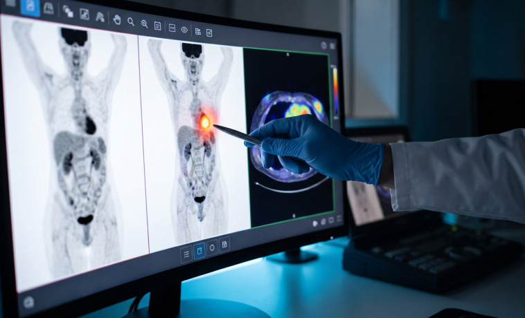
PET-CT combines metabolic and anatomical imaging. It is widely used for staging and assessing treatment response in FDG-avid lymphomas, including Hodgkin lymphoma and many aggressive non-Hodgkin lymphomas. Not every lymphoma is strongly visible on PET-CT, and PET-CT is not routinely required for most leukemias.
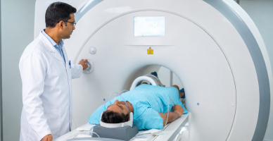
MRI provides detailed images of the brain, spinal cord, bone marrow, and soft tissues. It may be used when myeloma or lymphoma affects the spine, nerves, brain, or another specific area. MRI is particularly useful for assessing spinal-cord compression, which requires urgent treatment.


Bone marrow aspiration removes a small amount of liquid marrow, usually from the back of the pelvic bone. The sample is examined to assess the number and appearance of blood-forming cells. It is commonly used in the diagnosis and monitoring of leukemia, myeloma, MDS, MPNs, and selected lymphomas.


A trephine biopsy removes a small core of bone and marrow. It shows the structure of the marrow, how cells are distributed, whether fibrosis is present, and whether lymphoma or another cancer has entered the marrow. Aspiration and trephine biopsy are often performed during the same procedure under local anesthesia, with additional pain or sedation support where appropriate.


An excisional biopsy removes an entire enlarged lymph node. It is often preferred when lymphoma is suspected because the pathologist needs to assess the node’s overall structure. The procedure is planned to obtain adequate tissue while avoiding unnecessary delay in people with rapidly progressive disease.
A core needle biopsy collects several cylinders of tissue from a lymph node or mass, usually under ultrasound or CT guidance. It may be used when an excisional biopsy is difficult or unsafe. A repeat or excisional biopsy may be required if the sample does not provide enough information to classify the lymphoma accurately.
Some lymphomas involve the skin, liver, spleen, intestine, brain, or another organ. A biopsy may be taken from the involved site when this provides the safest and most informative tissue.
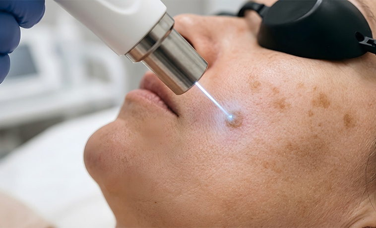
A lumbar puncture collects cerebrospinal fluid from the lower back. It may be used to determine whether acute leukemia or lymphoma has reached the brain or spinal fluid. Medicines can also be administered through the same route to prevent or treat central nervous system involvement.
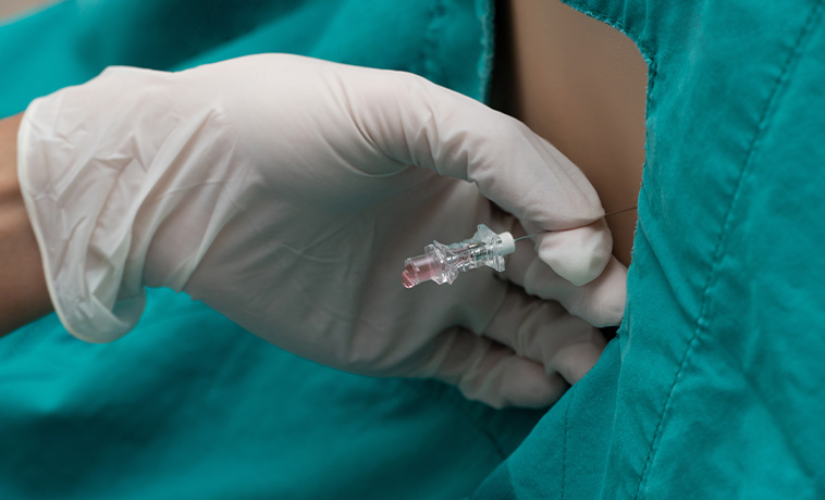
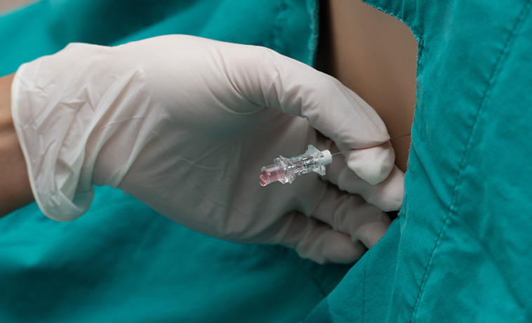
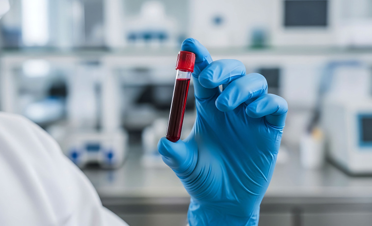
A complete blood count measures red blood cells, white blood cells, hemoglobin, and platelets. It may show abnormal cell numbers, anemia, low platelets, or very high or low white-cell counts. A normal blood count does not exclude every lymphoma or early plasma-cell disorder.


A peripheral smear allows blood cells to be examined under a microscope. It may show immature cells, abnormal lymphocytes, changes in red-cell shape, or other features that guide further testing.
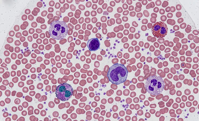
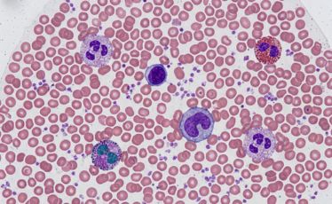
Flow cytometry identifies proteins on or within individual cells. It helps determine whether an abnormal population is present and whether it arises from B cells, T cells, myeloid cells, plasma cells, or another cell line. It is essential for classifying many leukemias and lymphomas and can also be used to measure very small amounts of residual disease.


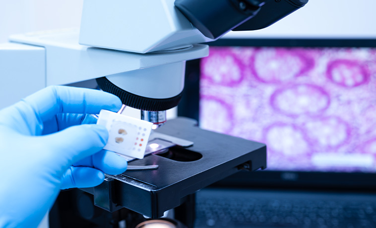
A pathologist examines bone marrow, lymph-node, or other tissue under a microscope. The pattern and appearance of abnormal cells help establish the diagnosis and subtype. Specialist hematopathology review is particularly important because treatment differs substantially between blood cancer subtypes.
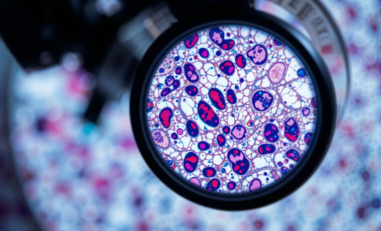
Immunohistochemistry uses specialised stains to identify proteins in abnormal cells. It helps classify lymphomas, plasma-cell cancers, and other hematological malignancies.


Cytogenetic testing examines the number and structure of chromosomes in cancer cells. It may identify changes that confirm the diagnosis, indicate prognosis, or guide treatment. Examples include the Philadelphia chromosome in CML and selected ALL cases.
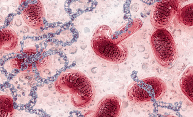
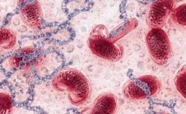
Fluorescence in situ hybridisation, or FISH, uses fluorescent probes to identify specific chromosomal changes. It can detect abnormalities associated with leukemia, lymphoma, myeloma, MDS, and MPNs.


Molecular tests detect gene variants, rearrangements, or other changes within cancer cells. Depending on the disease, these may include:
BCR::ABL1 in CML and selected ALL
PML::RARA in acute promyelocytic leukemia
FLT3, NPM1, IDH1, IDH2 and other changes in AML
TP53, IGHV and other markers in CLL
JAK2, CALR, MPL in MPNs
Disease-specific changes in lymphoma and myeloma
These results may confirm the diagnosis, define risk, select targeted treatment, and monitor response.


Tests may include kidney and liver function, calcium, uric acid, lactate dehydrogenase, electrolytes, and other markers. They help assess disease effects, tumour burden, and the safety of treatment.
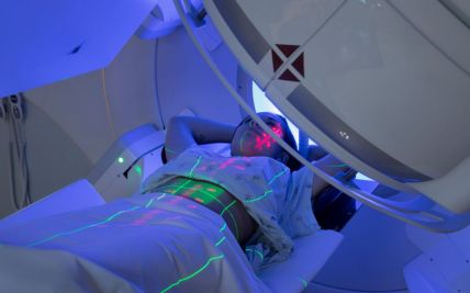
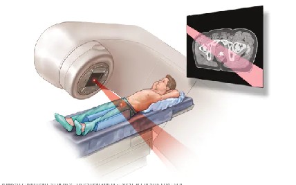
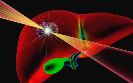
External beam radiation may be used to treat selected lymphomas, solitary plasmacytomas, painful myeloma bone lesions, enlarged organs, or areas causing pressure on the spinal cord or another structure. Radiation is usually directed at a defined site rather than the whole body.
Involved-site radiation therapy targets lymph-node regions or tissues known to contain lymphoma while limiting exposure to surrounding organs. It may be used alone for selected localised indolent lymphomas or combined with systemic treatment in Hodgkin and other lymphomas.
Total body irradiation delivers radiation to the entire body and may be used as part of conditioning before selected allogeneic stem-cell transplants. It is not required for every transplant. The conditioning plan depends on the cancer, age, previous treatment, and transplant protocol.

Proton therapy uses proton beams that release most of their radiation within the planned treatment area, with little radiation continuing beyond it. This may reduce exposure to some nearby healthy tissues.
It may be considered for selected lymphomas, particularly when disease in the chest lies close to the heart, lungs, or breast tissue and comparative planning shows a meaningful reduction in radiation exposure. It may also be considered in selected younger patients or people requiring treatment near previously irradiated areas.
Proton therapy is not required for every lymphoma and does not replace systemic treatment when systemic therapy is indicated.

Chemotherapy destroys rapidly dividing cancer cells and remains an important treatment for acute leukemia, lymphoma, myeloma, and other blood cancers. It may be given in phases, such as induction, consolidation, and maintenance, or combined with targeted medicines, antibodies, or immunotherapy.

Targeted medicines act on specific proteins or molecular changes within cancer cells. Examples include medicines directed against BCR:ABL1 in CML, FLT3 or IDH changes in AML, and signalling pathways in CLL or lymphoma. The relevant target must be demonstrated or supported by the specific diagnosis.
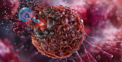
Monoclonal antibodies recognise proteins on the surface of cancer cells. They may destroy cells directly, recruit the immune system, or deliver another treatment to the cancer cell. They are widely used in lymphoma, CLL, myeloma, and selected leukemias.
Antibody–drug conjugates combine an antibody with a cancer-killing medicine. The antibody directs the medicine towards cells carrying a particular target. These treatments are used in selected lymphomas and leukemias according to the cancer subtype and previous therapy.
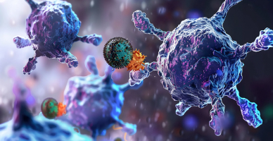
Immunomodulatory medicines affect both cancer cells and the immune environment around them. They form an important part of treatment for multiple myeloma and selected other disorders.

Immunotherapy uses or modifies the immune system to attack cancer cells. It includes antibodies, bispecific medicines, immune-checkpoint inhibitors, and cellular therapies. The type used depends on the cancer subtype, target expression, prior treatment, and individual clinical situation.
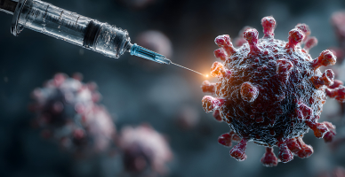
Flow cytometry, cytogenetics, FISH, and molecular testing are used to define the disease and select therapy. Results can also help estimate risk and monitor treatment response.
Precision testing does not replace the need to consider age, organ function, infections, previous therapy, and treatment goals.
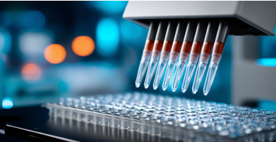
CAR T-cell therapy collects a patient’s T cells and modifies them in a laboratory so that they recognise a particular protein on cancer cells. The cells are then returned to the patient after preparatory chemotherapy.
It may be used for selected relapsed or refractory B-cell leukemias, lymphomas, and multiple myeloma. Treatment requires specialised monitoring for complications such as cytokine-release syndrome, neurological effects, low blood counts, and infection.
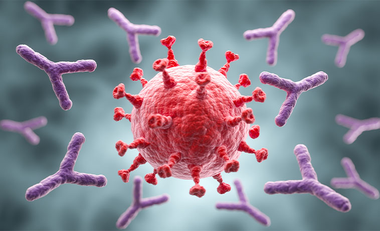
An autologous transplant collects and stores the patient’s own stem cells. High-dose chemotherapy is then given, after which the stored cells are returned to restore blood-cell production.
It is commonly used in multiple myeloma and selected lymphomas. The transplant itself does not attack the cancer; it allows high-dose treatment to be delivered safely.
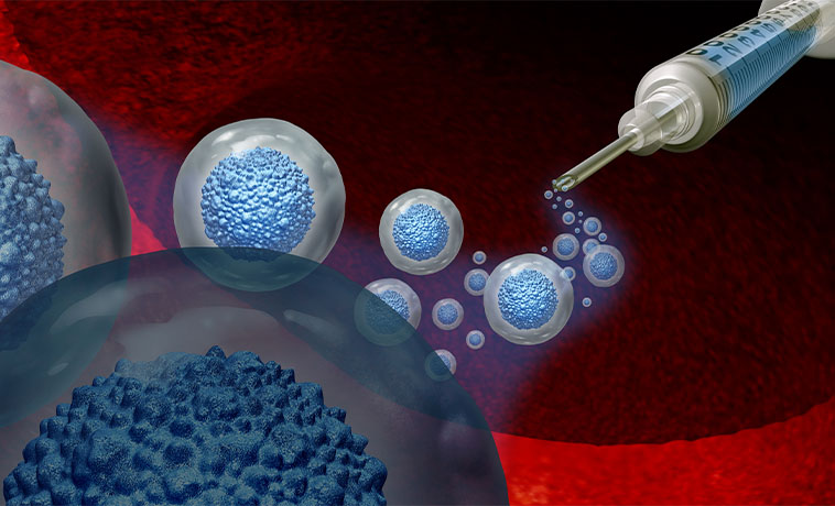
An allogeneic transplant uses blood-forming stem cells from a matched related, unrelated, or partially matched donor. Conditioning treatment prepares the body before the donor cells are infused.
In addition to restoring blood formation, donor immune cells may attack remaining cancer cells. This can provide long-term disease control or cure in selected leukemias, MDS, MPNs, and lymphomas.
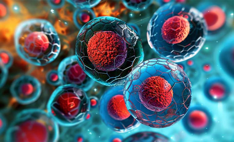
A haploidentical transplant uses a partially matched family donor. Advances in conditioning and immune control have made this an option for selected patients who do not have a fully matched donor.

Umbilical cord blood contains blood-forming stem cells and may be used as a donor source for selected patients. It can be particularly useful when a matched adult donor is unavailable.
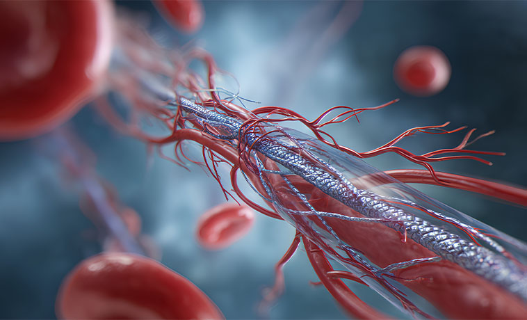
Donor lymphocytes may be given after an allogeneic transplant if the cancer persists or returns. The aim is to strengthen the donor immune response against the cancer. It is suitable only in selected situations because it can also increase the risk of graft-versus-host disease.
Prognosis for Hematological Cancer
The outlook varies substantially between blood cancers. Some can be cured, some can be controlled for many years, and others may require repeated or long-term treatment.
Factors influencing prognosis include:
The exact cancer type and subtype
Age and overall health
Blood counts and organ function
Disease stage, phase, or risk group
Cytogenetic and molecular findings
Central nervous system or other organ involvement
Response to initial treatment
Minimal residual disease
Duration of remission
Availability of targeted, cellular, or transplant options
Ability to tolerate and complete treatment
Many people with Hodgkin lymphoma, selected non-Hodgkin lymphomas, acute leukemias, and other blood cancers achieve long-term remission or cure. CML can often be controlled for many years with targeted tablets, while CLL, indolent lymphoma, myeloma, MDS, and MPNs may follow a chronic or relapsing course.
Prognosis should be discussed according to the individual diagnosis rather than using survival figures for blood cancer as a single group.


Why Choose ACC for Hematological Cancer Treatment
Dedicated hemato-oncology care for leukemia, lymphoma, myeloma, MDS, MPNs, and related disorders
Specialist hematopathology supported by flow cytometry, cytogenetics, FISH, and molecular testing
Disease-specific targeted therapy, immunotherapy, and precision-oncology services
CAR T-cell therapy for eligible blood cancers
Autologous, allogeneic, haploidentical, and cord-blood transplant capabilities
Donor matching, HLA typing, transfusion medicine, and transplant laboratory support
Advanced radiation therapy, including proton therapy for selected lymphomas
Minimal residual disease testing for appropriate cancers
Multidisciplinary review by hemato-oncologists, transplant physicians, pathologists, radiation oncologists, and supportive-care specialists
Infection prevention, blood-product support, intensive monitoring, and critical-care backup
Long-term surveillance following chemotherapy, cellular therapy, and stem-cell transplantation
Health Blogs