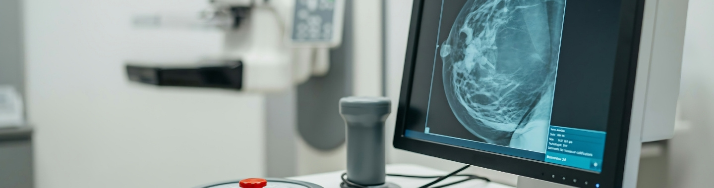
Digital Breast Tomosynthesis, commonly called DBT or 3D mammography, is an advanced form of mammography that uses low-dose X-rays to create a series of thin images of the breast. These images allow the radiologist to examine breast tissue in layers rather than as a single two-dimensional image.
DBT may be used for breast cancer screening or to investigate a symptom or abnormality. By reducing the effect of overlapping breast tissue, it can help reveal some abnormalities that may be less visible on standard 2D mammography.
DBT does not detect every breast cancer or confirm whether an abnormality is cancerous. Additional mammographic views, ultrasound, contrast-enhanced mammography, MRI or biopsy may still be required.
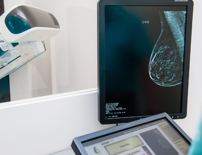


Role in Cancer Care





What to Expect
Before the Test
No fasting or medicine changes are usually required. If your breasts are particularly tender before your menstrual period, you may prefer to schedule routine screening when they are less sensitive, but a concerning symptom should not be left unevaluated for this reason.
Before the examination, inform the team if you:
Are pregnant or may be pregnant
Have a breast lump, nipple discharge, skin change, focal pain or another symptom
Have previously undergone breast surgery, biopsy or radiation therapy
Have breast implants
Have a personal or family history of breast or ovarian cancer
Are using hormone therapy
Have difficulty standing or moving your shoulders or arms
Have previous mammograms or DBT images from another centre
Bring previous breast images and reports whenever possible. Comparison with earlier examinations can help identify subtle changes and reduce unnecessary additional testing.
Do not apply deodorant, antiperspirant, talcum powder, lotion or cream to the breasts or underarms on the day of the examination. These products may appear as spots on the images.

During the Test
You will stand in front of the mammography unit while the technologist positions one breast at a time on the imaging platform. The breast will be gradually compressed with a clear plastic paddle.
The X-ray tube will move in an arc over the breast and capture multiple images. You will need to remain still and may be asked to hold your breath briefly. The process is repeated for the required views of each breast.
Compression may feel tight or uncomfortable, but it lasts only a few seconds for each view. Tell the technologist if you experience significant pain.
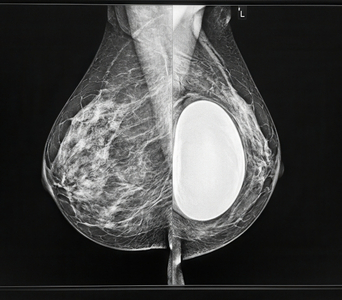
People with breast implants may require additional implant-displacement views. Inform the centre about your implants when scheduling the appointment.
The examination commonly takes approximately 20 to 30 minutes. Diagnostic DBT may take longer if additional or magnified views are required.

After the Test
No recovery period is required, and you can return to normal activities immediately. Temporary breast tenderness may occur but usually settles quickly.
For a diagnostic examination, you may be asked to wait while the radiologist reviews the images. Additional mammographic views or ultrasound may sometimes be performed during the same visit.
The report will usually include a BI-RADS assessment category and information about breast density. Depending on the findings, routine screening, short-interval follow-up, additional imaging or biopsy may be recommended.

Potential Benefits
- Improved tissue separation: Thin image sections can make abnormalities less likely to be hidden by overlapping breast tissue.
- Increased cancer detection: DBT may identify some breast cancers that are less visible on standard 2D mammography.
- Fewer screening recalls: It may reduce some callbacks caused by normal overlapping tissue.
- Better assessment of distortion: DBT can improve the visibility of architectural distortion and some masses.
- Useful in dense breasts: It may provide clearer assessment in people with dense breast tissue, although limitations remain.
- Screening and diagnostic use: DBT can be used for routine screening and focused investigation of an abnormality or symptom.
- Precise localisation: Image slices can help determine the position and extent of certain abnormalities.
- Procedure guidance: Abnormalities visible primarily on DBT may be targeted for biopsy or localisation where appropriate technology is available.