




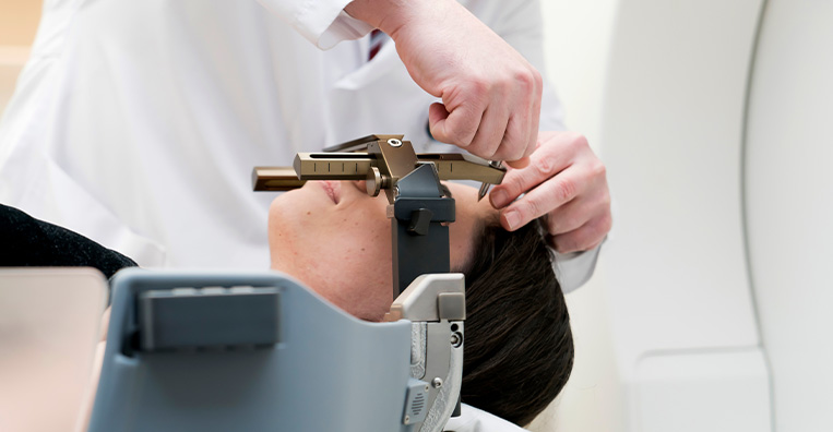
CyberKnife Radiosurgery is a non-invasive form of radiation treatment used for selected tumours and other abnormalities in the brain, spine and different parts of the body. Despite its name, CyberKnife treatment does not involve a knife, surgical incision or removal of tissue.
The system combines a compact linear accelerator with a robotic arm that delivers radiation beams to the target from multiple angles. Imaging is used throughout treatment to monitor the target’s position and adjust radiation delivery for patient or tumour movement. This is particularly useful for tumours that can move with breathing.
CyberKnife may be used to deliver stereotactic radiosurgery to the brain and stereotactic body radiation therapy to other parts of the body. Treatment is generally completed in one to five sessions, although the schedule depends on the tumour and treatment plan.
- Primary brain tumours and brain metastases
- Meningiomas, vestibular schwannomas and pituitary adenomas
- Selected tumours involving the spine or spinal region
- Early-stage lung cancers in patients who are not suitable for surgery
- Selected lung, liver, kidney, pancreatic, adrenal and prostate cancers
- A limited number of metastatic tumours in selected organs
- Tumours located close to sensitive organs or other critical structures
- Tumours that move with breathing, such as those in the lung, liver or pancreas
- Residual or recurrent tumours following surgery or other treatment
- Selected tumours in areas that have previously received radiation
- Certain non-cancerous conditions, such as arteriovenous malformations and trigeminal neuralgia

How CyberKnife Radiosurgery Is Performed
Before Treatment
The radiation oncologist reviews the diagnosis, biopsy findings where applicable, imaging, previous treatments and overall health.
A planning CT scan is performed with the patient in the position that will be used during treatment.
MRI, PET-CT or other imaging may be combined with the planning CT to define the tumour and surrounding organs.
A customised mask, body mould or other support may be prepared to help the patient remain comfortably positioned.
Small markers called fiducials may be placed in or near certain tumours to help the system track their position. These are not required for every patient.
Breathing-related tumour movement may be assessed for tumours in the chest or abdomen.
The radiation oncologist, medical physicist and treatment-planning team calculate the dose and determine the beam directions.
Patients receive instructions about food, medicines and other preparation, particularly if fiducial placement, sedation or contrast imaging is required.

During Treatment
The patient lies on the treatment table with the customised mask or support in place.
Imaging is used to locate the target and monitor its position throughout the session.
The robotic arm moves around the patient and delivers multiple radiation beams from carefully planned angles.
When treating certain moving tumours, the system can adjust the direction of the radiation beam in response to the tumour’s movement.
The machine does not touch the patient, and the radiation cannot be seen or felt.
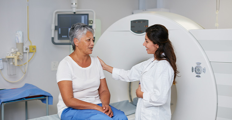
The patient remains awake and can communicate with the treatment team throughout the session.
Most adults do not require anesthesia. Sedation or anesthesia may be considered for children or patients who cannot remain still.
A session may take approximately 30 to 90 minutes, depending on the location and complexity of the target.
Treatment is commonly delivered in one to five sessions, although some conditions may require a different schedule.

After Treatment
Most patients can return home after each treatment session.
Normal activities can often be resumed within a short period, depending on the treatment area and how the patient feels.
The patient is not radioactive and does not need to avoid contact with family members.
Side effects may appear gradually during the treatment course or in the days and weeks that follow.
Medicines may be prescribed to manage swelling, nausea, pain or other symptoms.
Patients who have undergone fiducial placement may receive separate instructions about caring for the insertion site.
Follow-up appointments are scheduled to monitor symptoms and treatment-related effects.
CT, MRI, PET-CT or blood tests may be used to assess the response, depending on the condition treated.
The full treatment response may take several weeks or months to become apparent.

Potential Benefits of CyberKnife Radiosurgery
- Non-invasive treatment without a surgical incision
- Precise delivery of radiation to the target
- Radiation delivery from multiple angles
- Continuous imaging to monitor the target during treatment
- The ability to adjust for certain types of tumour or patient movement
- Reduced radiation exposure to surrounding healthy tissue
- Treatment of selected tumours close to sensitive structures
- Treatment completed in fewer sessions than conventional radiation therapy in many cases
- Outpatient treatment with little interruption to routine activities for many patients
- An option for selected patients who cannot undergo surgery
- The ability to treat certain residual, recurrent or previously irradiated tumours

Side Effects of CyberKnife Radiosurgery
- Tiredness or fatigue
- Temporary skin redness, dryness, irritation or darkening over the treated area
- Temporary hair loss limited to the treatment area
- Headache, nausea or swelling-related symptoms following treatment to the brain
- Cough, breathlessness, chest discomfort or inflammation following treatment to the chest
- Nausea, reduced appetite or abdominal discomfort following treatment to the abdomen
- Changes in bowel or bladder habits following treatment to the pelvis or prostate
- Temporary pain or discomfort near the treated area
- Swelling or inflammation that may temporarily worsen existing symptoms
- Delayed changes in the treated tissue, including radiation necrosis
- Site-specific effects involving nearby nerves, organs, bones or blood vessels
- A small long-term risk of developing another cancer related to radiation exposure



