icon
icon
Overview

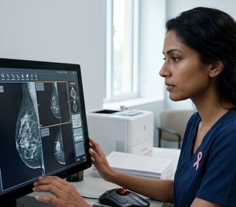
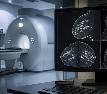
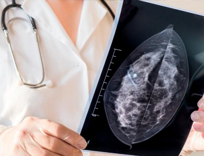
Contrast-Enhanced Mammography, or CEM, is a breast-imaging examination that combines digital mammography with an intravenous iodine-based contrast agent. It provides standard mammographic information while also showing areas of increased blood flow or contrast uptake within the breast.

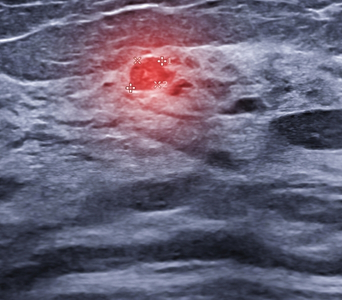
Cancers often develop new blood vessels and may therefore enhance after contrast administration. However, enhancement is not specific to cancer and can also occur in benign breast conditions.

CEM may be used to investigate an abnormal finding, assess the extent of known breast cancer or provide additional imaging in selected patients. It does not replace biopsy when tissue confirmation is required.
Contrast-Enhanced Mammography (CEM)
How the Technology Works
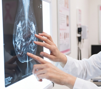
CEM combines low-dose X-ray imaging with information about contrast uptake in breast tissue. The examination produces both conventional mammographic images and contrast-enhanced images.
work-icon
Iodinated Contrast
An iodine-based contrast agent is injected into a vein before the breast images are acquired. The contrast circulates through the bloodstream and may accumulate in areas with increased or abnormal blood supply.
work-icon
Dual-Energy Imaging
Each compressed breast is imaged using low- and higher-energy X-ray exposures. The low-energy images resemble standard 2D mammograms, while the additional exposure captures information related to the iodine-based contrast.
work-icon
Recombined Images
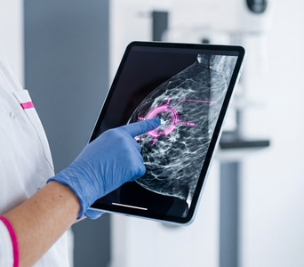
Computer processing subtracts much of the background breast tissue and produces recombined images highlighting areas of contrast enhancement. The radiologist evaluates these alongside the low-energy mammographic images to assess both breast structure and blood-flow patterns.

Role in Cancer Care

Evaluating an Abnormal Finding
CEM may help assess an abnormality found on mammography, digital breast tomosynthesis, ultrasound or clinical examination. Enhancement patterns can provide additional information about the location and extent of a suspicious area. Biopsy may still be required to establish the diagnosis.
Evaluating an Abnormal Finding
Assessing Dense Breasts
Dense breast tissue can make abnormalities more difficult to see on standard mammography because both dense tissue and many cancers appear white. CEM adds information about contrast uptake that may reveal abnormalities obscured by overlapping tissue. Its use as supplemental screening is considered selectively according to individual risk, local practice and contrast suitability.
Assessing Dense Breasts
Assessing the Extent of Known Cancer
After breast cancer has been diagnosed, CEM may help assess the size of the tumour and look for additional areas in the same or opposite breast. This information may contribute to surgical and treatment planning. Additional ultrasound, MRI or biopsy may be needed to evaluate some findings.
Assessing the Extent of Known Cancer
Planning Biopsy or Surgery
CEM can help identify the most suspicious area for tissue sampling and assess whether more than one site requires evaluation. CEM-guided biopsy may be available when an enhancing abnormality is not clearly visible on standard mammography or ultrasound. The surgical plan is based on the combined imaging and pathology findings.
Planning Biopsy or Surgery
Monitoring Treatment Response
CEM may be used in selected patients receiving systemic treatment before surgery. Changes in tumour size and enhancement can provide information about response. The findings are interpreted alongside clinical assessment and other imaging examinations.
Monitoring Treatment Response
Alternative or Additional Imaging
CEM may be considered when breast MRI is unavailable, unsuitable or cannot be completed. It is not identical to MRI and is not automatically interchangeable with it. The choice depends on the clinical question, kidney function, contrast history, pregnancy status and other patient factors.
Alternative or Additional Imaging

What to Expect

Before the Test 

Preparation is similar to mammography, with additional screening for intravenous iodinated contrast. You will receive instructions about eating, drinking and regular medicines from the imaging centre.

Before the examination, inform the team if you:

  • Are pregnant or may be pregnant

  • Are breastfeeding

  • Have kidney disease or reduced kidney function

  • Have previously reacted to iodinated contrast material

  • Have asthma, significant allergies or thyroid disease

  • Have diabetes

  • Take prescription medicines, non-prescription medicines or supplements

  • Have breast implants

  • Have undergone previous breast surgery, biopsy or radiation therapy

  • Have a breast lump, nipple discharge, skin change or another symptom

A kidney-function blood test may be required before contrast is administered, particularly if you have known kidney disease or relevant risk factors. Do not stop or alter medicines unless specifically instructed.

Bring previous mammograms, tomosynthesis images, ultrasound, MRI or CEM studies and reports whenever possible. Comparison with earlier examinations can help identify changes.

Do not apply deodorant, antiperspirant, talcum powder, lotion or cream to the breasts or underarms on the day of the examination. These products may appear on the mammographic images.

Before the Test

During the Test 

A cannula will be inserted into a vein in the hand or arm, and the iodinated contrast agent will be administered. You may briefly experience warmth, flushing or a metallic taste.

After the contrast has circulated, the technologist will position and compress one breast at a time in the mammography unit. Images are usually acquired from standard mammographic angles using dual-energy exposures.

You will need to remain still and may be asked to hold your breath briefly during each image. Compression may feel tight or uncomfortable but lasts only a few seconds for each view.

The examination commonly takes approximately 15 to 30 minutes, although the appointment may be longer because of preparation, contrast administration and image review. Tell the technologist immediately if you develop itching, hives, breathing difficulty, swelling, dizziness or another unusual symptom.

During the Test

After the Test

The cannula will usually be removed before you leave. You may be observed briefly after the contrast injection, depending on the imaging centre’s protocol and your medical history.

You can generally return to normal activities immediately. Drinking water may be advised to help eliminate the contrast unless you are on fluid restriction or have received different instructions.

Seek urgent medical attention if you develop breathing difficulty, facial or throat swelling, severe dizziness or another significant reaction after leaving the imaging centre. Contact the care team if a delayed rash or other persistent symptoms develop.

The radiologist will assess the low-energy and recombined images and prepare a report. Additional ultrasound, MRI, mammographic views or biopsy may be recommended depending on the findings.

After the Test

Potential Benefits

CEM combines structural mammographic imaging with information about blood flow and contrast enhancement.
  • Dual information: It provides conventional mammographic images and contrast-enhanced images during the same examination.
  • Improved lesion visibility: Enhancement may reveal abnormalities obscured by dense or overlapping tissue.
  • Extent assessment: CEM may help evaluate tumour size and identify additional suspicious areas.
  • Bilateral imaging: Both breasts can be assessed during the examination.
  • Treatment planning: Findings may contribute to decisions about biopsy, surgery and other treatment.
  • Response monitoring: Changes in tumour enhancement may help assess response to preoperative systemic treatment.
  • Alternative in selected patients: CEM may provide additional information when breast MRI is unsuitable or unavailable.
  • Procedure guidance: Enhancing abnormalities may be targeted for biopsy where CEM-guided systems are available.

Potential Benefits
Frequently Asked Questions
01 Is CEM used for routine breast cancer screening?
icon icon
Standard mammography or digital breast tomosynthesis remains the usual basis of routine breast screening. CEM may be considered as supplemental screening in selected people, such as some individuals with dense breasts or increased risk. Its use depends on local protocols, individual risk and suitability for intravenous contrast.
02 How is CEM different from standard mammography?
icon icon
Standard mammography shows the structure of breast tissue using low-dose X-rays. CEM also uses an intravenous iodine-based contrast agent and dual-energy imaging to show areas of increased contrast uptake. This adds functional information about blood flow within the breast.
03 How is CEM different from breast MRI?
icon icon
CEM uses X-rays and iodine-based contrast, whereas breast MRI uses a magnetic field, radiofrequency signals and usually gadolinium-based contrast. Both can show contrast enhancement but differ in technique, radiation exposure, contraindications and diagnostic capabilities. The most appropriate test depends on the clinical situation.
04 Does CEM involve more radiation than standard mammography?
icon icon
CEM generally involves a higher radiation dose than standard 2D mammography because dual-energy images are obtained. The dose varies by equipment and protocol and is kept within accepted mammography limits. No radiation remains in the body after the examination.
05 Can I undergo CEM if I have kidney disease or a contrast allergy?
icon icon
Possibly, but individual assessment is required. Kidney function may need to be checked, and a previous reaction to iodinated contrast must be discussed with the radiology team. The team may recommend precautions, a different imaging examination or CEM without proceeding, depending on the level of risk.
06 Can CEM confirm breast cancer?
icon icon
No. Both cancerous and non-cancerous conditions can enhance after contrast administration. CEM can identify and characterise suspicious areas, but biopsy is required to confirm whether an abnormality is cancerous.
image image
Book an Appointment
Please Enter Your Name
Please Enter Your Mobile Number
Please Enter OTP