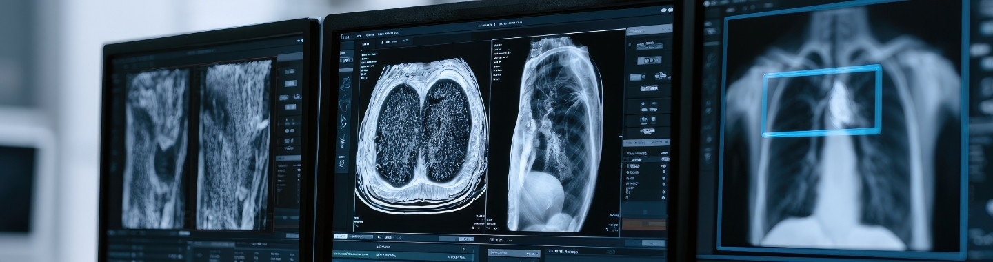
Low-Dose Computed Tomography, commonly called LDCT, is a specialised CT scan that uses a lower radiation dose than a conventional diagnostic chest CT. It produces detailed cross-sectional images of the lungs and can detect small lung nodules or other abnormalities that may not be visible on a standard chest X-ray.
LDCT is principally used to screen for lung cancer in selected people who have a higher risk of developing the disease, particularly because of a significant history of cigarette smoking. Screening is intended for people who do not have symptoms. Eligibility is determined using factors such as age, smoking history, current health, previous cancer history and the screening guidelines followed by the treating centre.
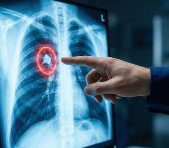
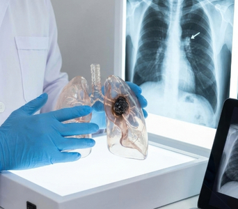
LDCT does not confirm whether an abnormality is cancer. Many lung nodules are non-cancerous and may result from previous infections, inflammation or scarring. If an abnormality is found, comparison with earlier scans, follow-up LDCT, diagnostic CT, PET-CT, bronchoscopy or biopsy may be recommended.


Role in Cancer Care
What to Expect
Before the Test
LDCT usually requires little preparation. You can generally eat, drink and take your regular medicines unless the care team provides different instructions.
Before the scan, inform the team if you:
Are pregnant or may be pregnant
Have undergone previous chest imaging
Have previously been diagnosed with cancer
Have symptoms such as persistent cough, coughing up blood, unexplained weight loss, chest pain or breathlessness
Have recently had a chest infection or other respiratory illness
Have undergone lung surgery, radiation therapy or treatment for a lung condition
Previous CT scans and chest X-rays should be made available whenever possible. Comparison with earlier imaging can help determine whether a nodule is new or has changed.
LDCT lung screening is generally performed without intravenous contrast. Therefore, fasting, kidney-function tests and contrast-related preparation are not usually required. You may be asked to remove jewellery or metal objects from the chest area and change into a hospital gown.

During the Test
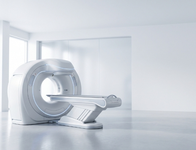
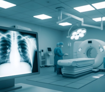
You will lie on your back on a motorised table. The table will move through the centre of the CT scanner, which is open at both ends.
The technologist will position you, usually with your arms raised above your head, and may ask you to hold your breath briefly while the images are taken. Remaining still helps produce clear images.
The scan is painless and usually takes only a few minutes. The actual image acquisition may be completed within a single breath-hold. The technologist can see, hear and communicate with you throughout the examination.

After the Test
No recovery period is usually required. You can generally return to your usual activities immediately after the scan.
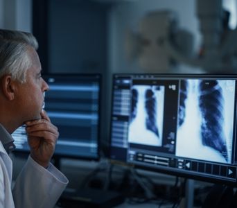
A radiologist will examine the images, compare them with any previous scans and prepare a report. Findings may be grouped according to their likelihood of being benign or clinically significant, with a recommendation for routine screening, earlier follow-up or further evaluation.
The doctor will explain what the result means and whether another scan or test is required. It is important to complete any recommended follow-up, even if you continue to feel well.

Potential Benefits
- Earlier detection: LDCT can identify some lung cancers at a smaller and potentially more treatable stage.
- Detailed lung imaging: It can detect small nodules that may not be visible on a conventional chest X-ray.
- Lower radiation dose: Screening protocols use less radiation than conventional diagnostic chest CT.
- Quick and non-invasive: The scan is painless, does not usually require contrast and can be completed within minutes.
- Structured monitoring: Nodules can be followed over time to determine whether they remain stable or change.
- Risk-based follow-up: The size, appearance and growth of a nodule can help determine whether routine screening, surveillance or further testing is appropriate.
- Reduced lung cancer mortality: Evidence shows that LDCT screening can reduce the risk of dying from lung cancer in appropriately selected high-risk populations when accompanied by suitable follow-up.