




Breast cancer is the most commonly diagnosed cancer among women in India. According to GLOBOCAN 2024 estimates, India recorded approximately 2.37 lakh new breast cancer cases, accounting for over 30% of all cancers diagnosed among women.
When breast cancer is detected early, treatment can often begin before the disease has spread and may be less extensive. Apollo Cancer Centres’ fast-track breast diagnosis program brings together breast specialists, radiologists and pathologists to evaluate breast symptoms or abnormal screening findings through a coordinated pathway. Depending on the individual’s needs, this may include a clinical breast examination, mammography, ultrasound, breast MRI and image-guided biopsy, as well as an expert oncologist's review.
Why Screening is Important
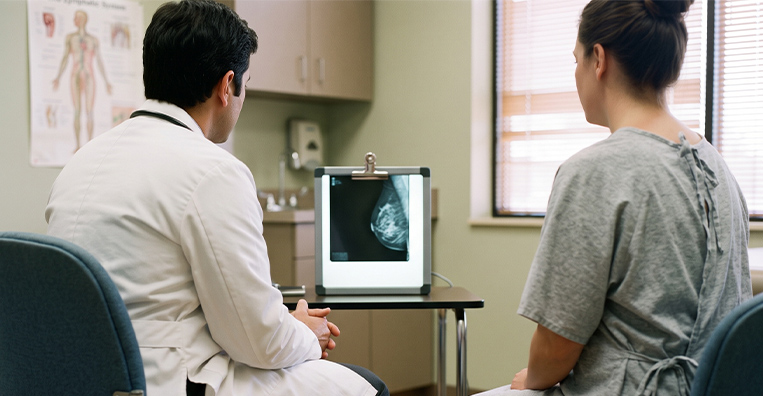
Breast cancer may not cause noticeable symptoms in its early stages. A mammogram, which is the gold standard for breast cancer screening, uses low-dose X-rays to identify suspicious breast changes, including small masses and microcalcifications, before they can be seen or felt.
Enables earlier detection: Mammography can identify breast cancer before symptoms develop, when it may be smaller and confined to the breast.
Supports better outcomes: Breast cancers detected at an earlier stage are generally associated with more treatment options and better outcomes.
May allow less extensive treatment: Early detection may increase the possibility of breast-conserving surgery and reduce the need for more intensive treatment in some patients.
Detects subtle breast changes: Mammograms can reveal small abnormalities and changes in breast tissue that may not be detected during a clinical examination.
Provides a basis for comparison: Regular mammograms allow radiologists to compare images over time and identify new or evolving changes.
A mammogram does not detect every breast cancer. Any new or persistent breast change should be evaluated promptly, even after a recent normal mammogram.


How It Works
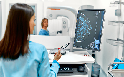
During a mammogram, each breast is positioned between two plates and briefly compressed while low-dose X-ray images are taken from different angles. The compression spreads the breast tissue to produce clearer images and reduce radiation exposure. A specialist breast radiologist then examines the images for lumps, calcifications or other changes that may require further evaluation.
Simple Steps for Self-Breast Exam
Take charge of your breast health by incorporating a monthly self-examination into your routine. Methodically assess for anomalies such as lumps, variations, or irregularities in your breast tissue. Please keep an eye out for any signs of dimpling or skin alterations while both upright and reclined.
Timely diagnosis plays a pivotal role in mitigating the elevated mortality rates and compromised quality of life associated with breast cancer.Early detection of breast cancer eliminates the risk of late-stage consequences and allows for personalized treatments, leading to better outcomes.
Who Should Be Screened
Your Screening Journey at Apollo Cancer Centres
- Registration
- Risk assessment and consultation
- Mammogram/ultrasound
- Biopsy (if required)
- Specialist review
- Results and next steps

Advantages of Breast Screening at ACC
Improved Accessibility

Greater Convenience

Reduced Anxiety

