Gastrointestinal Cancer
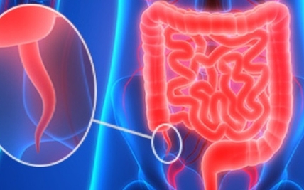
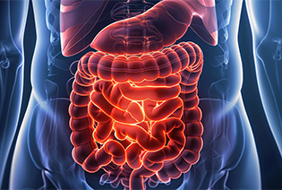
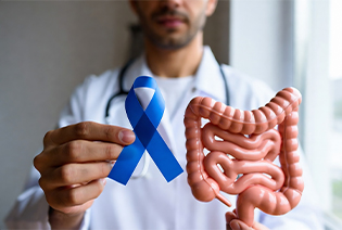
Gastrointestinal cancers develop in the digestive system. They include cancers of the esophagus, stomach, small intestine, colon, rectum, anus, liver, gallbladder, bile ducts, and pancreas. Tumours such as gastrointestinal stromal tumours and neuroendocrine tumours may also arise within the digestive system.
These cancers differ considerably in their causes, symptoms, behaviour, and response to treatment. Some produce early symptoms, while others may remain difficult to detect until they have grown or spread. Persistent digestive changes, bleeding, difficulty swallowing, jaundice, or unexplained weight loss should therefore be evaluated.
Treatment often requires collaboration between gastroenterologists, GI and hepatopancreatobiliary surgeons, medical oncologists, radiation oncologists, interventional radiologists, nuclear medicine specialists, pathologists, and clinical nutrition teams. The treatment plan is based on the organ involved, cancer subtype, stage, molecular features, overall health, and nutritional status.
Types of GI Cancers
Gastrointestinal cancer is not a single disease. The organ in which it begins and the type of cell involved determine its behaviour, staging, and treatment.
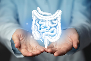
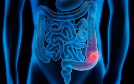
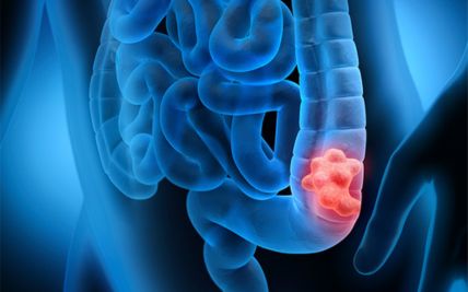
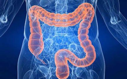
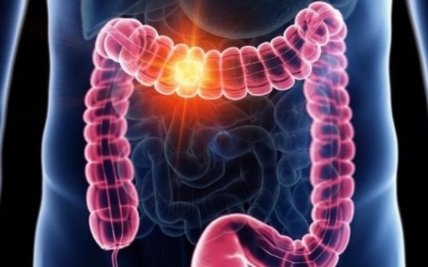
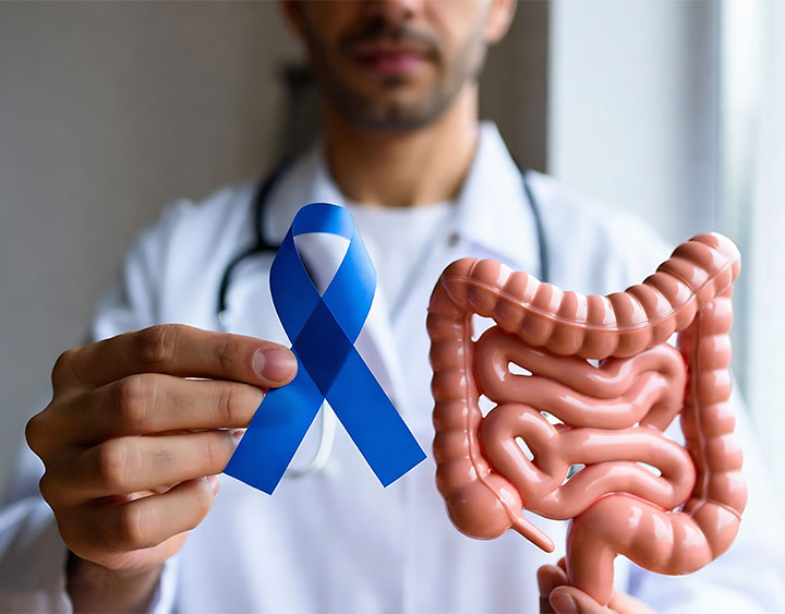
Colorectal cancer develops in the colon or rectum. Most cases begin as abnormal growths called polyps, although not every polyp becomes cancerous. Screening can identify precancerous polyps or cancer before symptoms develop. Treatment differs between colon and rectal cancer because their anatomy and patterns of spread are not the same.
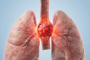
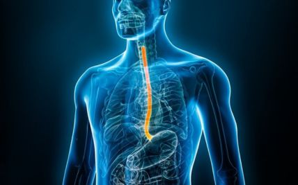
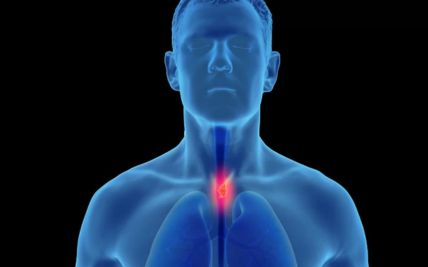
Esophageal cancer develops in the tube that carries food from the throat to the stomach. The two main types are squamous cell carcinoma and adenocarcinoma. Difficulty swallowing is a common symptom, but early disease may not cause noticeable changes. Treatment may involve endoscopic therapy, surgery, chemotherapy, radiation therapy, immunotherapy, or a combination.
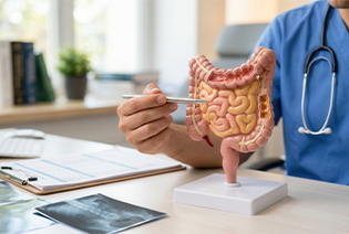
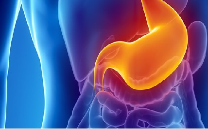
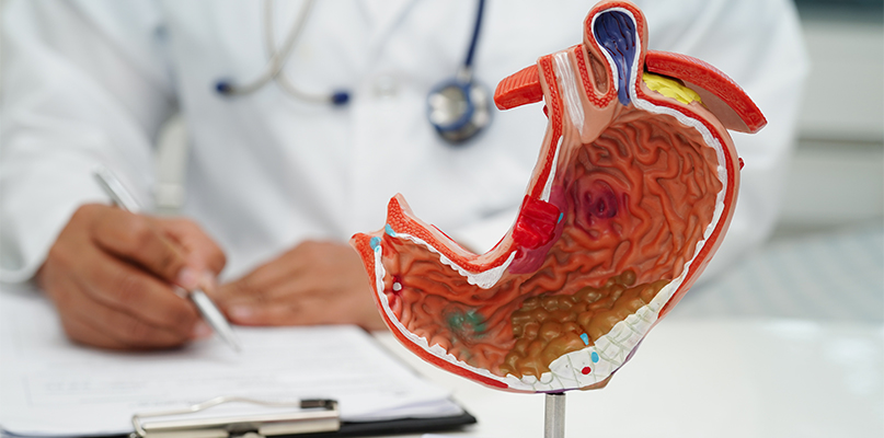
Stomach, or gastric, cancer usually begins in the glandular cells lining the stomach. Some cancers develop slowly from long-standing changes in the stomach lining. Symptoms may include indigestion, upper abdominal discomfort, reduced appetite, early fullness, anaemia, or weight loss. Treatment depends on the tumour’s location, depth, lymph-node involvement, and molecular features.
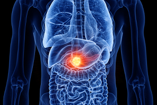
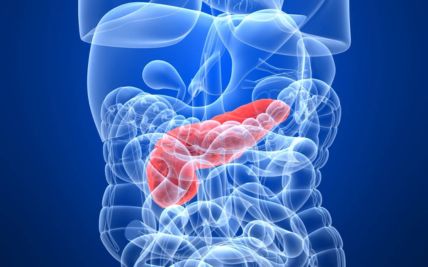
Pancreatic cancer begins in the pancreas, an organ involved in digestion and blood-sugar regulation. Pancreatic ductal adenocarcinoma is the most common type, while pancreatic neuroendocrine tumours form a separate group. Pancreatic cancer may not cause symptoms until it affects nearby structures. The relationship of the tumour to major blood vessels is important in determining whether surgery is possible.
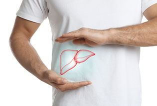
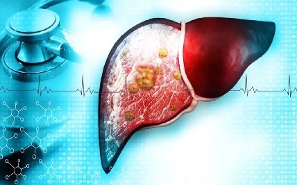
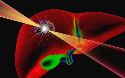
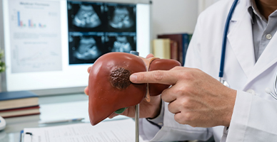
Primary liver cancer begins in the liver. Hepatocellular carcinoma is the most common type in adults and often develops in a liver affected by cirrhosis or long-standing hepatitis B or C. Liver function is considered alongside tumour size and spread when planning treatment. Options may include surgery, liver transplantation, ablation, embolisation, radiation, or systemic therapy.
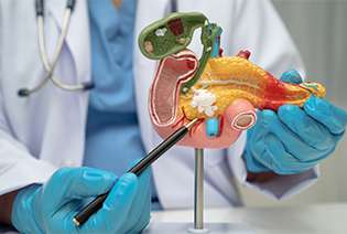
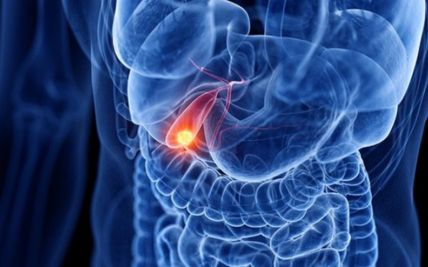
Gallbladder cancer begins in the wall of the gallbladder and is often an adenocarcinoma. It may be found unexpectedly after surgery for gallstones or diagnosed after symptoms such as abdominal pain or jaundice develop. Treatment depends on how deeply the tumour has entered the gallbladder wall and whether the liver, bile ducts, lymph nodes, or other organs are involved.
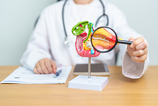
Bile duct cancer, or cholangiocarcinoma, develops in the tubes that carry bile from the liver to the intestine. It may arise within the liver, near the junction of the right and left bile ducts, or farther down the bile duct. The tumour’s location strongly influences surgery and other treatments. Jaundice is a common symptom of cancers that block bile flow.
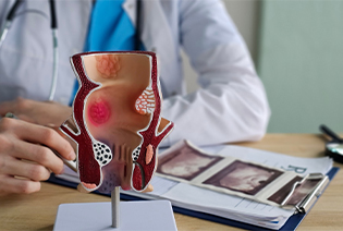
Anal cancer develops in the anal canal or skin around the anus. Most cases are squamous cell carcinomas and many are associated with persistent high-risk HPV infection. Unlike rectal cancer, most anal cancers are treated primarily with combined chemotherapy and radiation. Surgery is generally reserved for persistent or recurrent disease.
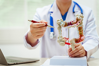
Small intestinal cancer can develop in the duodenum, jejunum, or ileum. It includes adenocarcinoma, neuroendocrine tumours, lymphomas, and gastrointestinal stromal tumours. Symptoms and treatment vary according to the tumour type and location. Diagnosis may require specialised endoscopic or imaging techniques because much of the small intestine is difficult to examine with routine endoscopy.
Gastrointestinal stromal tumours, or GISTs, arise from specialised cells in the wall of the digestive tract. They most commonly develop in the stomach or small intestine. Treatment is guided by the tumour’s size, location, mitotic rate, risk of recurrence, and molecular changes such as alterations in the KIT or PDGFRA genes.
Neuroendocrine tumours may arise in the stomach, intestine, rectum, appendix, or pancreas. Some produce hormones and cause specific symptoms, while others are found incidentally. Their behaviour ranges from slow-growing, well-differentiated tumours to aggressive neuroendocrine carcinomas. Grade, Ki-67 index, hormone production, and disease extent are important in treatment planning.
Appendiceal cancers begin in the appendix and may include adenocarcinoma, mucinous tumours, or neuroendocrine tumours. Some mucinous tumours spread within the abdomen and produce a condition called pseudomyxoma peritonei. Selected patients with disease confined mainly to the abdominal lining may be considered for cytoreductive surgery, sometimes combined with heated intraperitoneal chemotherapy.
Risk factors differ considerably between GI cancers. Having a risk factor does not mean that cancer will develop, and some people have no identifiable risk factors.
Modifiable Risk Factors
Tobacco use: Increases the risk of esophageal, stomach, pancreatic, colorectal, liver, and several other cancers.
Alcohol consumption: Heavy or prolonged alcohol use increases the risk of esophageal and liver cancer and may contribute to colorectal and other GI cancers.
Excess body weight: Associated with colorectal, esophageal adenocarcinoma, liver, pancreatic, and gallbladder cancers.
Physical inactivity: Associated particularly with an increased risk of colorectal cancer.
Processed and red meat: Frequent consumption is associated with increased colorectal cancer risk.
Low-fibre diet: May contribute to colorectal cancer risk, particularly when accompanied by low intake of whole grains, fruits, and vegetables.
Helicobacter pylori infection: Persistent infection can increase the risk of stomach cancer. Testing and treatment may reduce this risk in appropriate individuals.
Hepatitis B and C: Long-standing viral hepatitis increases liver cancer risk. Hepatitis B vaccination and appropriate treatment of hepatitis B or C can reduce risk.
Unsafe food storage: Long-term exposure to aflatoxins in contaminated food can increase liver cancer risk.
HPV infection: Persistent high-risk HPV infection is a major risk factor for anal cancer.
Poorly controlled metabolic disease: Obesity, type 2 diabetes, and metabolic liver disease may increase the risk of liver and some other GI cancers.
Non-Modifiable Risk Factors
Increasing age
A family history of colorectal, gastric, pancreatic, or other GI cancers
Hereditary diffuse gastric cancer and other inherited gastric cancer syndromes
Inherited pancreatic cancer syndromes or pathogenic variants involving genes such as BRCA1, BRCA2, PALB2, CDKN2A, or STK11
Long-standing inflammatory bowel disease
Barrett’s esophagus
Cirrhosis from any cause
Primary sclerosing cholangitis
Chronic pancreatitis, including hereditary pancreatitis
Certain bile duct or gallbladder abnormalities
Previous abdominal or pelvic radiation
A personal history of colorectal polyps or GI cancer
Certain inherited GIST or neuroendocrine tumour syndromes
People with a strong family history, multiple related cancers, cancer at an unusually young age, or features of an inherited syndrome may benefit from genetic counselling.
Signs and Symptoms
Symptoms depend on the organ involved. Some early GI cancers may not cause noticeable symptoms.
Possible signs and symptoms include:
These symptoms can also be caused by ulcers, reflux, hemorrhoids, gallstones, infection, inflammatory bowel disease, or other non-cancerous conditions. However, bleeding, progressive difficulty swallowing, jaundice, persistent vomiting, or unexplained weight loss should be assessed promptly.
Diagnosis begins with a review of symptoms, bowel and digestive health, medical history, family history, and risk factors. The doctor may examine the abdomen, lymph nodes, skin and eyes for jaundice, and the rectum where relevant. Imaging is used to locate and stage the tumour, while biopsy and laboratory testing help establish the exact cancer type.
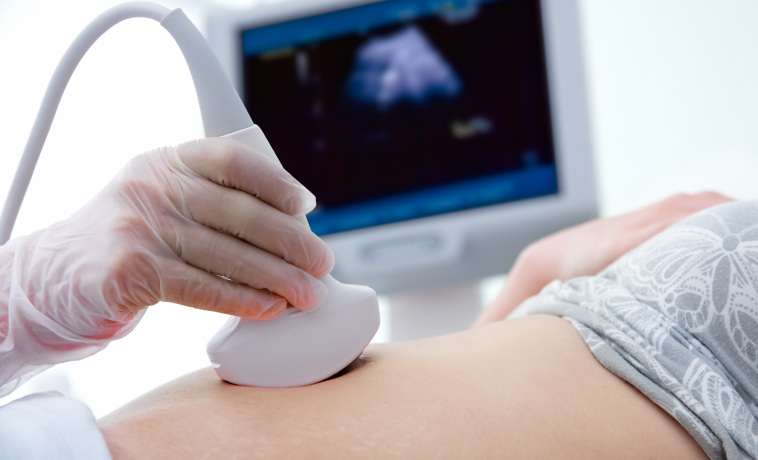
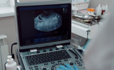
Ultrasound uses sound waves to examine the liver, gallbladder, bile ducts, pancreas, and abdominal organs. It may be the first test performed when jaundice, liver disease, gallstones, or an abdominal mass is suspected. Contrast-enhanced ultrasound may be used in selected cases to characterise liver lesions.
Endoscopic ultrasound combines endoscopy with a small ultrasound probe. It provides detailed images of the wall of the oesophagus, stomach, rectum, and nearby organs. It may help determine how deeply a tumour has grown, assess nearby lymph nodes, and guide needle biopsy of pancreatic, bile duct, upper GI, or rectal lesions.


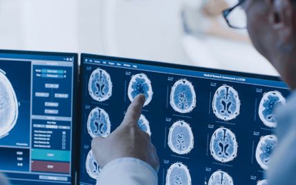
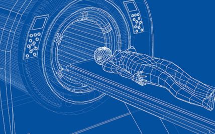
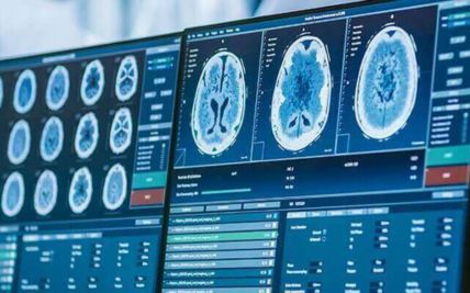
A contrast-enhanced CT scan produces detailed images of the chest, abdomen, and pelvis. It is widely used to assess tumour size, involvement of nearby structures, lymph nodes, and distant spread. Specialised pancreatic-protocol or liver-protocol CT scans may be used to examine the relationship of a tumour to blood vessels and determine whether surgery may be possible.
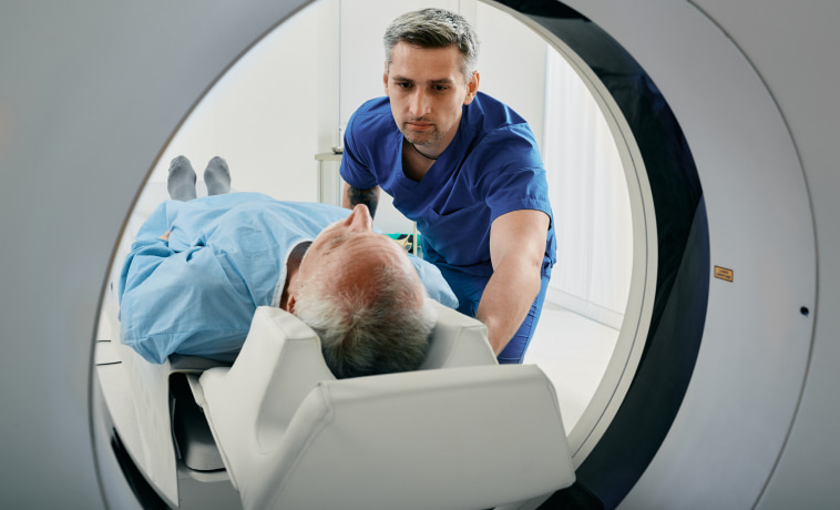
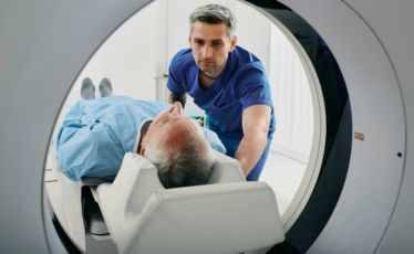
MRI provides detailed images of soft tissues and is particularly useful for assessing cancers of the liver, bile ducts, pancreas, and rectum. MRI of the pelvis helps determine the depth and local extent of rectal cancer. Liver MRI can help characterise lesions, while pancreatic MRI can provide further information about the pancreatic and bile ducts.
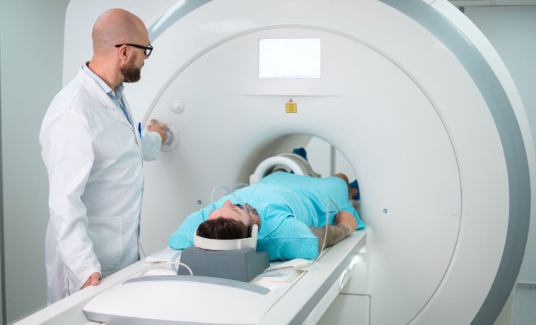
Magnetic resonance cholangiopancreatography, or MRCP, is a specialised MRI technique that produces detailed images of the bile ducts and pancreatic duct. It can identify narrowing, obstruction, stones, and tumours without inserting an instrument into the ducts.
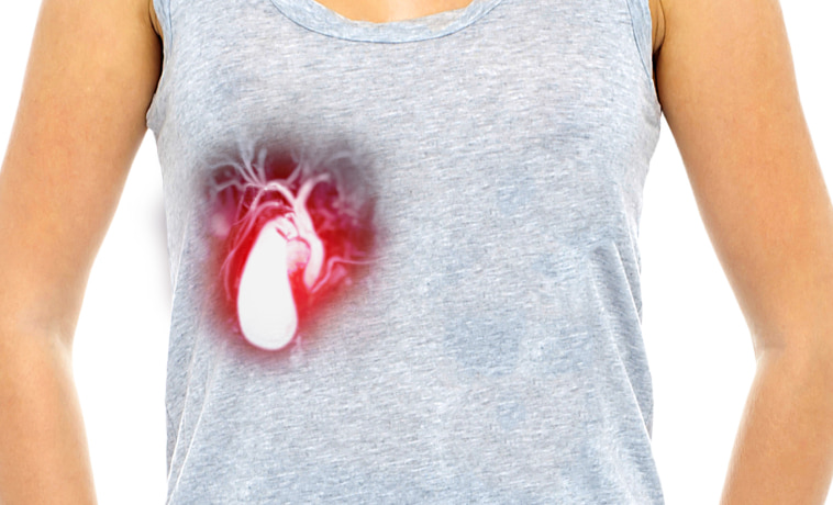
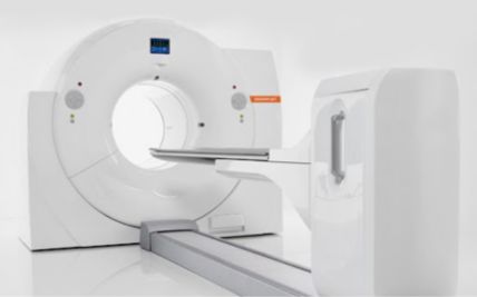
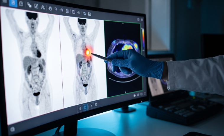
PET-CT combines metabolic and anatomical imaging. It may be used to assess disease spread, clarify uncertain findings, plan radiation, or investigate recurrence in selected GI cancers. It is not required for every GI cancer. Its usefulness depends on the tumour type, because some cancers and low-grade tumours may not show strong uptake with standard FDG PET.
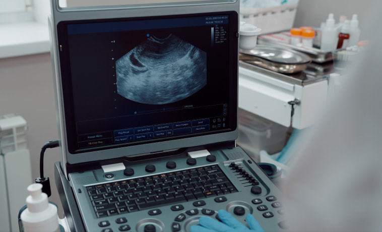
Endorectal ultrasound uses a probe placed within the rectum to examine a rectal or anal tumour. It may help assess how deeply an early tumour has grown and whether nearby tissues are involved.
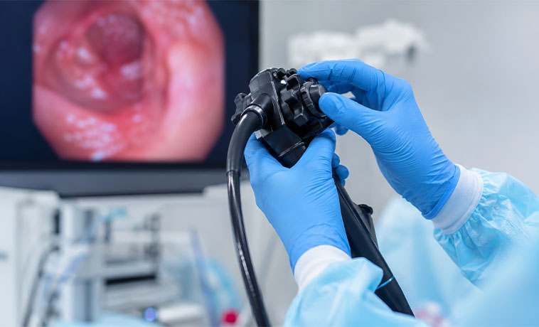
Upper GI endoscopy passes a thin camera through the mouth to examine the esophagus, stomach, and first part of the small intestine. Suspicious areas can be photographed and sampled. Very early lesions may sometimes be removed completely through the endoscope.
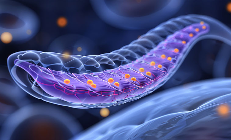
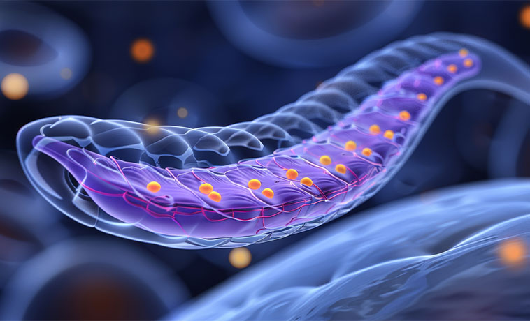
Colonoscopy examines the colon and rectum using a flexible camera. Polyps can be removed, and tissue samples can be collected from suspicious areas. If a large tumour prevents the instrument from passing through the colon, additional imaging may be needed to examine the remaining bowel.
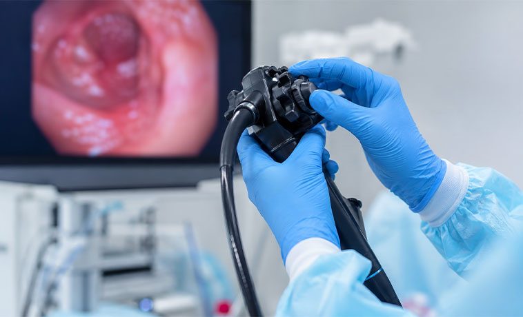
During endoscopic ultrasound, a needle can be guided into a pancreatic, bile duct, upper GI, rectal, or lymph-node lesion. Fine-needle aspiration collects cells, while fine-needle biopsy obtains a small core of tissue. The technique is chosen according to the tumour’s location and the amount of tissue required.
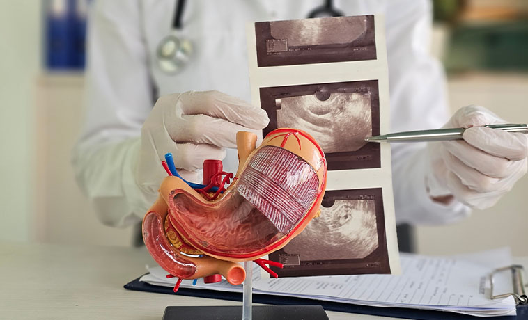
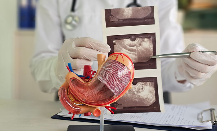
Ultrasound or CT guidance may be used to collect tissue from a liver lesion, abdominal mass, lymph node, or site of suspected metastatic disease. The biopsy pathway is planned carefully when surgery may be possible, particularly for potentially resectable pancreatic or hepatobiliary tumours.
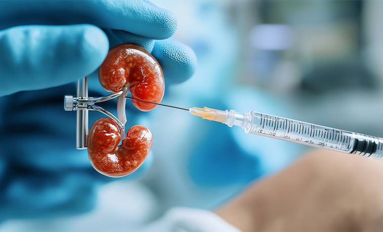
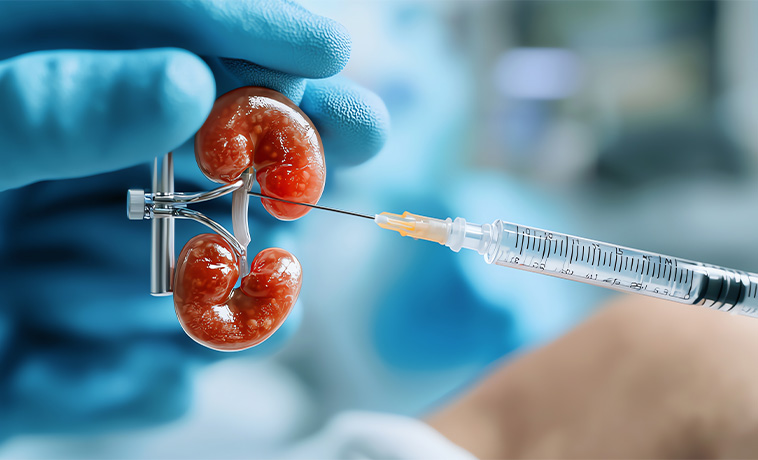
Endoscopic retrograde cholangiopancreatography, or ERCP, passes an endoscope to the opening of the bile duct. Dye is injected into the ducts, allowing areas of narrowing or blockage to be assessed. Brushings or biopsy samples may be collected. A stent can also be placed during the procedure to relieve bile duct obstruction. ERCP is generally used for tissue sampling or treatment rather than routine diagnostic imaging alone.
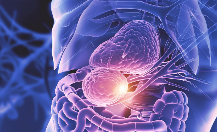
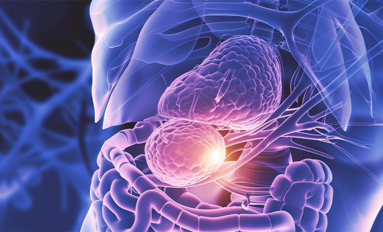
Endoscopic mucosal resection and endoscopic submucosal dissection can remove selected early cancers or precancerous lesions from the esophagus, stomach, colon, or rectum. The removed tissue is examined to determine the cancer’s depth, grade, margins, and risk features and whether additional surgery is needed.
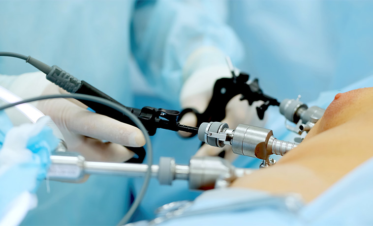
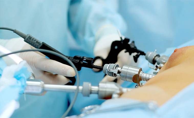
In selected cases, tissue may be obtained through laparoscopy or surgery when endoscopic or image-guided biopsy is unsuitable or has not provided a diagnosis. Diagnostic laparoscopy may also identify small deposits on the abdominal lining that are not clearly visible on scans.
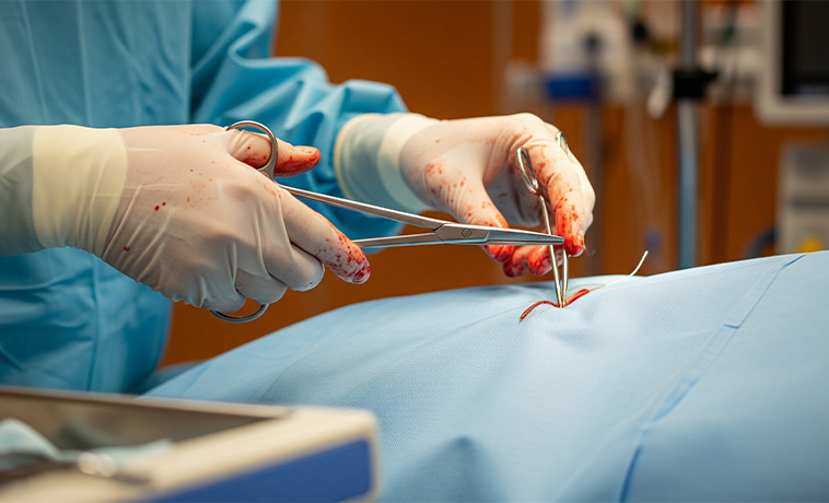
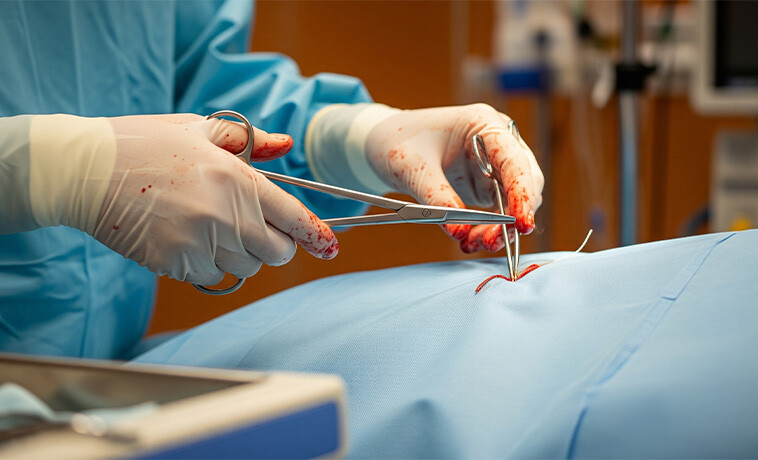
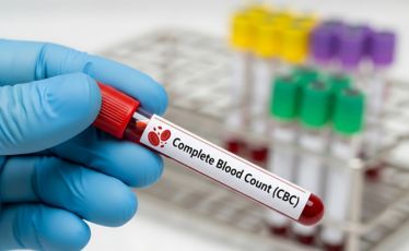
A complete blood count can identify anaemia caused by chronic blood loss, poor nutrition, or advanced disease. It also measures white blood cells and platelets before surgery or systemic treatment.
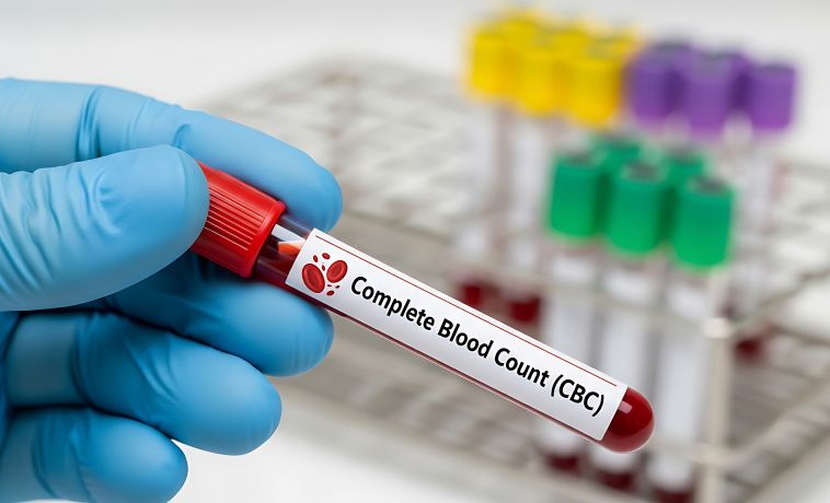
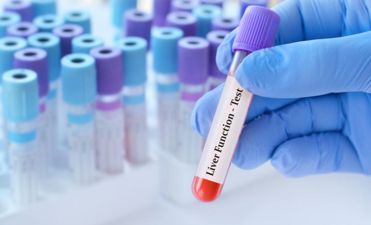
Liver function tests measure bilirubin, liver enzymes, proteins, and other indicators of liver and bile duct function. Abnormal results may suggest obstruction, inflammation, liver damage, or tumour involvement but are not specific to cancer.
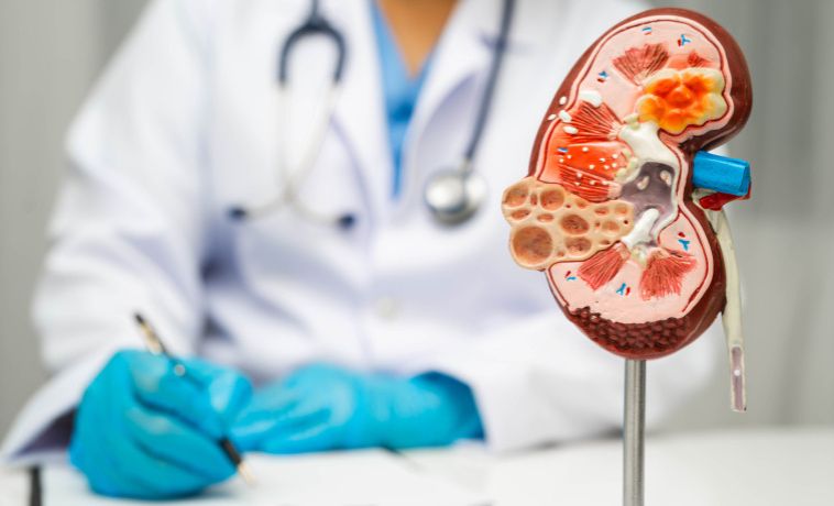
These tests assess hydration, kidney function, and electrolyte balance. They are important before contrast imaging, surgery, chemotherapy, and other treatments.
Fecal immunochemical testing and other stool-based tests can detect small amounts of blood and may be used for colorectal cancer screening. A positive result does not confirm cancer and usually requires colonoscopy. Stool tests are screening tools and should not replace diagnostic evaluation in someone with visible bleeding or other concerning symptoms.


Carcinoembryonic antigen, or CEA, may be measured in colorectal and selected other GI cancers. It is mainly used to establish a baseline, assess response, and monitor for recurrence. A normal CEA does not exclude cancer, and an elevated result can occur for reasons other than cancer.
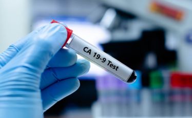
CA 19-9 may be elevated in pancreatic and bile duct cancers. It can also rise because of bile duct obstruction, pancreatitis, or other non-cancerous conditions. It is generally used alongside imaging and pathology rather than as a screening or stand-alone diagnostic test.
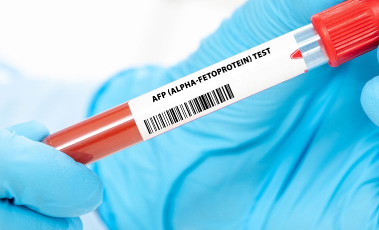
Alpha-fetoprotein may be measured when hepatocellular carcinoma is suspected and during follow-up. Some liver cancers do not produce AFP, while elevated levels can occur in other liver conditions.
A pathologist examines biopsy or surgically removed tissue to confirm the cancer type, grade, depth of invasion, lymph-node involvement, margins, and other clinically important features.
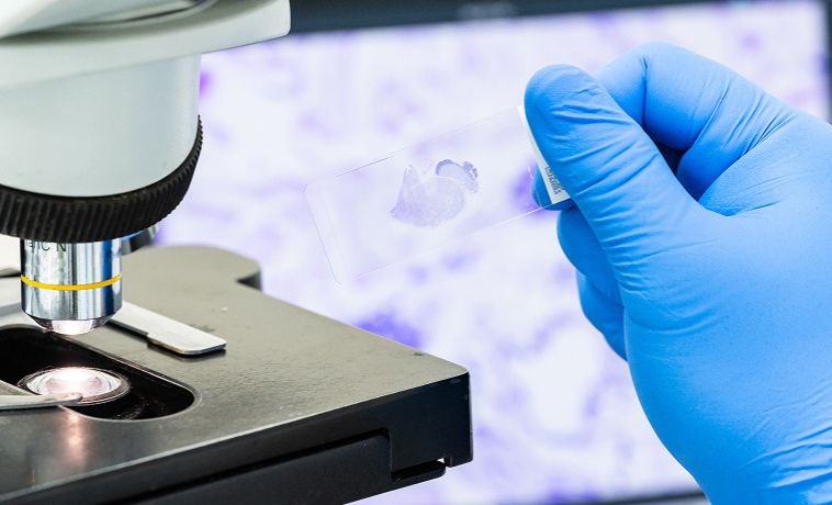
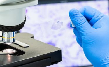
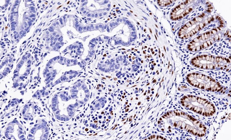
Immunohistochemistry uses specialised stains to identify proteins within tumour cells. It can help determine where a cancer began, classify GISTs and neuroendocrine tumours, and distinguish between cancers with a similar microscopic appearance.
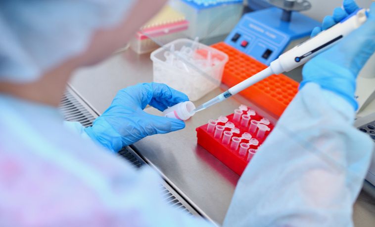
Molecular testing may identify biomarkers that guide treatment. Depending on the cancer, testing may include:
KRAS, NRAS, BRAF, HER2, MSI, and mismatch-repair status in colorectal cancer
HER2, PD-L1, MSI, and selected other markers in gastric or oesophageal cancer
BRCA1, BRCA2, PALB2, and other alterations in pancreatic cancer
FGFR2, IDH1, HER2, BRAF, and other changes in bile duct cancer
KIT and PDGFRA in GIST
Somatostatin receptor status and Ki-67 index in neuroendocrine tumours
Not every patient requires every test. Testing is selected according to the cancer type, stage, and treatment being considered.
Staging describes the extent of cancer and helps guide treatment and prognosis. Most GI cancers use the TNM system, which considers the primary tumour, regional lymph nodes, and distant spread. Exact stage definitions differ by organ.
Stage 0: Abnormal or cancerous cells are confined to the innermost lining and have not invaded deeper tissues.
Stage I: The cancer is generally confined to the organ where it began and has not spread to distant sites.
Stage II: The cancer has grown more deeply into or through the organ wall or nearby tissue but has limited or no lymph-node involvement, depending on the cancer type.
Stage III: The cancer has spread more extensively into nearby structures or regional lymph nodes but has not spread to a distant organ.
Stage IV: The cancer has spread to distant organs or distant lymph nodes. Common sites include the liver, lungs, abdominal lining, bones, or distant lymph nodes.
Endoscopic mucosal resection and endoscopic submucosal dissection may be used to remove selected early cancers or precancerous lesions from the oesophagus, stomach, colon, or rectum without external incisions. Further surgery may be required if pathology shows deep invasion, involved margins, lymphatic or vascular invasion, or other high-risk features.
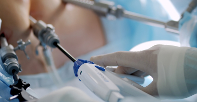
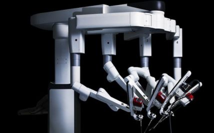
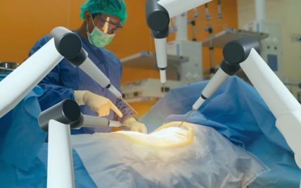
Esophagectomy removes part or most of the oesophagus along with nearby lymph nodes. The digestive tract is reconstructed, commonly by bringing the stomach upward to replace the removed section. Surgery may be performed through open, minimally invasive, or robotic-assisted approaches depending on the tumour and individual clinical situation.
Gastrectomy removes part or all of the stomach together with regional lymph nodes. A partial gastrectomy may be possible for selected tumours, while others require removal of the entire stomach.The remaining digestive tract is reconnected to allow food to pass into the intestine.

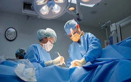
Colectomy removes the section of colon containing the cancer together with nearby lymph nodes. The healthy ends of the bowel are usually reconnected. A temporary or permanent stoma may be required when immediate reconnection is not safe or possible.
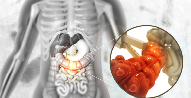
Rectal cancer surgery removes the tumour and surrounding mesorectal tissue. Depending on the tumour’s location and response to treatment, the anal sphincter may be preserved. Very low or extensive tumours may require removal of the anus and rectum with creation of a permanent colostomy. Selected patients with a complete clinical response after treatment may be considered for structured non-operative surveillance.
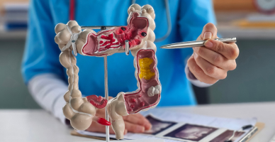
Liver resection removes the part of the liver containing the tumour while preserving sufficient functioning liver. It may be used for selected primary liver cancers and liver metastases, particularly from colorectal cancer. Preoperative procedures may sometimes be used to increase the volume of the liver that will remain after surgery.
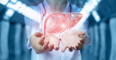
Liver transplantation replaces the diseased liver with a donor liver. It may be considered for carefully selected patients with hepatocellular carcinoma or certain other hepatobiliary cancers who meet defined criteria. Suitability depends on tumour extent, liver function, overall health, and transplant-specific requirements.
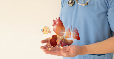
Pancreaticoduodenectomy, or the Whipple procedure, removes the head of the pancreas, duodenum, gallbladder, part of the bile duct, and sometimes part of the stomach. The digestive and biliary pathways are then reconstructed. It is used for selected cancers of the pancreatic head, lower bile duct, ampulla, or duodenum.
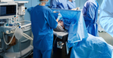
Distal pancreatectomy removes the body and tail of the pancreas and commonly includes removal of the spleen. It may be performed for selected pancreatic cancers, neuroendocrine tumours, or other malignant pancreatic lesions.
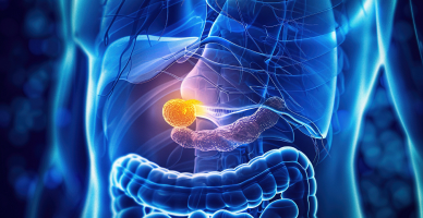
Early gallbladder cancer found after routine gallbladder removal may require additional surgery involving part of the liver and nearby lymph nodes. Bile duct cancer surgery varies by location and may include liver resection, bile duct removal, or pancreaticoduodenectomy. The remaining bile ducts and intestine are reconstructed to restore bile drainage.\
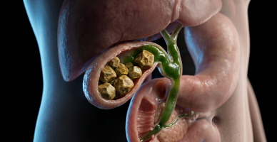
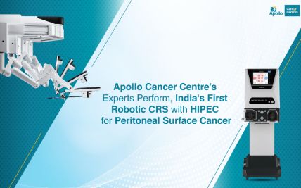
Cytoreductive surgery removes visible cancer from the surfaces of abdominal organs and the abdominal lining. Heated intraperitoneal chemotherapy, or HIPEC, circulates heated chemotherapy within the abdomen during surgery. This intensive treatment may be appropriate for selected appendiceal cancers, pseudomyxoma peritonei, colorectal peritoneal metastases, or other carefully selected conditions. It is not suitable for all cancers that have spread within the abdomen.
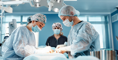
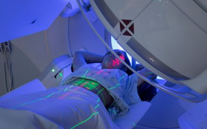
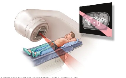
External beam radiation may be used before or after surgery, combined with chemotherapy, as the main treatment, or to relieve symptoms.
It has an established role in oesophageal, rectal, anal, pancreatic, gastric, and selected hepatobiliary cancers. Its role in colon cancer is more limited.
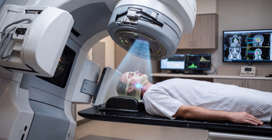
Intensity-modulated radiation therapy and volumetric-modulated arc therapy shape the radiation dose around the tumour. Image guidance checks the tumour and nearby organs during treatment. These techniques can help limit radiation exposure to the bowel, stomach, liver, kidneys, spinal cord, and other nearby tissues.\
Stereotactic body radiation therapy, or SBRT, delivers a highly focused radiation dose in a small number of sessions. It may be used for selected liver or pancreatic tumours, limited metastatic disease, or cancers that cannot be treated surgically. Suitability depends on tumour size, location, organ movement, and proximity to the stomach or bowel.
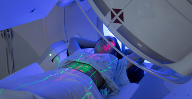
Proton therapy uses proton beams that release most of their radiation within the planned treatment area, with little radiation continuing beyond it. This may reduce radiation exposure to some nearby organs.
It may be considered for selected liver, pancreatic, oesophageal, gastric, recurrent, or paediatric GI cancers when comparative treatment planning demonstrates a meaningful advantage. The benefit depends on tumour location, organ movement, previous radiation, and the treatment dose required.
Proton therapy is not routinely required for every GI cancer and does not remove the need to account for the movement of the liver, pancreas, stomach, and bowel during treatment.

Chemotherapy may be given before surgery to shrink a tumour, after surgery to reduce recurrence risk, with radiation, or as the main treatment for advanced disease. The medicines and duration vary significantly between colorectal, oesophageal, gastric, pancreatic, biliary, anal, and other GI cancers.

Targeted medicines act on specific proteins or molecular changes within cancer cells. They may be used in selected colorectal, gastric, oesophageal, pancreatic, bile duct, liver, GIST, and neuroendocrine tumours. Treatment requires the relevant biomarker or molecular change to be identified. A medicine effective in one GI cancer may not have the same role in another.

Immunotherapy helps the immune system recognise and attack cancer cells. It may be used for selected GI cancers based on factors such as mismatch-repair deficiency, microsatellite instability, PD-L1 expression, tumour type, and stage. Not every GI cancer responds to immunotherapy, so biomarker assessment and cancer-specific evidence are important.
Pathology and molecular testing may identify treatments for selected cancers with actionable biomarkers. It can also clarify inherited cancer risk and support clinical-trial selection. Precision oncology complements—not replaces—cancer type, stage, overall health, and established treatment principles.
After esophagectomy, the digestive tract is commonly reconstructed by shaping the stomach into a tube and joining it to the remaining esophagus. In selected cases, a segment of colon or small intestine may be used when the stomach is unsuitable.
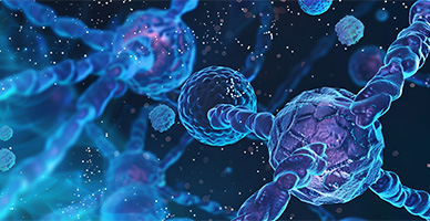
After partial gastrectomy, the remaining stomach may be connected to the small intestine. After total gastrectomy, the esophagus is connected directly to the small intestine, commonly using a Roux-en-Y reconstruction. The method depends on the extent and location of the removed stomach.
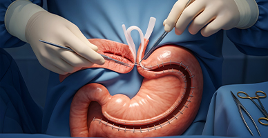
After part of the small intestine, colon, or rectum is removed, the healthy ends may be surgically joined. The location and type of join depend on the section removed and whether adequate blood supply and healing can be achieved safely.
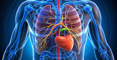
A colostomy or ileostomy brings part of the bowel through an opening in the abdomen so that stool can pass into an external pouch. A stoma may be temporary while a bowel join heals or permanent when the remaining bowel cannot be safely connected to the anus.
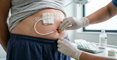
After removal of part of the bile duct, the remaining duct may be connected to the small intestine to restore bile flow. This is commonly performed as a hepaticojejunostomy. The complexity of reconstruction depends on the tumour’s location and the number and size of the remaining bile ducts.
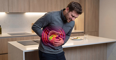
After pancreaticoduodenectomy, the remaining pancreas, bile duct, and stomach or duodenum are reconnected to the intestine. These connections restore the passage of pancreatic enzymes, bile, and food. Different reconstruction techniques may be selected according to pancreatic texture, duct size, and surgical findings.
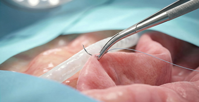
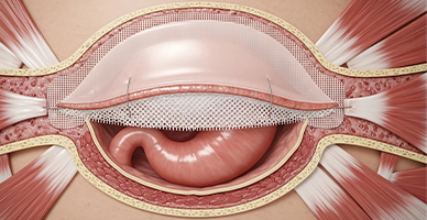
Extensive or recurrent cancers may require removal of part of the abdominal wall. Mesh, tissue flaps, or a combination may be used to close the defect, protect internal organs, and restore abdominal support.
Ablation destroys tumour tissue using heat, cold, or other energy delivered through a needle. Radiofrequency or microwave ablation may be considered for selected small liver tumours or metastases. Suitability depends on the number, size, and location of tumours and their relationship to blood vessels, bile ducts, and other organs.

Transarterial chemoembolisation, or TACE, delivers chemotherapy into the artery supplying a liver tumour and then blocks the blood flow. It may be used for selected liver cancers that cannot be removed surgically.
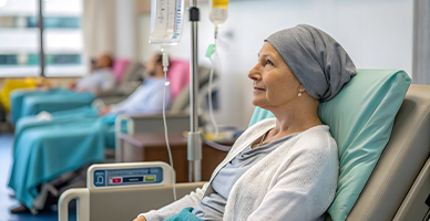
Radioembolisation delivers small radioactive particles through the arteries supplying a liver tumour. It may be considered for selected primary or metastatic liver cancers.
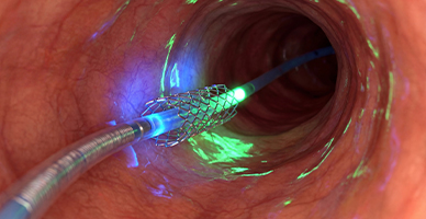
A stent may be placed endoscopically or through the skin to relieve blockage of a bile duct, esophagus, stomach outlet, colon, or intestine. Stenting may relieve jaundice, improve swallowing, restore bowel passage, or allow other treatments to proceed.
Prognosis for Gastrointestinal Cancer
The outlook varies considerably between GI cancers. Several early-stage cancers can be treated effectively, while some pancreatic, liver, biliary, and other cancers may be difficult to detect before they have advanced.
Factors influencing prognosis include:
The organ in which the cancer began
Cancer type, subtype, and grade
Stage at diagnosis
Lymph-node or distant-organ involvement
Whether the tumour can be completely removed
Liver function in people with liver cancer
Molecular and genetic features
Response to treatment
Nutritional status and overall health
Ability to complete treatment and follow-up
Stage IV does not have the same meaning for every GI cancer. Selected colorectal cancers with limited liver or lung spread, some neuroendocrine tumours, and certain peritoneal cancers may still be treated with combinations of systemic and local therapies.
Regular follow-up is important because the pattern, timing, and likelihood of recurrence differ by cancer type and treatment.


Screening for Gastrointestinal Cancer
There is no single screening test for all GI cancers. Screening is established for colorectal cancer and may be recommended for liver cancer in defined high-risk groups. Most other GI cancers do not have routine population screening tests.
Colorectal Cancer Screening
Colorectal screening can identify precancerous polyps and early cancers before symptoms develop. Options may include:
Fecal immunochemical testing
Other approved stool-based tests
Colonoscopy
CT colonography in selected settings
The age to begin, testing interval, and preferred method depend on national guidance and individual risk. People with a family history, inflammatory bowel disease, previous polyps, or an inherited syndrome may need earlier and more frequent colonoscopy.
Why Choose ACC for Gastrointestinal Cancer Treatment
Multidisciplinary GI and hepatopancreatobiliary cancer teams
Organ-specific care for esophageal, gastric, colorectal, pancreatic, liver, biliary, anal, and other GI cancers
Advanced endoscopy for diagnosis and selected early-stage treatments
Open, laparoscopic, and robotic-assisted surgery selected according to clinical need
Expertise in complex liver, pancreatic, esophageal, colorectal, and peritoneal cancer surgery
Access to liver transplantation for appropriately selected cancers
Interventional oncology, including ablation, embolisation, and biliary procedures
Comprehensive chemotherapy, targeted therapy, immunotherapy, and precision-oncology services
Advanced radiation techniques, including IMRT, IGRT, VMAT, SBRT, and proton therapy for selected patients
Molecular pathology and genetic assessment to support personalised treatment
Reconstructive expertise for digestive, biliary, pancreatic, bowel, and abdominal-wall procedures
Coordinated nutrition, stoma, pain, and supportive-care services

Health Blogs