icon
icon
Overview

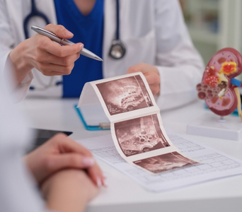
Ultrasound, also called sonography, is a non-invasive imaging test that uses high-frequency sound waves to produce real-time images of structures inside the body. It does not use ionising radiation.

Ultrasound can examine organs, soft tissues, blood vessels and superficial lymph nodes. In cancer care, it may help detect and characterise abnormalities, guide biopsies and treatments, assess blood flow and monitor selected findings.

Its usefulness depends on the organ and location being examined. Air, bone and deep tissues can limit image quality, so CT, MRI, mammography, PET-CT, endoscopy or another test may also be required.
Ultrasound
How the Technology Works
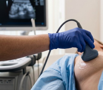
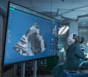
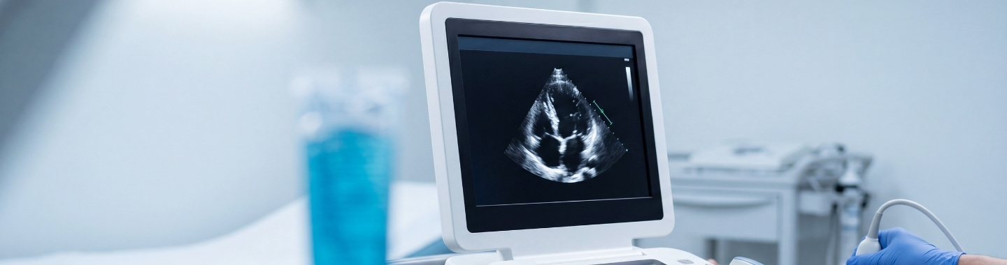
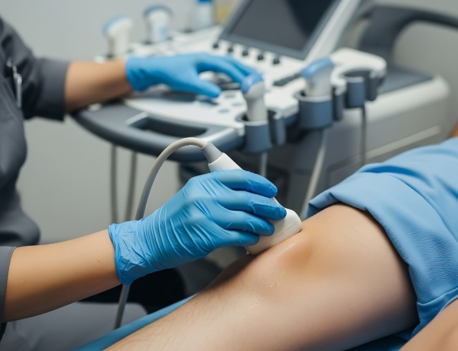
Ultrasound uses a handheld device called a transducer to send sound waves into the body and receive the returning echoes. A computer converts these echoes into images displayed on a monitor.
work-icon
Sound-Wave Imaging
A layer of gel is placed on the skin to help sound waves travel between the transducer and the body. Different tissues reflect the waves in different ways, allowing organs, fluid collections and some abnormal growths to be visualised.
work-icon
Real-Time Images
Ultrasound produces images as the examination is being performed. This allows the operator to assess movement, compressibility and the relationship of an abnormality to nearby structures, as well as guide a needle during biopsy or treatment.
work-icon
Specialised Ultrasound Techniques
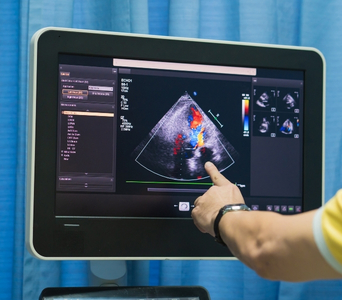
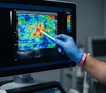
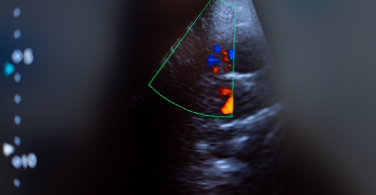
Doppler ultrasound can assess the direction and pattern of blood flow, while elastography can provide information about tissue stiffness. Contrast-enhanced ultrasound uses injected microbubbles to show blood flow within organs or abnormalities more clearly. The techniques used depend on the body area, clinical question and local availability.

Role in Cancer Care

Detecting and Characterising Abnormalities
Ultrasound may be used to evaluate a lump, swelling, pain or abnormality found on examination or another scan. It can help assess the thyroid, breast, liver, kidneys, pancreas, pelvis, testes, soft tissues and superficial lymph nodes. It may also help determine whether an abnormality is solid, fluid-filled or has a combination of features.
Detecting and Characterising Abnormalities
Supporting Cancer Diagnosis
Ultrasound can identify features that help doctors decide whether an abnormality needs follow-up or biopsy. However, benign and cancerous growths can sometimes appear similar on ultrasound. A tissue sample is generally required to confirm cancer.
Supporting Cancer Diagnosis
Assessing Local Disease
Ultrasound may help assess a tumour’s size, location and relationship to nearby tissues or blood vessels. It can also evaluate selected lymph nodes and look for abnormalities in accessible organs. CT or MRI may be required when a broader or more detailed assessment is needed.
Assessing Local Disease
Guiding Biopsy
Real-time ultrasound can guide a needle into an abnormality while helping the doctor avoid nearby blood vessels and other structures. It is commonly used for biopsies of accessible organs, masses and lymph nodes. The tissue obtained is examined by a pathologist to establish or refine the diagnosis.
Guiding Biopsy
Guiding Treatment
Ultrasound may be used to guide selected procedures such as fluid drainage, line placement or tumour ablation. Real-time imaging allows the doctor to monitor the position of the needle or treatment device throughout the procedure. Suitability depends on whether the target can be clearly visualised and safely reached.
Guiding Treatment
Monitoring and Follow-up
Ultrasound may be repeated to monitor selected tumours, lymph nodes or organ abnormalities over time. It can also assess some treatment-related changes or complications, such as fluid collections or blood-flow abnormalities. Follow-up plans depend on the cancer type, treatment received and clinical circumstances.
Monitoring and Follow-up

What to Expect

Before the Test

Preparation depends on the body area being examined. Some ultrasound examinations require no special preparation, while others may require fasting or a full bladder.

Before the scan, inform the care team if you:

  • Are pregnant or may be pregnant

  • Have pain or tenderness in the area being examined

  • Have undergone recent surgery, biopsy or treatment

  • Have dressings, wounds, tubes or drains near the area

  • Take blood-thinning medicines, particularly if a biopsy or procedure is planned

  • Have previously reacted to an ultrasound contrast agent

  • Have relevant previous imaging or reports

For an abdominal ultrasound, you may be asked not to eat or drink for several hours to reduce bowel gas and improve visualisation. For some pelvic examinations, you may need to drink water and avoid emptying your bladder beforehand.

Ultrasound guidance for a biopsy or treatment requires separate preparation. This may include blood tests, temporary adjustment of certain medicines and instructions about eating or drinking. Do not stop any medicine unless specifically instructed.

Before the Test

During the Test 

You will usually lie on an examination table with the area being examined exposed. The sonographer or radiologist will apply a water-based gel to the skin and move the transducer over the area.

Gentle pressure may be applied to obtain clear images. You may be asked to change position, hold your breath briefly or remain still. The examination is generally painless, although pressure over a tender area may cause temporary discomfort.

Some pelvic, prostate or other examinations may require an internal transducer to obtain clearer images. The procedure and the reason for it will be explained beforehand, and consent will be obtained.

If contrast-enhanced ultrasound is required, a cannula will be inserted into a vein and a small amount of microbubble contrast will be administered. If ultrasound is guiding a biopsy or treatment, local anesthesia and sterile preparation may also be required.

A routine diagnostic ultrasound commonly takes approximately 15 to 45 minutes. Specialised, contrast-enhanced or image-guided procedures may take longer.

During the Test

After the Test

After a routine external ultrasound, the gel is wiped away and you can generally return to normal activities immediately. No recovery period is required.

If an internal examination, contrast injection, biopsy or treatment was performed, you will receive specific aftercare instructions. Observation may be required after some procedures.

A radiologist will interpret the images and prepare a report for the doctor who requested the examination. Further imaging or biopsy may be advised if an abnormality cannot be fully characterised by ultrasound.

After the Test

Potential Benefits

Ultrasound provides real-time imaging without exposing the patient to ionising radiation.
  • No ionising radiation: Ultrasound uses sound waves rather than X-rays.
  • Real-time assessment: Structures, movement and blood flow can be observed during the examination.
  • Soft-tissue imaging: It can evaluate many organs, superficial masses and lymph nodes.
  • Solid and fluid differentiation: Ultrasound can often distinguish a solid mass from a cyst or fluid collection.
  • Blood-flow assessment: Doppler techniques can assess blood vessels and vascularity within or around an abnormality.
  • Procedure guidance: Ultrasound can guide biopsies, drainage and selected minimally invasive treatments.
  • Repeatable imaging: It can be repeated when clinically necessary without cumulative radiation exposure.
  • Accessible examination: Ultrasound equipment is widely available and may be brought to the bedside when required.
Ultrasound
Frequently Asked Questions
01 Does ultrasound use radiation?
icon icon
No. Ultrasound uses high-frequency sound waves and does not involve ionising radiation. It can therefore be repeated when clinically required and is generally suitable during pregnancy, although every examination should still have a valid medical indication.
02 Can ultrasound confirm whether a lump is cancerous?
icon icon
Not usually. Ultrasound can show whether a lump is solid or fluid-filled and identify features that may be reassuring or suspicious. However, imaging appearances can overlap, and a biopsy may be needed to confirm the diagnosis.
03 Can ultrasound examine every part of the body?
icon icon
No. Ultrasound waves do not pass well through air or bone, which limits evaluation of structures such as the lungs, bowel and adult brain. Deep structures may also be difficult to assess, particularly when bowel gas or body size affects image quality.
04 Is ultrasound contrast the same as CT or MRI contrast?
icon icon
No. Contrast-enhanced ultrasound generally uses gas-filled microbubbles rather than the iodine-based contrast used for CT or the gadolinium-based contrast commonly used for MRI. It is used selectively to assess blood flow and enhancement within organs or abnormalities.
05 Is an internal ultrasound painful?
icon icon
An internal ultrasound may cause pressure or temporary discomfort but should not usually be painful. The procedure will be explained beforehand, and you should tell the operator if you experience pain. Not every patient or clinical situation requires an internal examination.
06 Is ultrasound used instead of mammography for breast cancer screening?
icon icon
Ultrasound does not generally replace mammography for routine breast cancer screening. It may be used to evaluate a breast symptom or mammographic finding and may provide additional information in selected patients. The appropriate combination of tests depends on age, breast density, symptoms and individual risk.
image image
Book an Appointment
Please Enter Your Name
Please Enter Your Mobile Number
Please Enter OTP