Gynaecological Cancer
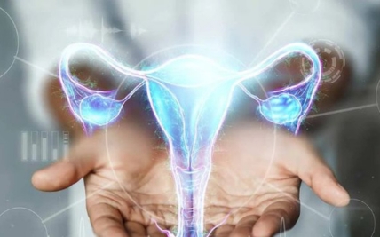
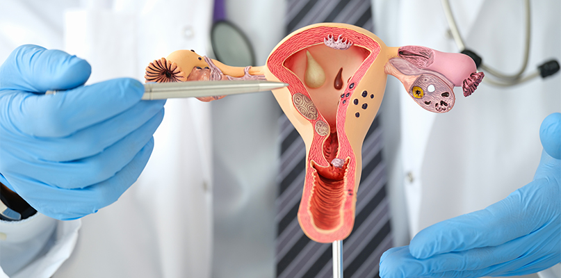
Gynecological cancers develop in the female reproductive system. They include cancers of the cervix, ovaries, fallopian tubes, uterus, vagina, and vulva. Primary peritoneal cancer and gestational trophoblastic disease are also commonly treated within gynecological oncology.
Each of these cancers behaves differently. Cervical cancer is commonly associated with persistent high-risk HPV infection, while some ovarian and endometrial cancers are linked to inherited genetic changes. Some produce early warning signs, such as abnormal bleeding, while others may initially cause vague symptoms such as bloating or pelvic discomfort.
Treatment often requires collaboration between gynecological oncologists, medical oncologists, radiation oncologists, radiologists, pathologists, fertility specialists, genetic counsellors, and reconstructive surgeons. Care is planned around the cancer type and stage while considering fertility, hormonal health, sexual function, and quality of life.
Types of Gynecological Cancer
Gynecological cancer is not a single disease. The organ where the cancer begins and the type of cell involved influence its symptoms, staging, treatment, and outlook.

Cervical cancer develops in the cervix, the lower part of the uterus that opens into the vagina. Most cases are caused by persistent infection with certain high-risk types of human papillomavirus, or HPV. The main types are squamous cell carcinoma and adenocarcinoma. Cervical screening can detect precancerous changes before they become cancer, while HPV vaccination can prevent infection with the HPV types responsible for most cervical cancers.
Ovarian cancer begins in the ovaries. Epithelial ovarian cancer is the most common type and includes high-grade serous, low-grade serous, endometrioid, clear-cell, and mucinous cancers. Less common ovarian cancers include germ-cell tumours and sex-cord stromal tumours. These often affect younger patients and may require different treatment approaches.

Fallopian tube cancer begins in the tubes connecting the ovaries to the uterus. Most are high-grade serous carcinomas and are treated similarly to epithelial ovarian cancer. Evidence suggests that many cancers previously classified as ovarian high-grade serous cancers may begin in the end of the fallopian tube.

Primary peritoneal cancer begins in the lining of the abdomen and pelvis. It closely resembles epithelial ovarian and fallopian tube cancer in its appearance, pattern of spread, and treatment. It can occur even after the ovaries have previously been removed, because it develops from cells within the peritoneal lining.

Endometrial cancer begins in the lining of the uterus. Abnormal uterine bleeding is a common early symptom, which means many cases are diagnosed while still confined to the uterus. Endometrial cancers are classified according to their microscopic and molecular features. These characteristics help determine the likelihood of recurrence and whether treatment beyond surgery is required.
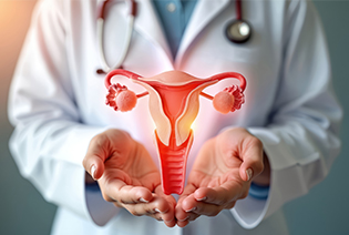
Uterine cancer arises from the muscle or supporting tissues of the uterus rather than its inner lining. They include leiomyosarcoma, endometrial stromal sarcoma, and other rare subtypes. These cancers behave differently from endometrial carcinoma and require sarcoma-specific pathology and treatment planning.
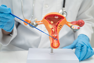
Vaginal cancer begins in the vagina and is rare. Most cases are squamous cell carcinomas, although adenocarcinoma, melanoma, and sarcoma can also occur. Some vaginal cancers are associated with persistent high-risk HPV infection. Cancer found in the vagina may also represent spread from the cervix, uterus, vulva, or another organ, so identifying the site of origin is important.

Vulvar cancer develops on the external genital area. Most cases are squamous cell carcinomas and may follow either an HPV-related pathway or a pathway associated with chronic vulval skin disease. Possible signs include a persistent lump, ulcer, thickened area, itching, pain, bleeding, or a change in the colour or texture of the skin.
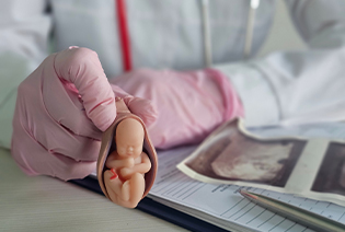
Gestational trophoblastic disease is a group of conditions arising from tissue formed during pregnancy. It includes molar pregnancy and malignant forms such as invasive mole, choriocarcinoma, and placental-site trophoblastic tumour. These conditions are assessed using pregnancy hormone levels and imaging. Many respond very well to specialised treatment, although the approach depends on the exact subtype and extent of disease.
Risk factors differ by cancer type. Having one or more risk factors does not mean that cancer will develop, and some people diagnosed with gynecological cancer have no identifiable risk factors.
Modifiable Risk Factors
High-risk HPV infection: Persistent infection is the main cause of cervical cancer and contributes to many vaginal and vulvar cancers.
Lack of HPV vaccination: Vaccination can prevent infection with the HPV types responsible for most cervical cancers and many other HPV-related cancers.
Smoking: Increases the risk of cervical cancer and some vulvar and vaginal cancers.
Excess body weight: A major risk factor for endometrial cancer and associated with some ovarian cancers.
Physical inactivity: May contribute to endometrial cancer risk, partly through its effect on body weight and metabolic health.
Unopposed oestrogen exposure: Oestrogen used without appropriate progesterone in a person with a uterus can increase endometrial cancer risk.
Delayed evaluation of precancerous changes: Follow-up and treatment of cervical, vaginal, vulvar, or endometrial precancer can reduce the likelihood of progression.
Non-Modifiable Risk Factors
Increasing age
A family history of ovarian, fallopian tube, endometrial, colorectal, or breast cancer
Inherited variants involving BRCA1, BRCA2, or other ovarian cancer-related genes
Lynch syndrome
A personal history of breast, ovarian, endometrial, or colorectal cancer
Endometriosis, which is associated with a small increase in certain ovarian cancer subtypes
Never having been pregnant or infertility, which is associated with ovarian and endometrial cancer risk
Early onset of menstruation or late menopause
Polycystic ovary syndrome and prolonged irregular ovulation
Chronic vulval conditions such as lichen sclerosus
Reduced immune function
Previous pelvic radiation
Exposure before birth to diethylstilbestrol, a medicine used in some countries several decades ago
Increasing age at first pregnancy or reproductive factors that prolong lifetime ovulation
People with a strong family history, cancer at a young age, multiple related cancers, or features of an inherited syndrome may benefit from genetic counselling.

Signs and Symptoms
Symptoms depend on the organ involved. Some symptoms, particularly abnormal bleeding, may help identify cancer at an earlier stage.
Possible signs and symptoms include:
These symptoms can also result from fibroids, endometriosis, infection, menopause, ovarian cysts, or other non-cancerous conditions. However, any bleeding after menopause, a persistent vulval lesion, or new symptoms that continue or worsen should be assessed promptly.
Diagnosis begins with a review of symptoms, menstrual and reproductive history, previous screening results, medical history, and family history. A pelvic examination may be performed to assess the cervix, vagina, vulva, uterus, ovaries, and nearby tissues. Imaging helps identify and stage the tumour, while biopsy and laboratory testing establish the exact cancer type.
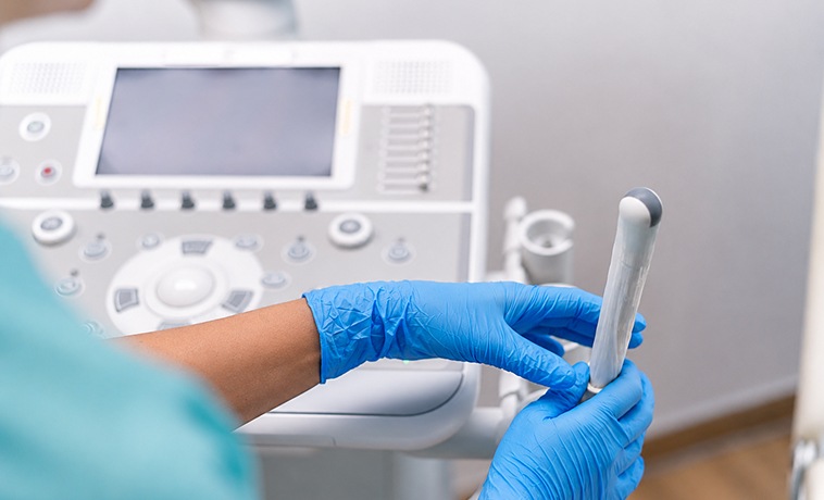
Pelvic ultrasound uses sound waves to examine the uterus, endometrium, ovaries, fallopian tubes, and surrounding tissues. It may be performed across the abdomen or through the vagina. It can identify an ovarian or pelvic mass, measure endometrial thickness, and show features that help determine whether further investigation is required.


Transvaginal ultrasound places a narrow ultrasound probe within the vagina to provide detailed images of the uterus, endometrium, ovaries, and pelvis. It is commonly used to investigate abnormal uterine bleeding or an ovarian mass. Ultrasound findings alone cannot reliably confirm or exclude cancer.
A CT scan produces detailed images of the abdomen, pelvis, chest, and other areas. It can assess tumour size, lymph nodes, abdominal fluid, peritoneal deposits, and spread to distant organs. CT is commonly used when ovarian, fallopian tube, peritoneal, advanced uterine, or other pelvic cancers are suspected.
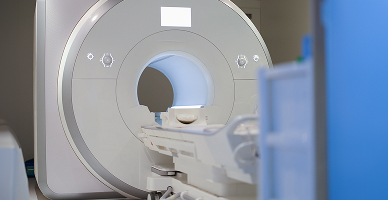
MRI provides detailed images of the uterus, cervix, vagina, vulva, ovaries, and surrounding pelvic structures. It is particularly useful for determining how deeply cervical or endometrial cancer has grown and whether nearby tissues are involved. MRI can also help assess complex ovarian masses and plan fertility-sparing or reconstructive surgery.
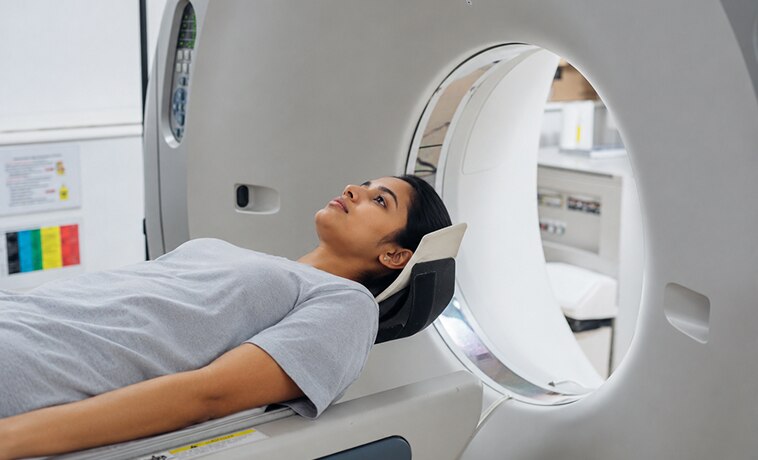

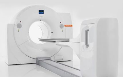
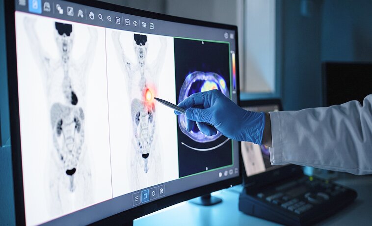
PET-CT combines metabolic and anatomical imaging. It may be used to assess lymph nodes or distant spread, plan radiation treatment, or investigate suspected recurrence. It is not required for every gynecological cancer and is selected according to the tumour type, stage, and findings on other tests.
Colposcopy uses a magnifying instrument to examine the cervix after an abnormal cervical screening result or when a suspicious lesion is seen. Small samples can be collected from abnormal areas. An endocervical sample may also be taken from the canal of the cervix.


A loop electrosurgical excision procedure or cone biopsy removes a larger area of abnormal cervical tissue. It may be used to diagnose and treat precancer or selected very early cervical cancers. The tissue is examined for the depth of invasion, margins, and other features that determine whether further treatment is needed.
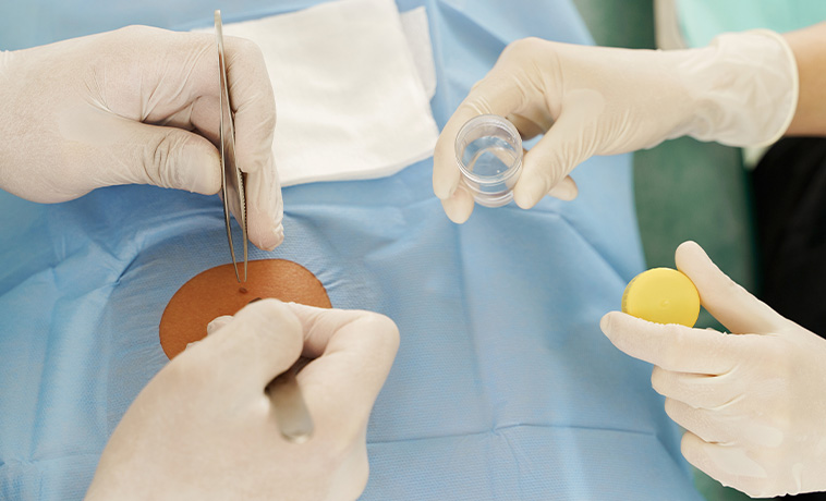
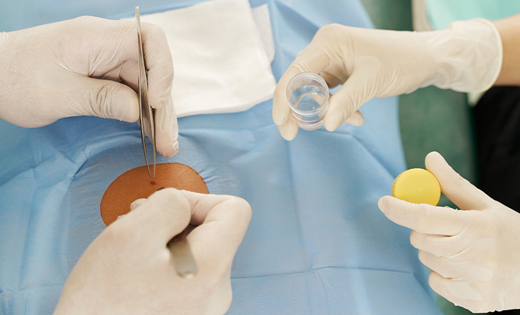
An endometrial biopsy uses a thin tube passed through the cervix to collect tissue from the uterine lining. It is commonly used to investigate postmenopausal bleeding or other abnormal uterine bleeding. If the sample is insufficient or symptoms continue, hysteroscopy or another procedure may be required.


Hysteroscopy passes a small camera through the cervix to examine the inside of the uterus. Suspicious areas can be sampled directly. Dilatation and curettage removes tissue from the uterine lining and may be used when an office biopsy does not provide enough information.
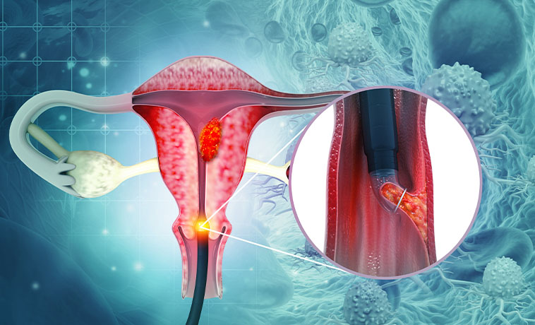
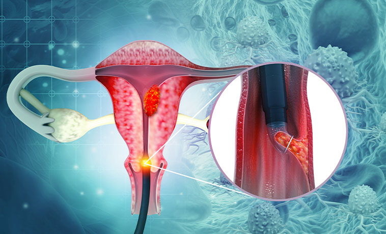
A small sample is removed from a persistent lump, ulcer, thickened area, or patch of altered vulval skin. A local anaesthetic is generally used. The biopsy confirms whether the change is cancerous, precancerous, inflammatory, or caused by another skin condition.


A tissue sample is taken from a suspicious area within the vagina, often during colposcopy. The biopsy helps determine whether the lesion began in the vagina or represents extension from another site.
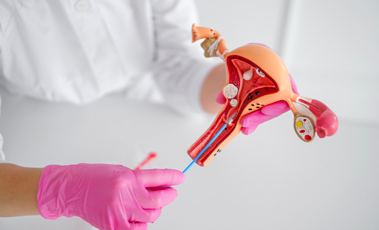
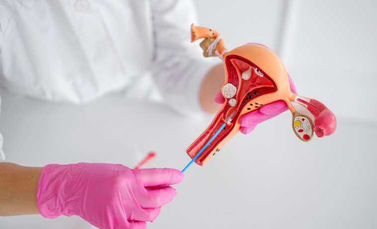
Ultrasound or CT guidance may be used to collect tissue from an ovarian or pelvic mass, lymph node, peritoneal deposit, or site of suspected metastatic disease. When an apparently early ovarian cancer may be removed surgically, a needle biopsy of the ovarian mass is not routinely performed because it could rupture the tumour. The biopsy pathway is therefore planned by the gynecological oncology team.
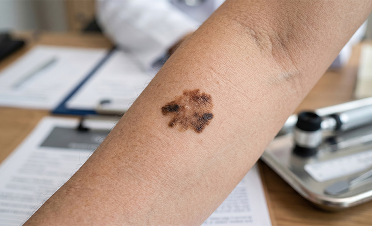
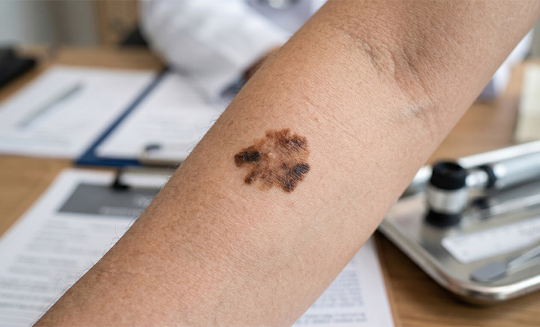
Laparoscopy or open surgery may be required when a diagnosis cannot be established safely through another method. It may also be used to assess disease within the abdomen and obtain tissue before systemic treatment.
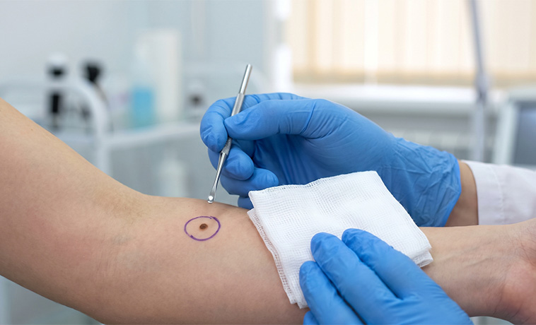
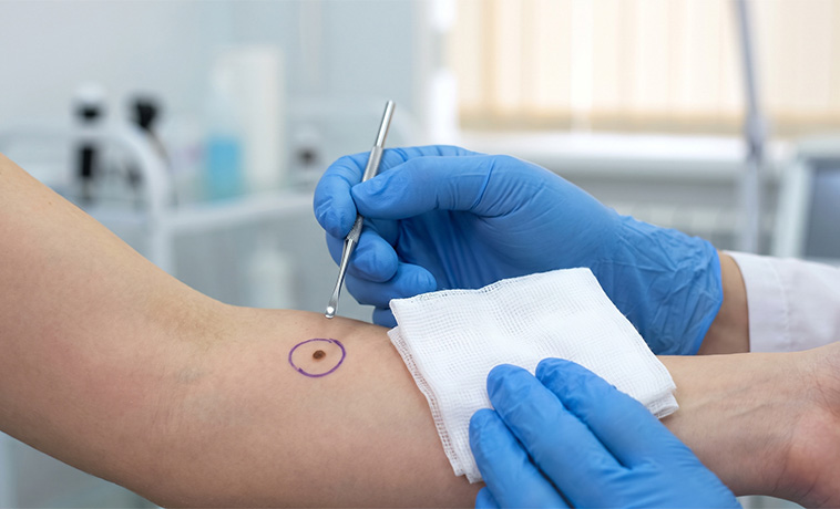
A cervical cytology test, commonly called a Pap test, examines cells collected from the cervix for precancerous or cancerous changes. An abnormal result does not necessarily mean cancer is present. Colposcopy and biopsy may be required for diagnosis.
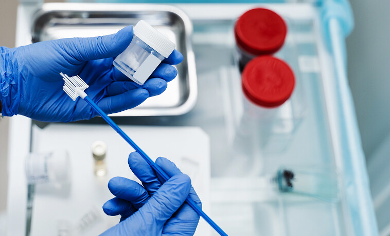

HPV testing checks cervical cells for high-risk HPV types associated with cervical cancer. It may be used as a primary screening test, alongside cytology, or to guide follow-up after an abnormal result. A positive HPV test means that a high-risk HPV type has been detected; it does not mean that cervical cancer is present.


CA-125 may be elevated in epithelial ovarian, fallopian tube, or primary peritoneal cancer. It can also rise because of endometriosis, menstruation, pregnancy, fibroids, liver disease, or other non-cancerous conditions. It is mainly used with imaging and clinical findings and to monitor treatment or recurrence. It is not an accurate stand-alone screening or diagnostic test.
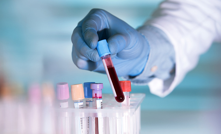
Depending on age and the type of ovarian mass, tests may include alpha-fetoprotein, beta-human chorionic gonadotropin, lactate dehydrogenase, inhibin, or other markers. These tests can help assess suspected germ-cell or sex-cord stromal tumours but must be interpreted with imaging and pathology.


Beta-human chorionic gonadotropin is central to diagnosing and monitoring gestational trophoblastic disease. Serial measurements help assess response to treatment and detect persistent or recurrent disease.


A pathologist examines biopsy or surgical tissue under a microscope to confirm the cancer type, subtype, grade, depth of invasion, lymphovascular involvement, and surgical margins.
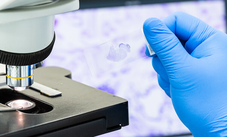
Immunohistochemistry uses specialised stains to identify proteins within tumour cells. It can help distinguish between gynecological cancer subtypes and determine whether a tumour began in the reproductive organs or spread there from another site.


Molecular testing may identify features that influence prognosis or treatment. Depending on the cancer, testing may include:
BRCA1, BRCA2 and homologous-recombination-related testing in ovarian cancer
Mismatch-repair and microsatellite-instability testing in endometrial and selected other cancers
POLE and p53-related classification in endometrial cancer
HER2 testing in selected uterine, ovarian, or cervical cancers
PD-L1 testing where immunotherapy is being considered
Not every patient requires every test. Testing is selected according to the cancer type, stage, and proposed treatment.


Inherited genetic testing may be recommended for people with epithelial ovarian, fallopian tube, or primary peritoneal cancer and for selected people with endometrial cancer or a strong family history. Genetic counselling helps patients and families understand the results, treatment implications, and whether relatives may also require testing or surveillance.


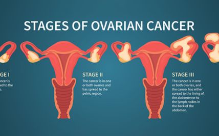
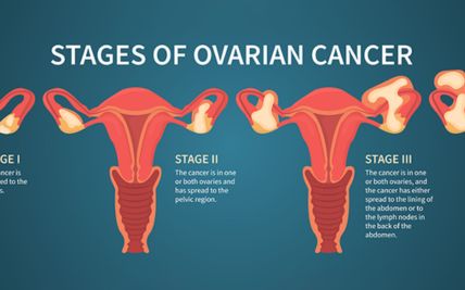
Staging describes how far the cancer has spread and guides treatment and prognosis. Most gynecological cancers use the FIGO system, which generally ranges from Stage I to Stage IV. Exact definitions differ by organ.
Stage I: The cancer is confined to the organ where it began.
Stage II: The cancer has spread beyond the organ where it began but remains within nearby reproductive structures or the pelvis.
Stage III: The cancer has spread more extensively within the pelvis or abdomen or to regional lymph nodes.
Stage IV: The cancer has invaded certain nearby organs or spread to distant sites.
Conisation removes a cone-shaped section of the cervix containing the abnormal area. It may be used to treat selected precancers and very early cervical cancers. The pathology result determines whether conisation alone is sufficient or further surgery is needed.
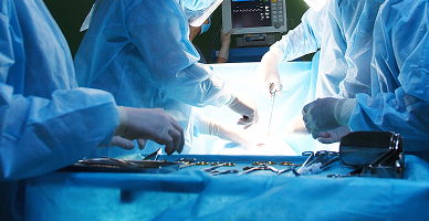
A trachelectomy removes the cervix while preserving the uterus. It may be considered for carefully selected people with early cervical cancer who wish to retain the possibility of pregnancy. A permanent stitch may be placed around the lower uterus, and future pregnancies require specialist obstetric care.

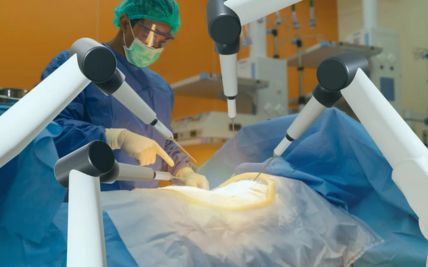
A hysterectomy removes the uterus and cervix. Depending on the cancer, surgery may also include the fallopian tubes, ovaries, nearby tissue, or lymph nodes. The procedure may be performed through an open, laparoscopic, or robotic-assisted approach according to the cancer type, stage, anatomy, and surgical requirements.
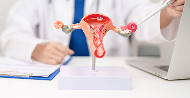
Radical hysterectomy removes the uterus, cervix, upper part of the vagina, and tissues surrounding the cervix. Pelvic lymph nodes are also assessed or removed. It may be used for selected early cervical cancers. More advanced cervical cancer is generally treated with chemotherapy and radiation rather than radical surgery.

Surgery for ovarian, fallopian tube, or primary peritoneal cancer aims to remove as much visible cancer as safely possible. It commonly includes removal of the ovaries, fallopian tubes, uterus, omentum, and selected lymph nodes or other involved tissues. Depending on the extent of disease, surgery may involve the bowel, spleen, diaphragm, liver surface, bladder, or peritoneum. In selected patients, chemotherapy may be given before surgery.

Selected early-stage ovarian germ-cell, sex-cord stromal, borderline, or carefully selected epithelial tumours may be treated by removing only the affected ovary and fallopian tube while preserving the uterus and opposite ovary. Suitability depends on the exact cancer type, stage, grade, and completeness of surgical staging.
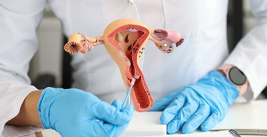
Surgery usually involves hysterectomy with removal of both fallopian tubes and ovaries. Sentinel lymph-node mapping or lymph-node assessment may be performed according to the cancer’s risk features. Fertility-sparing hormone treatment may be considered for a very select group with low-grade, early endometrioid cancer under close specialist surveillance.

Small vulvar cancers may be treated with wide local excision. Larger tumours may require removal of more extensive vulval tissue. Sentinel lymph-node biopsy or groin lymph-node dissection may be performed according to the tumour’s size, depth, and location.

Surgery may be used for selected small, localised vaginal cancers. Procedures range from local excision to removal of part or all of the vagina. Many vaginal cancers are treated primarily with radiation, sometimes combined with chemotherapy, because extensive surgery can significantly affect nearby organs.

Pelvic exenteration is an extensive operation that may be considered for selected locally recurrent cancers confined to the pelvis. It may involve removal of reproductive organs together with the bladder, rectum, or both. Urinary, bowel, vaginal, and pelvic reconstruction may then be required.

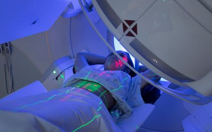
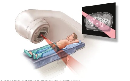
External beam radiation delivers radiation from a machine outside the body. It may target the primary tumour, surrounding tissues, and regional lymph nodes. It has an important role in cervical, endometrial, vaginal, and vulvar cancers and may be used in selected ovarian or recurrent cancers.

Intensity-modulated radiation therapy shapes the radiation dose around the tumour, while image-guided radiation therapy confirms the target’s position during treatment. These techniques can help reduce radiation exposure to the bowel, bladder, rectum, kidneys, and pelvic bone marrow.
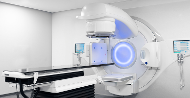
Brachytherapy places a radioactive source within or close to the tumour. It is a central part of curative treatment for many locally advanced cervical cancers and may also be used for endometrial or vaginal cancer. Intracavitary brachytherapy places applicators within the vagina or uterus. Interstitial brachytherapy places treatment needles directly into or around the tumour when required.

Chemoradiation combines radiation with chemotherapy that helps make cancer cells more sensitive to radiation. It is commonly used for locally advanced cervical cancer and selected vaginal or vulvar cancers. The chemotherapy dose and schedule differ from those used when chemotherapy is given as the main systemic treatment.
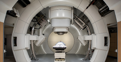
Proton therapy uses proton beams that release most of their radiation within the planned treatment area, with little radiation continuing beyond it. This may reduce radiation exposure to some nearby tissues.
It may be considered for selected pelvic or abdominal tumours when comparative planning shows a meaningful advantage, particularly in re-irradiation, extensive nodal treatment, or selected younger patients. The benefit depends on tumour location, internal-organ movement, previous radiation, and the required treatment area.
Proton therapy is not routinely required for every cervical, endometrial, ovarian, or other gynecological cancer. Brachytherapy remains an essential component of treatment for many cervical cancers and should not be replaced by proton therapy when it is clinically indicated.
Chemotherapy may be given before surgery, after surgery, with radiation, or as the main treatment for advanced or recurrent disease. It has an important role in ovarian, cervical, endometrial, vaginal, vulvar, and gestational trophoblastic cancers. The medicines and treatment schedule depend on the cancer type and stage.

Targeted medicines act on particular pathways or proteins used by cancer cells. These may include anti-angiogenic medicines and other biomarker-directed treatments. Their use depends on the cancer type, molecular findings, previous treatment, and stage.

PARP inhibitors interfere with cancer cells’ ability to repair DNA. They are used mainly as maintenance or later-line treatment in selected ovarian, fallopian tube, and primary peritoneal cancers. Suitability may be influenced by BRCA status, homologous-recombination deficiency, response to platinum-based chemotherapy, previous treatment, and the specific medicine being considered.

Immunotherapy may be used for selected advanced or recurrent cervical, endometrial, ovarian, vulvar, or vaginal cancers. Suitability may depend on factors such as mismatch-repair deficiency, microsatellite instability, PD-L1 expression, tumour type, and previous treatments.

Hormone therapy may be used for selected low-grade or hormone-sensitive endometrial, ovarian, or uterine stromal cancers. It may also be considered as fertility-sparing treatment for carefully selected early endometrial cancers under close surveillance.

Tumour profiling and inherited genetic testing may identify treatment options, estimate inherited risk, or support clinical-trial selection. Precision oncology complements the cancer’s histology, stage, response to treatment, and overall clinical circumstances.

Vulvar reconstruction may be performed after removal of a large vulvar tumour. Local tissue flaps, skin grafts, or tissue transferred from another area may be used to close the surgical defect and restore the vulval contour. The technique depends on the tumour’s location, the amount of tissue removed, and whether the urethra, vagina, or anus is involved.

Vaginal reconstruction may be considered when part or all of the vagina is removed. Skin grafts, local flaps, or tissue from the intestine or another part of the body may be used to create or restore the vaginal passage. The procedure is individualised according to the extent of surgery and the person’s treatment priorities.
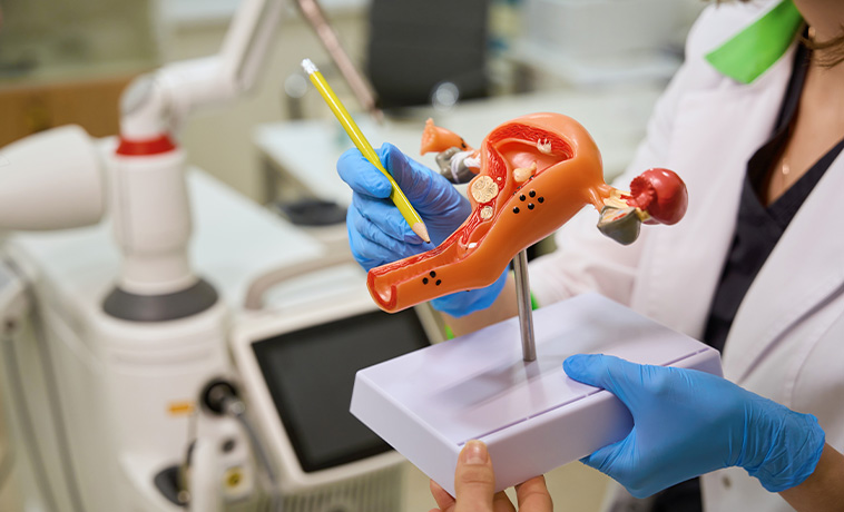
Pelvic-floor reconstruction may be required after extensive pelvic surgery. Mesh or tissue flaps may be used to fill and support the pelvic space, separate organs, close surgical defects, and reduce the risk of internal organ displacement.

When cancer surgery requires removal of the bladder or part of the urinary tract, urine may be diverted through an ileal conduit, continent reservoir, or another reconstructive procedure. The method is selected according to kidney function, anatomy, previous treatment, and the extent of surgery.
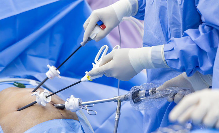
When part of the bowel or rectum is removed, the remaining ends may be reconnected where possible. A temporary or permanent colostomy or ileostomy may be required when a safe connection cannot be made.

If cancer surgery removes part of the abdominal wall, mesh, tissue flaps, or a combination may be used to close the defect, protect internal organs, and restore abdominal support.
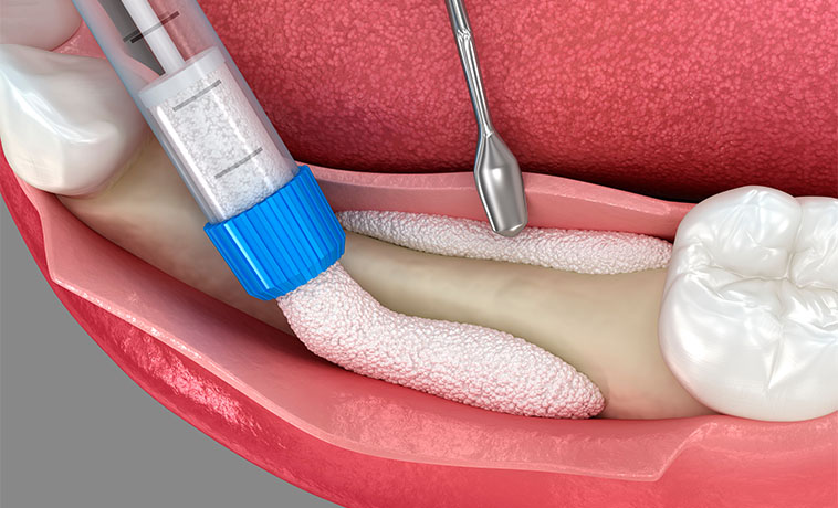
Prognosis for Gynecological Cancer
The outlook differs considerably between gynecological cancers. Many cervical, endometrial, ovarian, vulvar, and vaginal cancers can be treated effectively when diagnosed while localised, but prognosis depends on more than stage alone.
Factors influencing prognosis include:
The organ in which the cancer began
Cancer type, subtype, and grade
Stage at diagnosis
Lymph-node or distant-organ involvement
Molecular and genetic features
Whether visible disease can be completely removed
Response to chemotherapy, radiation, or other treatment
Overall health and nutritional status
Ability to complete treatment and follow-up
Endometrial cancer is often diagnosed earlier because abnormal bleeding prompts evaluation. Ovarian cancer may be diagnosed after it has spread within the abdomen, but outcomes vary by subtype, stage, molecular features, and response to treatment.
Gestational trophoblastic neoplasia is often highly responsive to treatment, although specialist monitoring of beta-hCG is essential. Regular follow-up is important after all gynecological cancers because recurrence patterns differ by disease and treatment.


Screening for Gynecological Cancer
There is no single screening test for all gynecological cancers. Routine population screening is established for cervical cancer, but not for ovarian, endometrial, vaginal, or vulvar cancer in people without symptoms.
Cervical Cancer Screening
Cervical screening can detect high-risk HPV infection and precancerous changes before invasive cancer develops. Depending on national guidance and available services, screening may use:
Primary HPV testing
Cervical cytology or Pap testing
HPV testing combined with cytology
Visual inspection with acetic acid in selected screening programmes
The age to begin, test used, and screening interval should follow national recommendations and take account of previous results and individual risk. An abnormal screening result does not necessarily mean cancer is present. Repeat testing, colposcopy, or biopsy may be recommended.
HPV Vaccination
HPV vaccination protects against the high-risk HPV types responsible for most cervical cancers and many vulvar, vaginal, anal, and other HPV-related cancers. It is most effective when given before exposure to HPV, but eligible older adolescents and adults may also benefit according to national recommendations. Vaccination does not replace cervical screening.
Why Choose ACC for Gynecological Cancer Treatment
Dedicated multidisciplinary gynecological oncology teams
Organ-specific care for cervical, ovarian, fallopian tube, uterine, vaginal, vulvar, and related cancers
Open, laparoscopic, and robotic-assisted surgery selected according to clinical need
Fertility-sparing and organ-preserving approaches for appropriately selected patients
Expertise in complex ovarian cancer cytoreduction and recurrent pelvic cancer surgery
Advanced radiation techniques, including external beam radiation, image guidance, and brachytherapy
Access to proton therapy for selected primary or recurrent cancers
Precision oncology supported by BRCA, mismatch-repair, microsatellite-instability, and other biomarker testing
Genetic counselling and inherited cancer-risk assessment
Reconstructive expertise for vulvar, vaginal, urinary, bowel, pelvic-floor, and abdominal-wall procedures
Coordinated fertility preservation, hormonal-health, and survivorship support
Multidisciplinary tumour-board review for individualised treatment planning

Health Blogs