Endocrine & Neuroendocrine Cancers
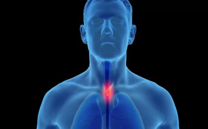
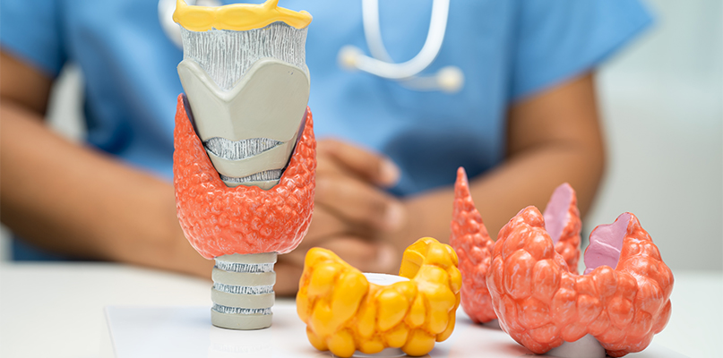
Endocrine and neuroendocrine cancers develop in hormone-producing glands or specialised neuroendocrine cells distributed throughout the body. This group includes thyroid cancer, malignant adrenal and parathyroid tumours, and neuroendocrine tumours arising most often in the gastrointestinal tract, pancreas or lungs.
These diseases differ greatly in their behaviour. Some remain slow-growing for years, while poorly differentiated neuroendocrine carcinomas, anaplastic thyroid cancer and adrenocortical carcinoma may progress rapidly. Some tumours release hormones and cause distinct clinical syndromes; others are non-functioning and are discovered because of a lump, pressure symptoms, imaging performed for another reason or metastatic disease.
Accurate classification requires specialist pathology, hormone testing and appropriate anatomical and functional imaging. Care may involve endocrinologists, surgical oncologists, medical oncologists, nuclear medicine physicians, radiation oncologists, interventional radiologists, radiologists, pathologists, genetic counsellors and other specialists. Treatment is individualised according to the organ of origin, tumour differentiation and grade, hormone activity, molecular findings, stage and the patient’s overall health.
Types of Endocrine & Neuroendocrine Cancers
Endocrine and neuroendocrine cancers are not a single disease. Identifying the site of origin, cell type, differentiation, grade, hormone activity and stage is essential because each subtype follows a different treatment pathway.
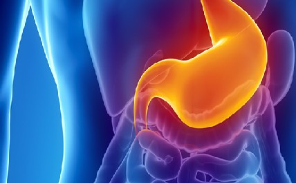
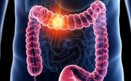
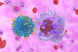
Neuroendocrine tumours arise from specialised cells that can receive signals from the nervous system and release hormones. They most commonly begin in the gastrointestinal tract, pancreas or lungs but may occur at other sites or present without an identifiable primary. Well-differentiated NETs are graded according to how actively their cells are dividing and may range from slow-growing to aggressive. Poorly differentiated neuroendocrine carcinomas are biologically distinct, usually high-grade cancers that require a different treatment approach. Some NETs are functioning and produce syndromes such as carcinoid syndrome, insulinoma syndrome, gastrinoma syndrome or glucagonoma syndrome; many do not cause hormone-related symptoms.

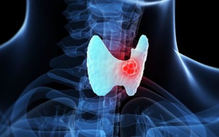
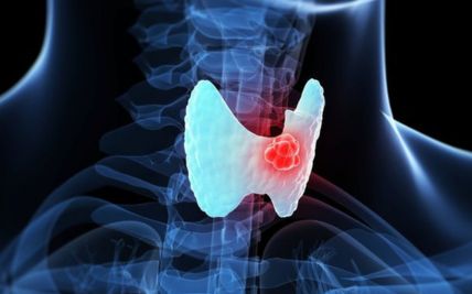
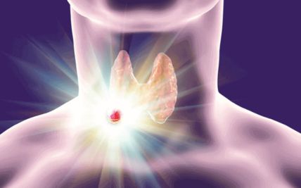
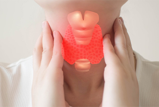
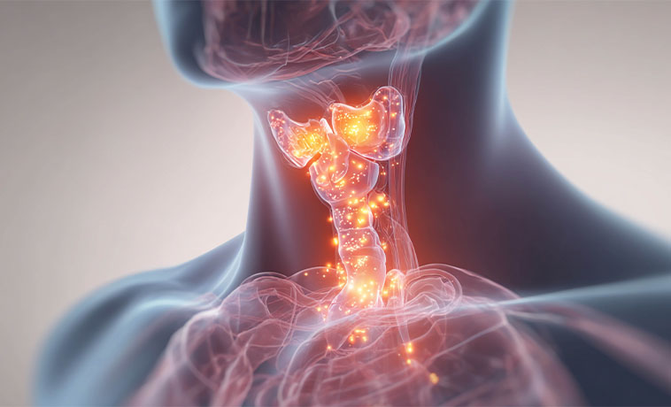
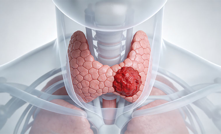
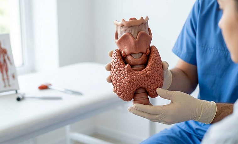
Thyroid cancer begins in the thyroid gland at the front of the neck. Papillary and follicular cancers are differentiated thyroid cancers and account for most cases. Other types include oncocytic thyroid carcinoma, medullary thyroid cancer arising from calcitonin-producing C cells, poorly differentiated thyroid cancer and anaplastic thyroid cancer. Each type differs in its pattern of spread, molecular features, treatment and expected course.

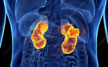
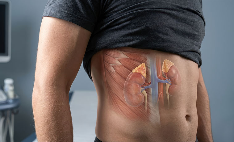
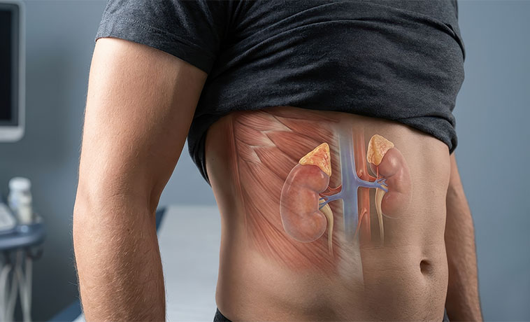
Adrenal tumours arise in glands located above the kidneys. Most adrenal masses are benign, but evaluation must determine whether a mass produces excess hormones and whether it has suspicious features. Malignant adrenal tumours include adrenocortical carcinoma and malignant pheochromocytoma. Pheochromocytomas arise from adrenal medullary cells and are considered malignant when they spread to sites where this tissue is not normally present; related tumours outside the adrenal glands are called paragangliomas. The adrenal gland may also be affected by lymphoma or cancer that has spread from another organ.
Parathyroid tumours arise from the small glands behind the thyroid that regulate calcium through parathyroid hormone. Most are benign adenomas. Parathyroid carcinoma is rare and commonly causes marked overproduction of parathyroid hormone and high calcium levels. Atypical parathyroid tumours have some concerning pathological features but do not meet all criteria for carcinoma. Diagnosis often depends on the clinical presentation, operative findings and evidence of invasion or spread rather than on a needle biopsy.
The causes of most endocrine and neuroendocrine tumours are not known. Having a risk factor does not mean that a tumour will develop, and many patients have no identifiable risk factor. Most cannot be prevented through lifestyle changes.
Established and Associated Risk Factors:
Previous ionising radiation to the neck: Exposure during childhood or adolescence can increase the later risk of differentiated thyroid cancer. The risk depends on the dose and age at exposure.
Family history: A family history may raise concern for an inherited predisposition, particularly when tumours occur at a young age, are multiple or bilateral, or affect several relatives.
Multiple endocrine neoplasia syndromes: MEN1 can be associated with pancreatic, duodenal and other NETs, parathyroid tumours and pituitary tumours. MEN2 is associated with medullary thyroid cancer and pheochromocytoma.
Inherited genetic variants: Inherited changes involving RET, VHL, NF1, SDHx, APC, PTEN, TP53, CDC73 and other genes can increase the risk of selected thyroid, adrenal, parathyroid or neuroendocrine tumours.
Certain thyroid conditions: Hashimoto thyroiditis is associated with thyroid lymphoma, while long-standing goitre or thyroid nodules may require assessment based on their clinical and ultrasound features. Most thyroid nodules are not cancerous.
Age and sex: Women are diagnosed with differentiated thyroid cancer more often than men. The age distribution varies between tumour types.
Underlying gastrointestinal conditions: Chronic atrophic gastritis and certain conditions that cause prolonged elevation of gastrin may be associated with selected gastric NETs.
People with a known hereditary syndrome may be offered genetic counselling and tumour-specific surveillance. Testing should be selected according to the individual and family history rather than used indiscriminately.
Signs and Symptoms
Symptoms depend on where the tumour begins, whether it produces hormones and whether it has spread. Small or non-functioning tumours may cause no symptoms initially.
Possible signs and symptoms include:
These symptoms can result from many non-cancerous conditions. Sudden severe headache, palpitations and very high blood pressure; severe confusion or dehydration associated with high calcium; breathing difficulty; or severe low blood sugar requires urgent medical assessment. Persistent or unexplained symptoms should be evaluated.
Diagnosis begins with a detailed history of symptoms, medicines, previous radiation exposure and personal and family history. The examination is directed by the suspected tumour and may include the neck, lymph nodes, blood pressure, skin and signs of hormone excess or deficiency. Investigations must establish the site of origin, hormone activity, pathology, grade and extent of disease.
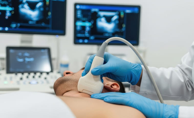
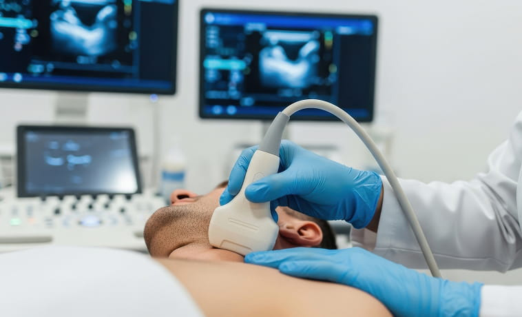
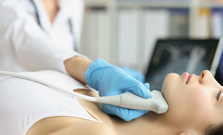
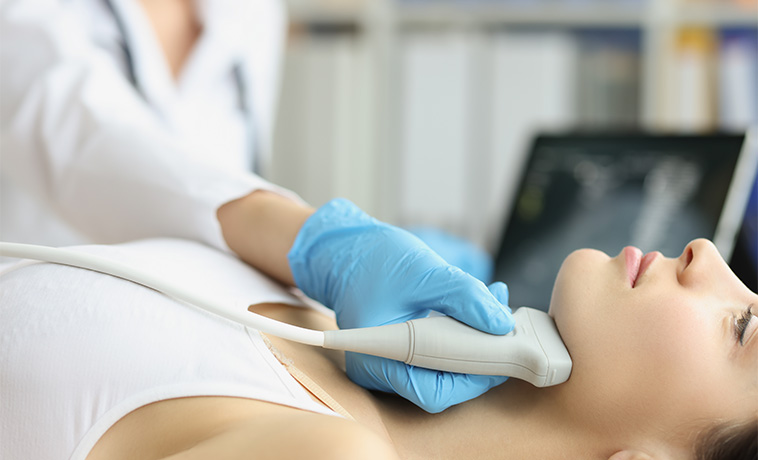
Neck ultrasound assesses thyroid and parathyroid-region abnormalities and cervical lymph nodes. It identifies features that guide whether a thyroid nodule requires fine-needle aspiration and helps target suspicious lymph nodes. Ultrasound can localise some enlarged parathyroid glands but cannot by itself determine whether a parathyroid tumour is cancerous.
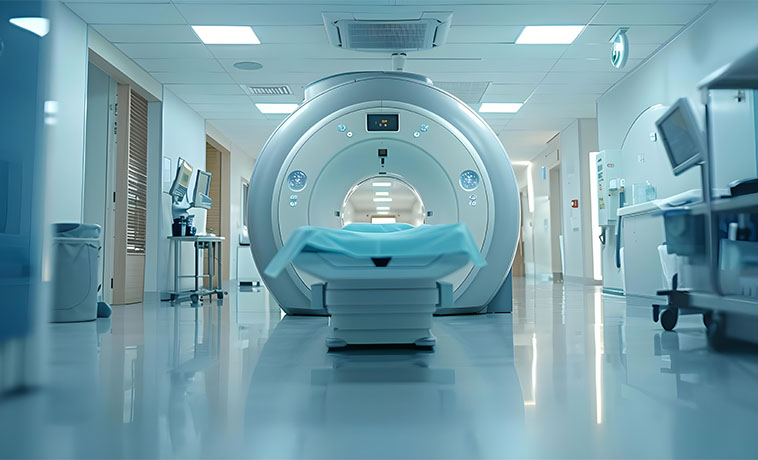
Contrast-enhanced CT provides detailed images of the neck, chest, abdomen and pelvis. It may assess a NET primary, lymph nodes, liver or lung metastases, an adrenal mass, local invasion and distant spread. A dedicated adrenal CT can evaluate density and contrast washout. When pheochromocytoma is suspected, biochemical assessment and careful procedural planning are important before intervention.
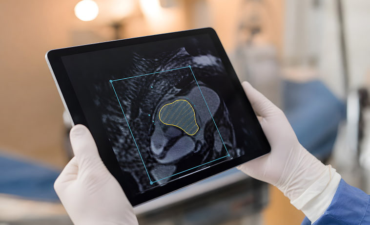
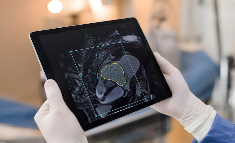
MRI offers detailed soft-tissue and vascular assessment without ionising radiation. It may characterise liver, pancreas, adrenal, head-and-neck or spinal lesions; assess local invasion; and evaluate disease close to major vessels. Chemical-shift MRI can help characterise selected adrenal masses.
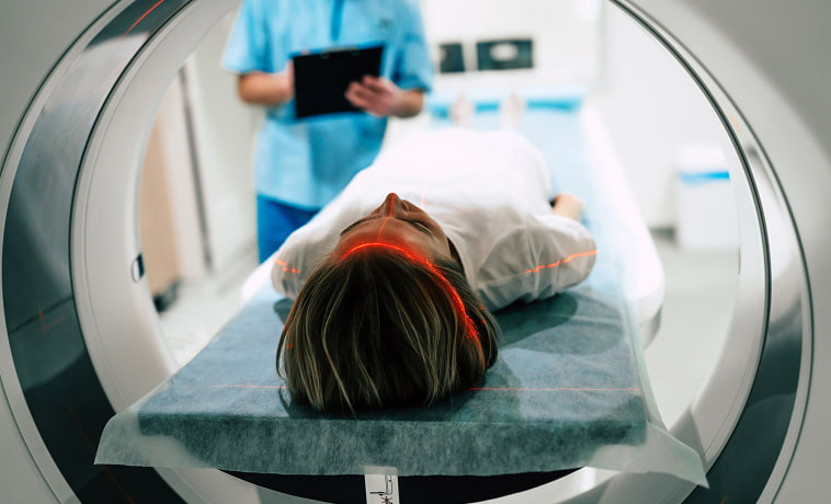
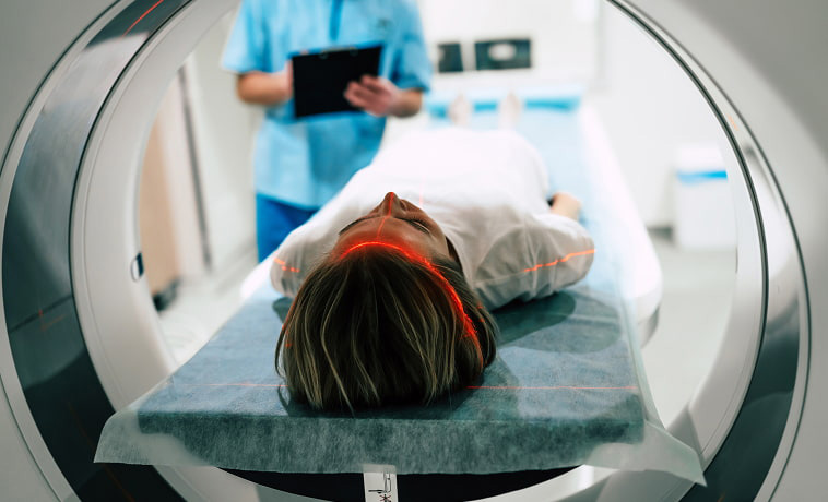
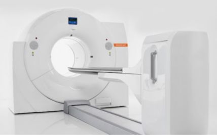
PET-CT using a somatostatin-receptor tracer can detect well-differentiated NET cells that express somatostatin receptors. It may help locate the primary tumour, stage disease, assess recurrence and determine whether somatostatin-receptor-targeted treatment such as peptide receptor radionuclide therapy may be appropriate. It is not equally useful for every high-grade neuroendocrine cancer.
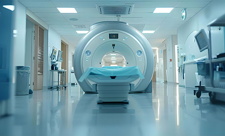
FDG PET-CT assesses glucose metabolism and may be useful for aggressive, poorly differentiated or higher-grade disease, selected thyroid cancers that do not take up iodine, adrenocortical carcinoma, treatment planning or suspected recurrence. Its role depends on the tumour biology and clinical question.
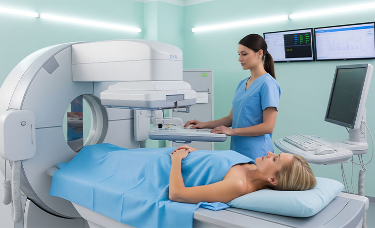
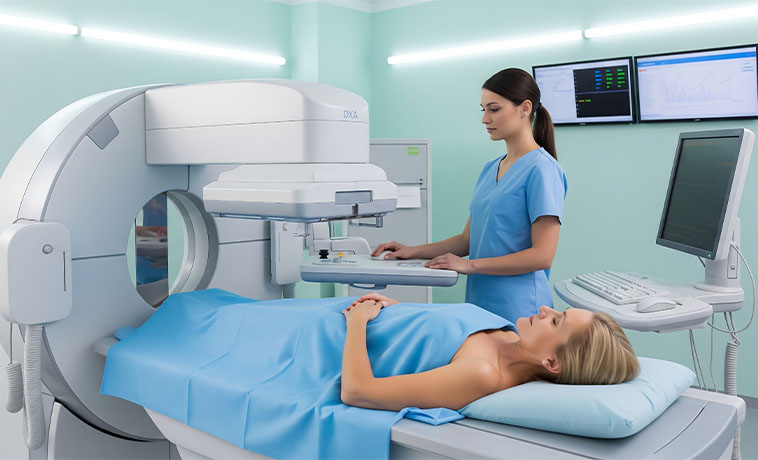
A radioactive iodine whole-body scan may be used after surgery or during follow-up for selected differentiated thyroid cancers. It identifies thyroid tissue or cancer that retains the ability to take up iodine. It is not useful for medullary or anaplastic thyroid cancer.
MIBG imaging may be used for selected pheochromocytomas and paragangliomas. It can help define disease distribution and assess eligibility for MIBG-targeted radionuclide therapy. Other functional imaging may be preferred depending on the tumour and genetic background.
FNAC is the main sampling method for thyroid nodules that meet clinical and ultrasound criteria. It can classify many nodules and may be combined with molecular testing when cytology is indeterminate. FNAC does not reliably distinguish every follicular-pattern thyroid cancer because capsular or vascular invasion must be assessed in surgically removed tissue.
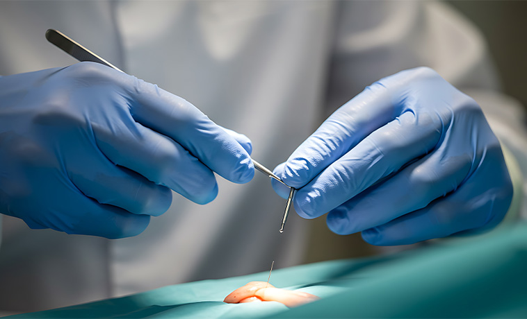
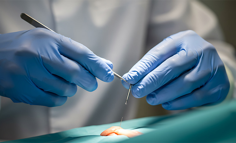
A core biopsy, endoscopic ultrasound-guided biopsy or bronchoscopic biopsy may be used to sample selected NETs or metastatic sites. The approach depends on the tumour location, vascularity and whether sufficient tissue is required for differentiation, grade and molecular testing.


A pathologist identifies the tumour type, differentiation, grade, invasion, margins, lymph-node findings and other prognostic features. NET pathology commonly includes mitotic activity and the Ki-67 proliferation index. Thyroid, adrenal and parathyroid tumours require organ-specific criteria and experienced endocrine pathology review.
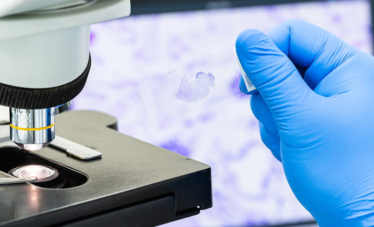
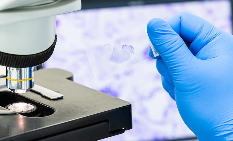
Specialised stains can confirm neuroendocrine differentiation, help identify a likely primary site and distinguish endocrine cancers from metastases or other tumour types. The panel is selected according to the morphology and clinical findings rather than applied identically to every case.


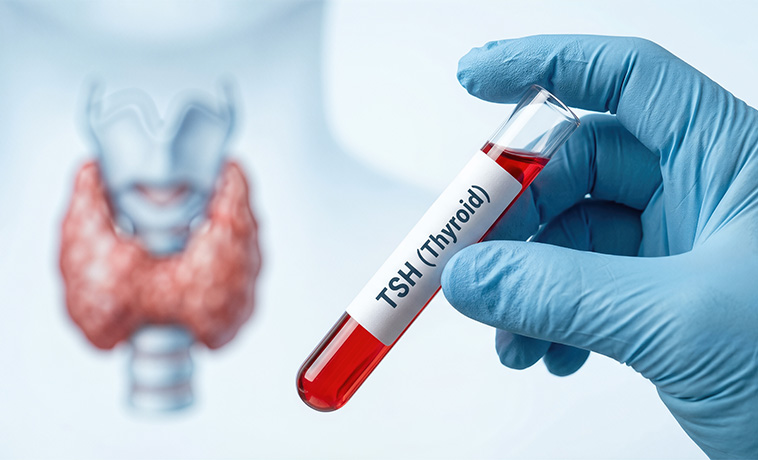
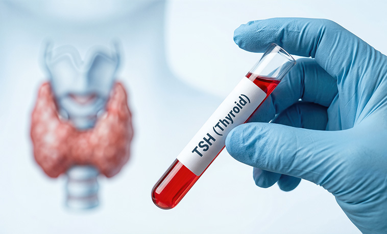
TSH and, when needed, free thyroid hormone levels assess thyroid function but do not confirm or exclude thyroid cancer. Calcitonin and CEA are used when medullary thyroid cancer is suspected or monitored. Thyroglobulin and anti-thyroglobulin antibodies are used mainly after treatment for differentiated thyroid cancer, not as screening tests for a thyroid nodule.
Corrected or ionised calcium, parathyroid hormone, phosphate, kidney function and vitamin D may be assessed when a parathyroid disorder is suspected. Markedly elevated calcium and PTH with a concerning neck mass may raise suspicion for parathyroid carcinoma, but no single blood test proves malignancy.


Testing may include plasma free or urinary fractionated metanephrines, cortisol assessment, ACTH, aldosterone and renin, androgens or other tests selected for the clinical presentation. Hormone evaluation is important even when an adrenal mass was found incidentally because treatment and perioperative safety depend on whether it is functioning.
Tumour testing may identify alterations that refine diagnosis or guide targeted therapy. Germline genetic testing may be recommended for medullary thyroid cancer, pheochromocytoma or paraganglioma, parathyroid carcinoma, multiple NETs, young-onset disease or a suggestive personal or family history. Genetic counselling helps select and interpret testing and guides family assessment.


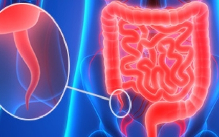
Surgery may remove a localised NET together with regional lymph nodes when appropriate. The operation depends on the primary site and may involve the bowel, appendix, pancreas, stomach, lung or another organ. Surgery can also be considered for selected liver metastases or to reduce hormone-producing tumour burden when meaningful benefit is expected.
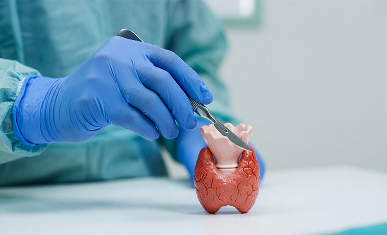
A lobectomy or total thyroidectomy may be recommended according to the thyroid cancer type, size, location, spread, molecular findings and patient factors. Central or lateral neck lymph nodes may be removed when involved or when indicated by the specific cancer. Active surveillance may be appropriate for carefully selected very small, low-risk papillary cancers.
Adrenalectomy is the main potentially curative treatment for localised adrenocortical carcinoma and for most resectable pheochromocytomas. Suspected malignant tumours should be removed intact without rupture. The open or minimally invasive approach depends on tumour size, invasion and surgical judgement. Functioning tumours require preoperative hormone control; pheochromocytoma generally requires alpha-blockade and careful anaesthetic planning.
When parathyroid carcinoma is suspected, surgery aims to remove the tumour intact, often with the involved parathyroid gland and adjacent thyroid tissue. Avoiding capsular rupture is important. Calcium may fall substantially after successful removal, so close postoperative monitoring and replacement may be required.
Repeat surgery or removal of limited metastatic disease may be considered for selected patients when it can control hormone symptoms, relieve pressure, delay progression or achieve durable disease control. The potential benefit must be balanced against operative risk and previous treatment.
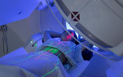
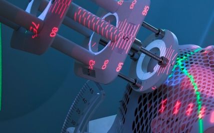
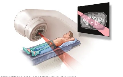
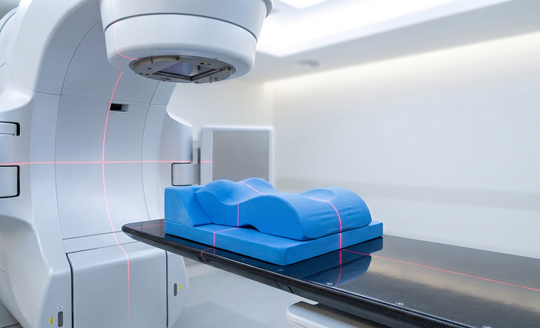
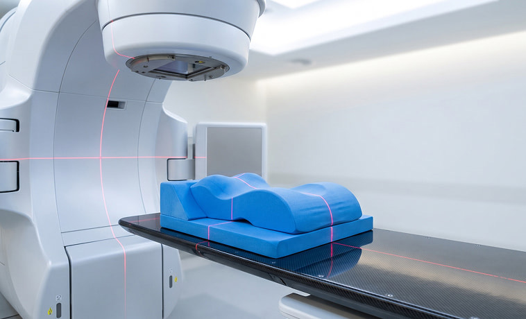
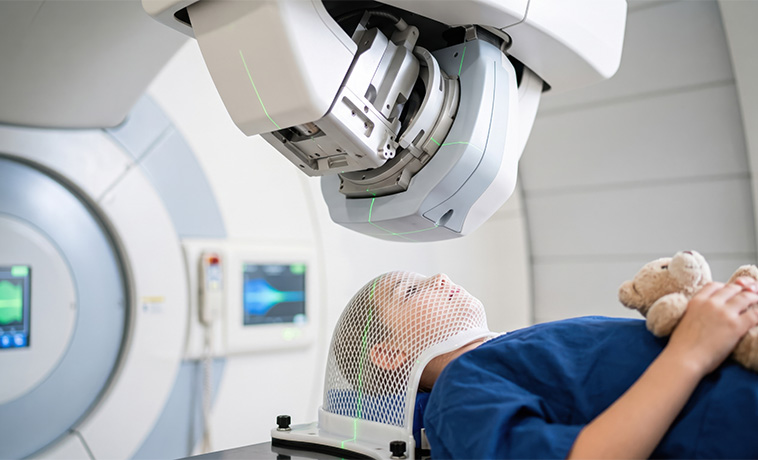
External beam radiation may be used after surgery, as definitive treatment when surgery is unsuitable, or to control local or metastatic symptoms. Its role includes selected thyroid cancers, unresectable or recurrent adrenal or parathyroid cancers, high-grade neuroendocrine carcinomas and painful or threatening metastases.
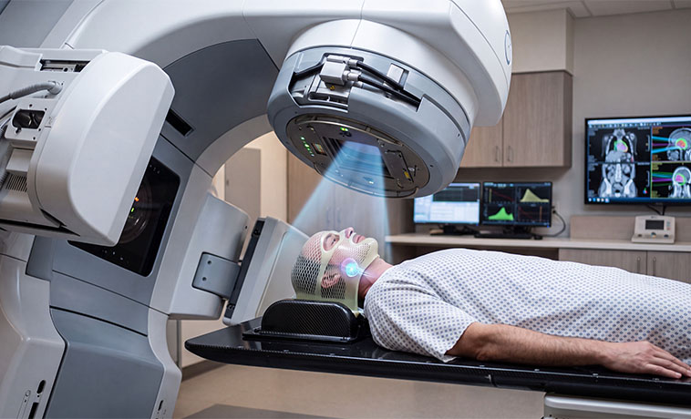
Stereotactic body radiation therapy or radiosurgery may deliver focused radiation to selected small lesions in the lung, liver, bone, brain or other sites. Suitability depends on the number, size and location of lesions and their relationship to nearby organs.
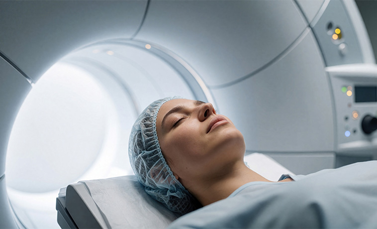
Proton therapy may be considered for selected complex, recurrent, paediatric or anatomically challenging tumours when comparative treatment planning shows a meaningful reduction in radiation exposure to nearby healthy tissues. It is not required for most endocrine or neuroendocrine cancers.
Somatostatin analogues can control hormone-related symptoms and slow tumour growth in many well-differentiated, somatostatin-receptor-positive NETs. Response and dosing are monitored clinically, biochemically and with imaging.

Targeted medicines may be used for selected progressive NETs, thyroid cancers or other endocrine cancers when supported by tumour type and molecular findings. Potential targets and medicines differ between diseases; treatment requires monitoring for tumour-specific and medicine-specific side effects.
Chemotherapy may be used for poorly differentiated neuroendocrine carcinoma, selected pancreatic or rapidly progressive NETs, advanced adrenocortical carcinoma, anaplastic thyroid cancer or other aggressive disease. The regimen depends on the diagnosis, grade, molecular profile, previous treatment and organ function.
Thyroid hormone replacement is required after total thyroidectomy and may be needed after lobectomy. In selected differentiated thyroid cancers, the dose is adjusted to suppress TSH according to recurrence risk and response. Excessive suppression is avoided when its risks outweigh the expected benefit.
Immunotherapy may be considered for selected advanced endocrine or neuroendocrine cancers based on the specific diagnosis, biomarkers, previous treatments and available evidence. It is not a standard treatment for every NET or endocrine cancer.
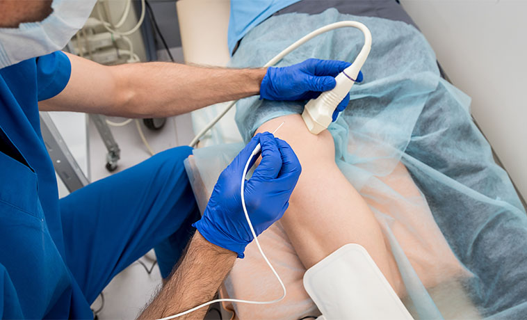
Radiofrequency, microwave or cryoablation may treat selected liver, lung, bone, adrenal or other lesions when surgery is not appropriate or when local control is clinically useful. The choice depends on lesion size, location, hormone activity and proximity to critical structures.
Arterial embolisation, chemoembolisation or radioembolisation may be used for selected liver-dominant NET metastases to reduce tumour burden, slow progression or control hormone-related symptoms. Liver function, vascular anatomy and the distribution of disease are assessed before treatment.
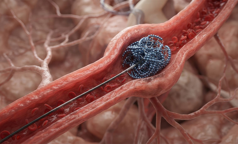
Radioactive iodine may be used after surgery for selected differentiated thyroid cancers to treat remaining thyroid tissue or iodine-avid cancer. It is not routinely required for every papillary or follicular cancer and does not treat medullary or anaplastic thyroid cancer.
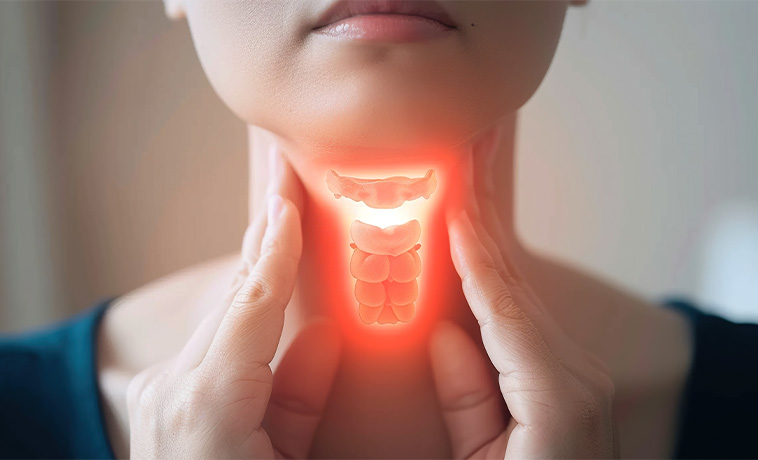
PRRT delivers a therapeutic radionuclide attached to a somatostatin analogue to somatostatin-receptor-positive NET cells. It may be used for selected unresectable or metastatic well-differentiated NETs after specialist assessment of receptor imaging, kidney and bone-marrow function, previous treatment and overall health.
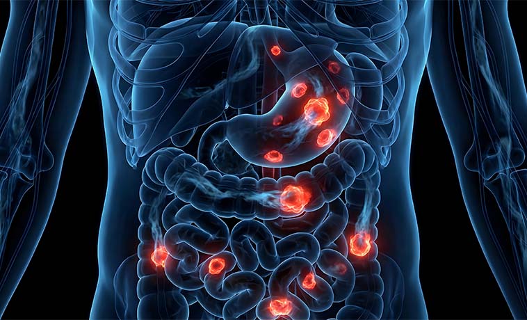
Radiolabelled MIBG therapy may be considered for selected unresectable or metastatic pheochromocytomas and paragangliomas that demonstrate adequate MIBG uptake. Availability and suitability depend on the tracer study, disease pattern, previous treatment and organ function.

Prognosis for Endocrine & Neuroendocrine Cancers
The outlook ranges from highly treatable, slow-growing disease to aggressive cancer requiring urgent combined treatment. Prognosis should be discussed for the exact tumour type rather than for endocrine and neuroendocrine cancer as one category.
Factors influencing prognosis include:
The organ and cell type in which the tumour began
Tumour differentiation and grade
Mitotic rate and Ki-67 index for NETs
Tumour size and invasion into nearby structures
Lymph-node involvement and distant spread
Whether the tumour can be completely removed
Hormone production and the ability to control hormone-related complications
Somatostatin-receptor or radioiodine uptake where relevant
Molecular and inherited genetic findings
Pathology findings, including vascular or capsular invasion and surgical margins
Response to treatment and the pattern or speed of recurrence
Age, general health, organ function and ability to receive treatment
Many differentiated thyroid cancers and localised, well-differentiated NETs have a favourable long-term outlook. Parathyroid carcinoma may recur after apparently complete removal and requires prolonged biochemical surveillance. Adrenocortical carcinoma, poorly differentiated neuroendocrine carcinoma and anaplastic thyroid cancer are generally more aggressive. Individual outcomes cannot be predicted from stage alone.


Why Choose ACC for Endocrine & Neuroendocrine Cancer Treatment
Multidisciplinary endocrine and neuroendocrine tumour boards
Integrated care across endocrinology, endocrine surgery, surgical oncology, medical oncology, nuclear medicine, radiation oncology, interventional radiology, pathology and genetics
Specialist management of gastrointestinal, pancreatic, lung and unknown-primary neuroendocrine tumours
Comprehensive thyroid, adrenal and parathyroid tumour evaluation and surgery
Advanced anatomical and functional imaging, including somatostatin-receptor PET-CT, FDG PET-CT, radioiodine and MIBG imaging for selected patients
Experienced endocrine and neuroendocrine pathology with tumour grading, immunohistochemistry and molecular testing
Hormone testing and coordinated perioperative management of functioning tumours
Radioactive iodine, peptide receptor radionuclide therapy and other molecularly targeted radionuclide treatments for selected cancers
Medical treatments including somatostatin analogues, chemotherapy, targeted therapy, immunotherapy and hormone-control medicines
Advanced radiation and interventional oncology options for selected primary and metastatic tumours
Genetic counselling and germline testing when an inherited cancer syndrome is suspected
Long-term surveillance focused on tumour control, hormone health and quality of life
Health Blogs