Breast Cancer
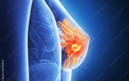
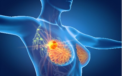
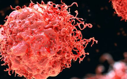
Breast cancer develops when abnormal cells in the breast grow uncontrollably. It predominantly affects women but can also occur in men, although much less commonly. Most breast cancers begin in the milk ducts or lobules. Over time, cancer cells may invade nearby tissues or spread to other parts of the body, if left untreated.
One of the most reassuring facts about breast cancer is that, when diagnosed at an early stage, five-year survival rates can be as high as 95%. This makes breast awareness, screening based on age and individual risk, and prompt evaluation of any breast changes especially important.
At Apollo Cancer Centres, breast cancer care is planned through a multidisciplinary approach. Breast surgeons, medical oncologists, radiation oncologists, radiologists, pathologists, and other specialists work together to review each patient’s diagnosis and recommend an appropriate treatment plan. Decisions are based on factors such as the stage of cancer, tumour size and grade, lymph-node involvement, hormone receptor and HER2 status, overall health, and individual needs. This coordinated approach helps personalise care for each patient.
Types of Breast Cancer
Breast cancer is not a single disease. It includes different types and biomarker-defined subtypes that may behave and respond to treatment differently:
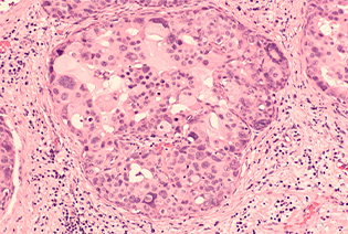
DCIS is a non-invasive or stage 0 breast cancer in which abnormal cells are confined to the milk ducts and have not spread into the surrounding breast tissue. It is highly treatable, although treatment is important because some cases may later develop into invasive cancer.
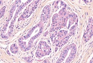
The most common type of invasive breast cancer, IDC begins in a milk duct and spreads into the surrounding breast tissue. It may also spread to nearby lymph nodes or other parts of the body.
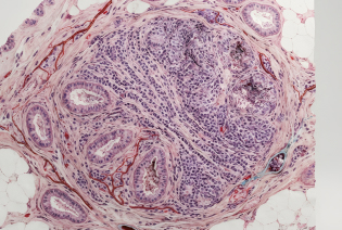
ILC begins in the lobules, the glands that produce milk, and spreads into the surrounding breast tissue. It is the second most common type of invasive breast cancer and can sometimes be more difficult to detect on examination or imaging.
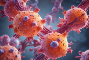
These cancers have receptors for estrogen, progesterone, or both, which can help them grow. Hormone or endocrine therapies that block these hormones or their effects are often an important part of treatment.

These cancers have high levels of the HER2 protein, which can promote cancer-cell growth. HER2-targeted therapies have significantly improved treatment options and outcomes for many patients with this subtype.
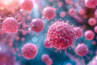
Triple-negative breast cancer does not have estrogen or progesterone receptors and does not have high levels of HER2. It may grow and spread more quickly than some other breast cancers. Treatment may include chemotherapy, surgery, radiation therapy, immunotherapy, or selected targeted treatments, depending on the stage and tumour features.
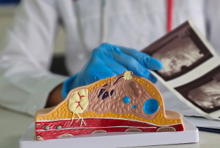
Inflammatory breast cancer is a rare, fast-growing form in which cancer cells block lymph vessels in the skin. The breast may become swollen, red, warm, heavy, or develop skin thickening, often without a distinct lump.
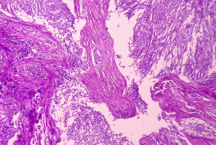
Paget disease is a rare form of breast cancer involving the skin of the nipple and areola. It may cause redness, scaling, itching, discharge, or changes in the nipple and is often associated with cancer inside the breast.

Breast cancer can also develop in men, although it is uncommon. It most often begins in the milk ducts and may present as a painless lump or thickening near or beneath the nipple. Nipple changes, discharge, or changes in the surrounding skin may also occur.
Accurate diagnosis requires more than identifying where the cancer began. Testing for estrogen and progesterone receptors, HER2, and other relevant biomarkers helps specialists understand the tumour and plan appropriate treatment.
Breast cancer can develop even when no specific risk factor is identified. However, certain lifestyle, medical, reproductive, and inherited factors may increase a person’s likelihood of developing the disease.
Modifiable Risk Factors
These are factors that may be reduced or managed through lifestyle changes or medical guidance:
Alcohol consumption
Tobacco use
Physical inactivity
Excess body weight, particularly after menopause
Use of combined menopausal hormone combined menopausal hormone therapy
Non-Modifiable Risk Factors
These are factors that cannot be changed:
Gender
Increasing age
A family history of breast or ovarian cancer
Inherited genetic variants, including BRCA1, BRCA2, and PALB2
Early onset of menstruation or later menopause
Older age at first full-term pregnancy
Never having carried a pregnancy to term
A personal history of breast cancer, DCIS, or certain high-risk breast conditions
Dense breast tissue
Previous radiation treatment to the chest, particularly at a young age
Having one or more risk factors does not mean that breast cancer will develop. Understanding individual risk can help guide decisions about screening, genetic counselling, and preventive care in consultation with a doctor.


Signs and Symptoms
Breast cancer may not cause noticeable symptoms in its early stages. Knowing how the breasts normally look and feel can make it easier to recognise a new or unusual change that needs medical attention.
Most breast changes are not caused by cancer. However, any new, unusual, or persistent change should be evaluated by a doctor rather than ignored or monitored without medical advice. Screening remains important because breast cancer may be present before symptoms appear.
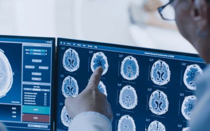
Breast cancer diagnosis usually begins with a review of symptoms, personal and family history, and a clinical breast examination. If an abnormality is found during examination or screening, the doctor may recommend breast imaging. A biopsy is then performed when necessary to determine whether the abnormal area is cancerous. It removes cells or tissue from an abnormal area for examination by a pathologist. It is the only definitive way to confirm whether an abnormality is cancerous.
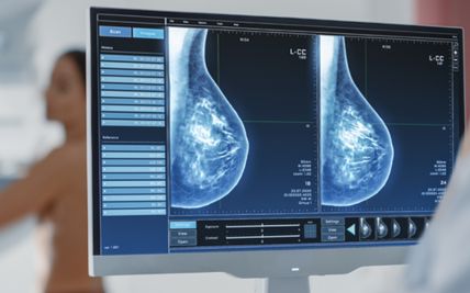
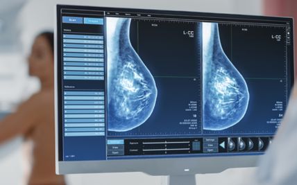
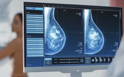
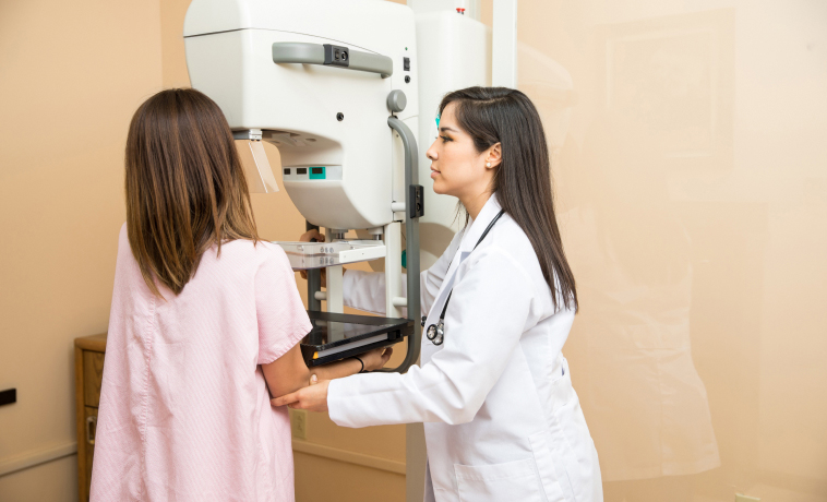
Mammography uses low-dose X-rays to create two-dimensional images of the breast. It can identify masses, calcifications, tissue distortion, and other changes, including some that cannot be felt during an examination.
A screening mammogram is performed in people without breast symptoms, while a diagnostic mammogram is used to evaluate a specific symptom or an abnormality found during screening. During the test, the breast is briefly compressed between two plates to spread the tissue, produce clearer images, and reduce the radiation dose required.
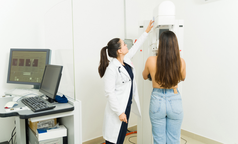
Digital breast tomosynthesis, commonly called 3D mammography, takes multiple low-dose X-ray images of the breast from different angles. These are reconstructed into thin sections that allow the breast tissue to be examined layer by layer.
By reducing the effect of overlapping breast tissue, tomosynthesis may help radiologists identify and assess abnormalities more clearly. Additional mammographic views or breast ultrasound may be recommended if further evaluation is required.
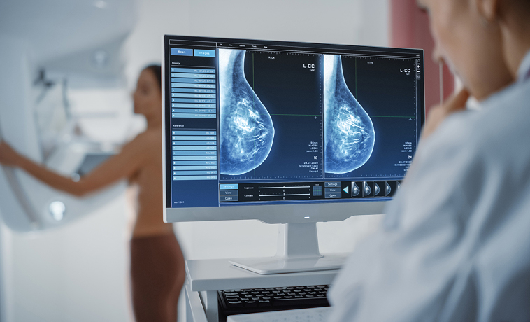
Breast ultrasound uses sound waves to create images of breast tissue. It can help determine whether a lump is solid or fluid-filled and assess abnormalities found during an examination or mammogram. Ultrasound is particularly useful for evaluating palpable lumps, examining dense breast tissue, and guiding a needle during biopsy. It does not use radiation.
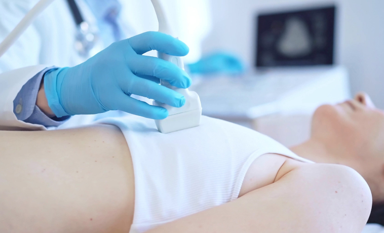
Contrast-enhanced mammography combines mammography with an iodine-based contrast agent injected into a vein. Areas with increased contrast uptake may indicate abnormal tissue requiring further assessment.
It may be used to examine the extent of a known cancer, look for additional suspicious areas, or provide further evaluation when standard imaging is inconclusive. It may also be considered when breast MRI is not suitable or available. Patients should inform the team about contrast allergies, kidney problems, or possible pregnancy.
Breast MRI uses a strong magnetic field, radio waves, and a dedicated breast coil to produce detailed images. A contrast dye is usually injected into a vein during the examination.
MRI may be recommended to:
Screen selected people at high risk of breast cancer
Assess the extent of a confirmed cancer
Look for additional areas of concern in either breast
Evaluate the response to treatment given before surgery
Examine certain abnormalities that remain unclear on other imaging
Breast MRI is not routinely required for every patient and does not replace mammography or biopsy.
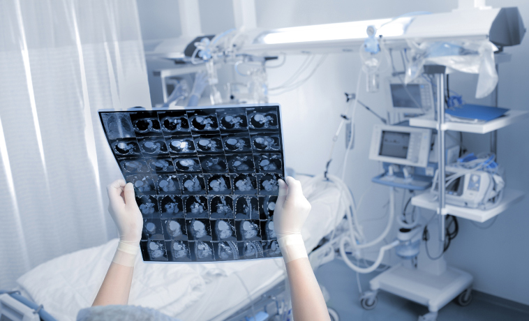
A thin needle is used to collect cells or fluid from a breast lump or lymph node, sometimes with ultrasound guidance. FNAC can provide useful information but may not collect enough tissue to determine all the features of a suspected cancer.
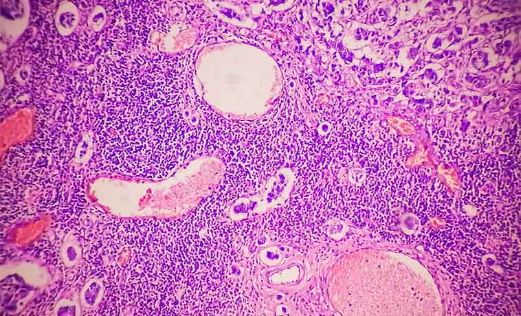
A hollow needle is used to collect small cores of breast tissue, usually under ultrasound, mammography, tomosynthesis, or MRI guidance. It is commonly preferred when breast cancer is suspected because it provides tissue to determine the cancer type, grade, and biomarker status.
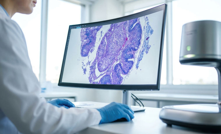
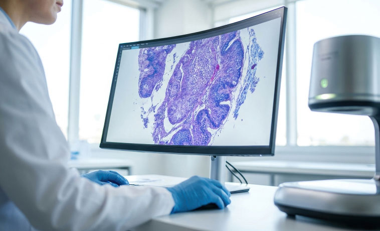
Using mammography, tomosynthesis, ultrasound, or MRI guidance, a vacuum-assisted device collects multiple, larger tissue samples through a single needle insertion. VABB may be recommended for small or non-palpable abnormalities, suspicious calcifications, or findings that require more tissue for assessment.
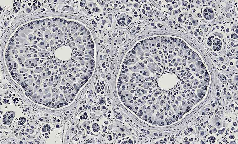
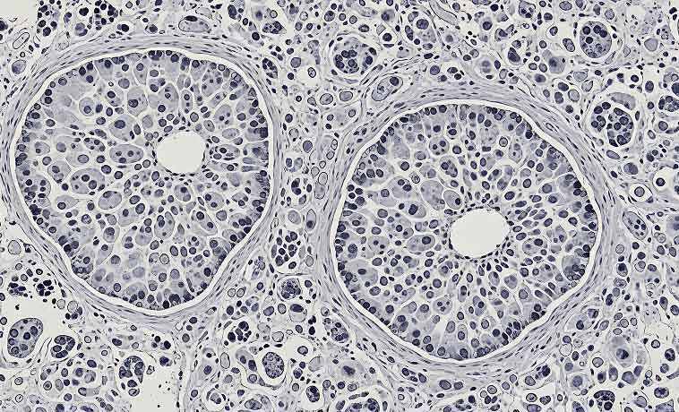
The biopsy sample is examined to determine whether cancer is present and, if so, identify:
The type of breast cancer
Whether it is invasive or non-invasive
The tumour grade
Estrogen receptor status
Progesterone receptor status
HER2 status
Selected patients may also undergo multigene or molecular testing to provide further information about prognosis or help guide treatment decisions. Testing for inherited genetic variants, such as BRCA1 and BRCA2, may be recommended separately based on age at diagnosis, cancer type, personal history, and family history.


Breast cancer staging (TNM system) is based on tumour size, lymph node involvement, and metastasis. Prognosis is based on the stage. Early detection (Stage 0 or I) has a 5‑year survival rate of over 90%. Advanced stages (Stage III or IV) have lower survival rates, but new therapies have improved outcomes.
Stage 0: DCIS, non‑invasive
Stage I: Early stage, confined to the breast
Stage II: Larger tumours or limited lymph node spread
Stage III: Locally advanced with extensive nodal involvement
Stage IV: Metastatic cancer, spread to other parts of the body
Also known as a lumpectomy, this procedure removes the tumour with a margin of surrounding healthy tissue while retaining most of the breast. Radiation therapy is usually recommended afterwards to reduce the risk of cancer returning in the breast. For tumours that cannot be felt, imaging-guided localisation may be used before surgery to help the surgeon identify the exact area that needs to be removed.
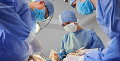
Oncoplastic surgery combines cancer removal with plastic-surgery techniques to reshape the remaining breast. It may allow larger tumours to be removed while helping preserve the breast’s shape and symmetry.

A mastectomy removes the entire breast and may be recommended when breast-conserving surgery is not suitable or preferred. Types include:
Simple mastectomy, which removes the breast tissue
Modified radical mastectomy, which removes the breast along with selected underarm lymph nodes
Skin-sparing mastectomy, which preserves most of the breast skin for reconstruction
Nipple-sparing mastectomy, which preserves the breast skin and nipple-areola complex in carefully selected patients
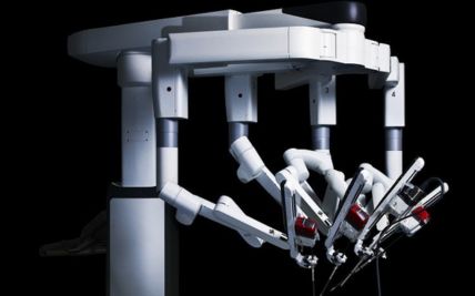
Endoscopic or robotic-assisted techniques, which allow mastectomies to be performed through smaller or less visible incisions, in carefully selected patients
Prophylactic mastectomy, which may be considered for people with a very high inherited risk, such as selected carriers of BRCA1 or BRCA2 variants

Chemotherapy, targeted therapy, hormone therapy, or immunotherapy may sometimes be given before surgery. The response to this treatment can help determine whether breast-conserving surgery or mastectomy is appropriate.

The lymph nodes under the arm may be assessed to determine whether cancer has spread beyond the breast.
Sentinel lymph-node biopsy removes the first lymph node or nodes most likely to contain cancer cells
Axillary lymph-node dissection removes a larger group of underarm lymph nodes and is recommended only in selected cases
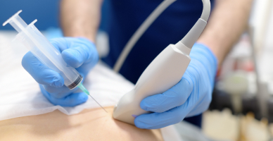
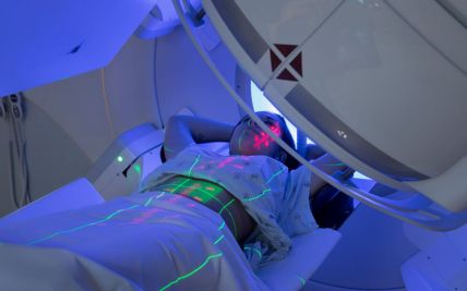
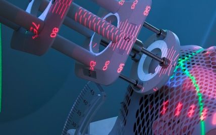
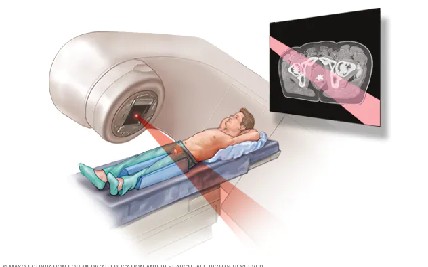
External beam radiation directs radiation from a machine towards the breast, chest wall, or nearby lymph nodes. Treatment is carefully planned to cover the required area while limiting exposure to surrounding healthy tissues.
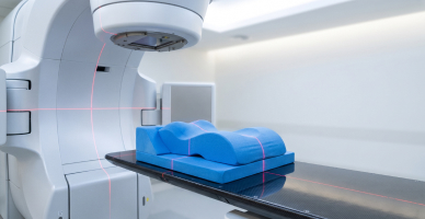
Three-dimensional imaging is used to shape radiation beams to the treatment area and reduce unnecessary exposure to nearby organs.
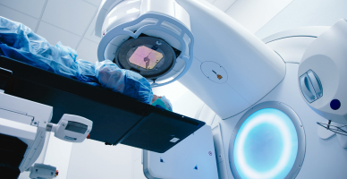
IMRT adjusts the intensity of individual radiation beams to improve dose distribution around the breast or chest wall. It may be recommended when treatment areas are complex or when greater dose shaping is required.
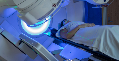
Hypofractionated radiation delivers treatment over fewer sessions using a slightly higher dose at each session. Shorter treatment schedules are appropriate for many patients and have been shown to provide outcomes comparable to longer conventional schedules.
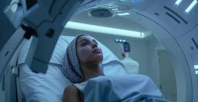
Proton therapy is an advanced form of radiation treatment that uses proton beams instead of X-rays to target cancer. Because protons release most of their radiation within the treatment area and deliver little radiation beyond it, they may help reduce exposure to nearby healthy tissues. For appropriate patients, Apollo Proton Cancer Centre offers proton therapy when treatment planning shows that it could meaningfully reduce radiation exposure to the heart, lungs, or other nearby tissues. It is not routinely required for every patient with breast cancer and suitability is determined through individual assessment.

Precision oncology uses information from pathology, biomarker, genetic, and molecular tests to identify treatments that may be appropriate for an individual cancer. Not every patient requires extensive molecular profiling; testing is recommended when it is likely to influence treatment.
Chemotherapy uses medicines to destroy cancer cells or slow their growth. It may be given intravenously before surgery to shrink the tumour, after surgery to reduce the risk of recurrence, or to treat recurrent or metastatic breast cancer. Not every patient with breast cancer requires chemotherapy. The decision depends on the stage, tumour subtype, recurrence risk, and overall health.

Hormone or endocrine therapy is used for cancers that have estrogen or progesterone receptors. It works by lowering hormone levels or blocking the effect of hormones on cancer cells. Depending on menopausal status and other clinical factors, treatment may include tamoxifen, aromatase inhibitors, ovarian suppression, or other endocrine medicines.

Targeted therapies act on specific proteins or genetic changes that help cancer cells grow. They may be used for HER2-positive, hormone receptor-positive, BRCA-associated, and certain other breast cancers. Biomarker testing helps determine whether a patient is likely to benefit from a particular targeted treatment.
Immunotherapy helps the immune system recognise and attack cancer cells. It may be used with chemotherapy for selected patients with triple-negative breast cancer, depending on the stage and biomarker findings.
Antibody-drug conjugates combine a targeted antibody with an anti-cancer medicine. The antibody attaches to a specific protein on the cancer cell and delivers the medicine more directly to it. These treatments may be used for selected HER2-positive, HER2-low, triple-negative, or advanced breast cancers.

An implant may be placed during mastectomy or after the skin has first been expanded using a temporary tissue expander. Implant reconstruction generally involves a shorter initial operation than tissue-flap reconstruction but may require future procedures.

DIEP flap reconstruction uses skin and fat from the lower abdomen to create a breast while preserving most of the abdominal muscle. Microsurgery is used to connect the flap’s blood vessels in the chest.

A TUG flap uses skin, fat, and part of a muscle from the inner upper thigh to reconstruct the breast. It may be considered when abdominal tissue is not suitable or preferred.
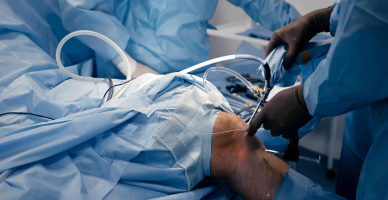
This procedure uses skin, fat, and muscle from the upper back to reconstruct the breast. An implant may also be used to provide additional volume.

After the reconstructed breast has healed, the nipple and areola may be recreated using minor surgery, medical tattooing, or a combination of both.
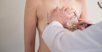
Procedures such as breast reduction, uplift, or reshaping of the opposite breast may be considered to improve balance and symmetry.

Patients who develop arm swelling after lymph-node surgery or radiation may receive physiotherapy, compression therapy, exercise guidance, and skin-care support. Selected patients may also be considered for specialised lymphatic surgery.

Prognosis for Breast Cancer
Breast cancer often has a favourable outlook when detected and treated at an early stage. For early-stage disease, five-year survival rates can be as high as 95%. Locally advanced breast cancer may also be treated effectively using a combination of medical treatment, surgery, and radiation therapy. Metastatic breast cancer is generally not considered curable, but modern treatments can often control the disease, relieve symptoms, and help maintain quality of life for extended periods.
Factors that influence prognosis include:
Stage at diagnosis, including tumour size, lymph-node involvement, and whether the cancer has spread
Type and grade of the cancer
Hormone receptor and HER2 status
Response to treatment, particularly treatment given before surgery
Completeness of surgery and other recommended treatment
Genomic or molecular findings, where relevant
Age, overall health, and coexisting medical conditions
Timely treatment and adherence to follow-up care


Screening for Breast Cancer
Breast cancer cannot always be prevented, but screening and timely evaluation of breast changes can support earlier diagnosis. The appropriate screening plan depends on age, family history, breast density, previous breast conditions, and inherited risk.
Breast Examination
Being familiar with how the breasts normally look and feel can help identify new changes. Any lump, nipple change, discharge, skin dimpling, or swelling should be evaluated promptly. A clinical breast examination may also form part of age-appropriate screening.
Screening Mammography
Mammography can detect breast abnormalities before they can be felt. When screening should begin and how often it is needed depend on individual risk. An abnormal result does not always mean cancer; further imaging or a biopsy may be required. People with a strong family history, inherited genetic variants such as BRCA1 or BRCA2, previous chest radiation, or certain high-risk breast conditions may require earlier or additional screening, including breast MRI.
Why Choose ACC for Breast Cancer Treatment
Multidisciplinary breast oncology team across surgical, medical, and radiation oncology
Advanced breast imaging, including 3D tomosynthesis, contrast-enhanced mammography, and dedicated breast MRI
Personalised treatment guided by hormone receptor, HER2, and selected genomic testing
Access to proton therapy for appropriately selected patients
Integrated rehabilitation, psychological support, and cosmetic care
Structured follow-up and survivorship support after treatment

Health Blogs