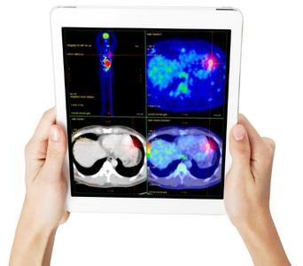
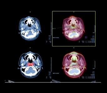
Positron Emission Tomography–Computed Tomography, commonly called PET-CT, is an imaging test that combines functional information from a PET scan with detailed anatomical images from a CT scan.
The PET component uses a small amount of a radioactive substance called a radiotracer. The tracer is designed to accumulate in or bind to particular tissues, allowing the scan to show patterns of metabolic or molecular activity. The CT component shows the size, shape and precise location of structures inside the body. The two sets of images are combined to help doctors determine where abnormal activity is occurring.
PET-CT can contribute to the diagnosis, staging, treatment planning and follow-up of many cancers. However, it is not required for every cancer, and not all cancers or cancer deposits are clearly visible on PET-CT. Its usefulness depends on the cancer type, tumour biology, radiotracer used and clinical questions being addressed.


Role in Cancer Care
What to Expect
Before the Test
Preparation depends on the radiotracer being used. You will receive instructions specific to your examination.
Before the scan, inform the care team if you:
Are pregnant or may be pregnant
Are breastfeeding
Have diabetes or problems controlling your blood glucose
Have kidney disease or reduced kidney function
Have previously reacted to CT contrast material
Have allergies, asthma or other significant medical conditions
Are taking prescription medicines, non-prescription medicines or supplements
Have recently had an infection, inflammation or vaccination
Have recently undergone surgery, biopsy, chemotherapy, immunotherapy or radiation therapy
Have difficulty lying still or experience anxiety in enclosed spaces
For an FDG PET-CT, you will usually be asked to avoid food and drinks containing calories for several hours before the scan. Plain water is generally permitted and may be encouraged. Strenuous physical activity may need to be avoided beforehand because muscular activity can affect FDG distribution.
Do not stop or alter medicines unless instructed. People with diabetes will receive individual advice about food, insulin or other diabetes medicines and appointment timing. Blood glucose is usually checked before an FDG injection because high glucose or insulin levels can reduce scan quality.
If intravenous contrast is planned for the CT component, kidney-function testing or additional preparation may be required. Remove jewellery and other metal objects as instructed.

During the Test
A cannula will usually be inserted into a vein in the hand or arm, and the radiotracer will be administered. The injection itself is similar to a routine blood test.
After the injection, you will rest quietly while the tracer distributes through the body. For an FDG PET-CT, this uptake period is commonly about 45 to 60 minutes. You may be asked to avoid talking, walking, chewing or using electronic devices during this period, as muscle and brain activity can influence tracer uptake.
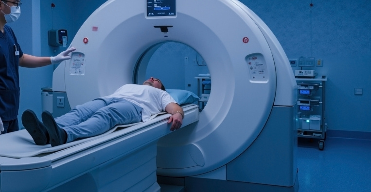
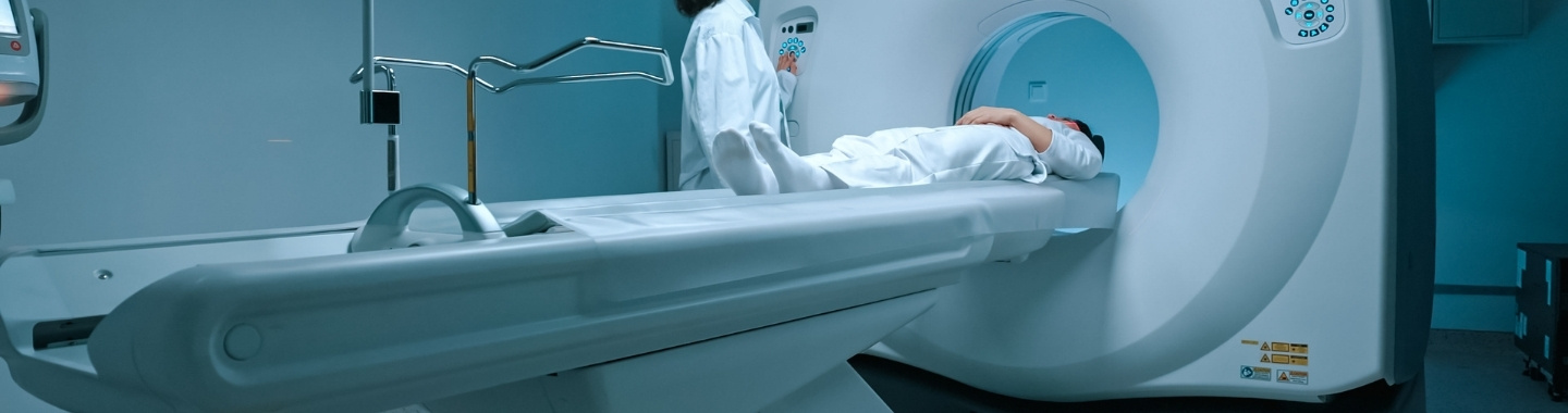
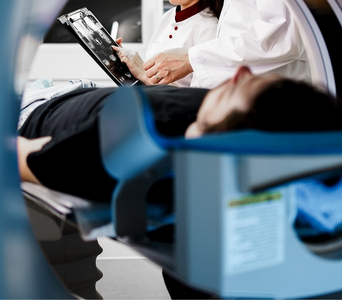
You will then lie on a motorised table that moves through the PET-CT scanner. The scanner is shaped like a large ring and is open at both ends. It is important to remain still, and you may occasionally be asked to hold your breath briefly.
The CT images are generally acquired first, followed by the PET images. The imaging portion commonly takes approximately 20 to 40 minutes, although the duration varies according to the area examined and whether additional images or a diagnostic CT are required. The overall appointment may take two to three hours, including preparation and tracer uptake time.
The scan is painless, apart from the intravenous injection. The technologist can see, hear and communicate with you throughout the examination.

After the Test
You can usually resume normal activities and eat normally after the scan unless given different instructions.
Drinking water may be advised to help the radiotracer pass out of the body through urine. Follow any hygiene or radiation-safety instructions provided by the nuclear medicine team. You may be advised to limit prolonged close contact with pregnant people and young children for a specified period, depending on the tracer and local protocol.
If you received CT contrast, follow the additional instructions given by the imaging team. Seek prompt medical attention if you develop breathing difficulty, facial or throat swelling, a widespread rash, severe dizziness or another significant reaction.
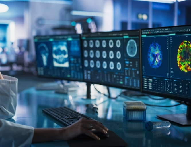
A nuclear medicine physician or appropriately trained radiologist will examine the PET, CT and fused images and prepare a report. The findings will be interpreted alongside previous imaging, pathology results, treatment history and other clinical information.

Potential Benefits
- Functional and anatomical information: PET shows biological activity, while CT identifies the precise location and structure of an abnormality.
- Whole-body assessment: Many PET-CT examinations can evaluate a large part of the body during one appointment.
- Cancer staging: PET-CT may identify disease in lymph nodes, organs or bones that affects the cancer stage and treatment plan.
- Treatment planning: Findings may help determine whether local or systemic treatment is appropriate and may contribute to radiation therapy planning.
- Response assessment: Changes in tracer uptake can provide information about treatment response, sometimes before major structural changes are visible.
- Detection of recurrence: PET-CT can help locate suspected recurrent disease when other findings are inconclusive.
- Tumour-specific imaging: Different radiotracers can assess particular metabolic processes, receptors or molecular targets in selected cancers.
- More precise localisation: Fused images help doctors match abnormal tracer uptake with the relevant organ, lymph node, bone or soft-tissue structure.