





Minimally invasive cancer surgery is an approach in which surgeons use small incisions, a camera and specialised instruments to examine, biopsy or remove a tumour. Depending on the part of the body being treated, the procedure may be described as laparoscopic surgery for the abdomen or pelvis, thoracoscopic surgery for the chest, or another type of video-assisted or endoscopic surgery.
The aim is to perform the required cancer operation while limiting disruption to the surrounding tissues. When appropriate, the surgeon may remove the tumour with a margin of healthy tissue, nearby lymph nodes, or part or all of an affected organ through small access points. One incision may be slightly enlarged to remove the surgical specimen safely.
At Apollo Cancer Centres, minimally invasive surgery is recommended only when it can meet the same oncological goals as an open operation. Surgical oncologists work with medical oncologists, radiation oncologists, radiologists, pathologists, anesthetists and rehabilitation specialists to select the most appropriate approach for each patient.
- Have a localised solid tumour that can be removed safely through small incisions
- Need a biopsy or surgical assessment to confirm the diagnosis or determine the extent of cancer
- Require removal of a tumour, part or all of an affected organ, or selected nearby lymph nodes
- Have a tumour in the abdomen, pelvis, chest or another area accessible through a minimally invasive approach
- Need staging surgery to assess whether cancer has spread within a body cavity or to nearby lymph nodes
- Have received chemotherapy, immunotherapy or radiation therapy and remain suitable for surgery
- Have selected recurrent disease that can be removed using a minimally invasive approach
- Are medically fit for anesthesia and the positioning or temporary gas insufflation required for the planned procedure
- Are expected to receive an oncologically complete and safe operation without the need for a larger incision
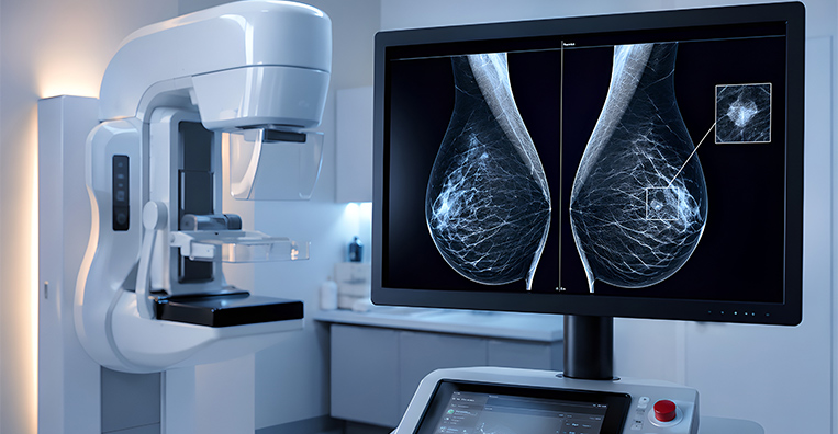
How the Procedure is Performed
Before the Procedure
Before minimally invasive cancer surgery:
The surgical team reviews the biopsy, imaging scans, laboratory results, medical history and current medications.
Additional imaging, endoscopy or other tests may be recommended to assess the extent of the cancer and plan the operation.
The patient’s fitness for surgery and anesthesia is evaluated. This may include blood tests, heart and lung assessments, and consultation with an anesthetist.
The case may be reviewed by a multidisciplinary tumour board to determine whether surgery should be performed first or combined with treatments such as chemotherapy, immunotherapy or radiation therapy.
The surgeon explains the goal and extent of the operation, expected benefits, possible risks, alternatives and anticipated recovery.
The possibility of removing nearby lymph nodes, part or all of an affected organ, or neighbouring structures is discussed when relevant.
The team explains that a larger incision or conversion to open surgery may be required if it is necessary to complete the operation safely.
The possible need for drains, tubes, blood transfusion or a temporary or permanent stoma is discussed whenever it can be anticipated.
Instructions are provided about fasting, medications, bowel preparation where required, smoking cessation and admission to the hospital.

During the Procedure
Minimally invasive cancer surgery is usually performed under general anesthesia.
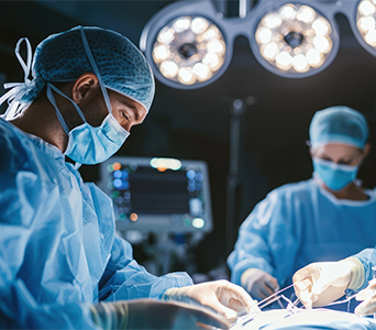
The surgeon makes one or more small incisions or uses an appropriate natural body opening, depending on the procedure and the organ being treated.
A thin camera or viewing instrument is introduced to provide a magnified view of the surgical area on a monitor.
Specialised instruments are inserted through separate access points to examine tissues and perform the planned operation.
The tumour is removed with an appropriate margin of surrounding tissue whenever feasible. Part or all of an affected organ may also need to be removed.
Nearby lymph nodes may be sampled or removed when required for cancer staging or treatment.
In selected cases, a pathologist may examine tissue during the operation to assess a margin or confirm the nature of a suspicious area.
The specimen is placed in a protective retrieval bag when appropriate and removed through an incision that may be slightly enlarged.
If safe access or complete tumour removal cannot be achieved through the minimally invasive approach, the surgeon may convert to open surgery. Drains, tubes or a temporary or permanent stoma may be placed when required.
At the end of the procedure, the instruments are removed, the incisions are closed and the patient is transferred to a recovery area for monitoring.
The duration of surgery varies according to the organ involved and the complexity of the procedure. A minimally invasive operation is not always shorter than open surgery. The surgeon will explain what to expect for the planned procedure.

After the Procedure
Recovery: Recovery varies according to the type and extent of surgery, the organ involved, the patient’s overall health and whether the procedure was completed through small incisions or converted to open surgery.
Hospital Stay: Some patients may go home on the same day, while others may remain in hospital for one or more days after major cancer surgery. The care team monitors pain, wound healing, breathing, circulation, bowel and bladder function, nutrition and mobility.
Pain Management: Pain is managed with a personalised combination of medicines. Discomfort is often concentrated around the incisions. After abdominal or pelvic laparoscopy, temporary shoulder-tip or abdominal discomfort may occur because of the gas used during the procedure.
Nutrition and Mobility: Fluids and food are restarted according to the operation and the return of normal body functions. Patients are encouraged to sit up, perform breathing exercises and begin walking as soon as it is safe to reduce the risk of chest infections and blood clots.
Incision and Drain Care: The team provides instructions for caring for the small incisions and for any drains, tubes or stoma. Patients are advised which symptoms are expected and which require prompt medical attention.
Resuming Routine: Many patients return to light activities sooner than after a comparable open operation, but recovery still depends on the extent of the cancer surgery. Heavy lifting, strenuous exercise, driving and return to work should follow the surgeon’s individual advice.
Follow-up Care: Follow-up appointments are scheduled to assess healing, review the histopathology report and discuss the next stage of treatment. Rehabilitation, nutrition, pain management, psychological support or stoma care may be arranged according to the patient’s needs.

Key Advantages

- Fever, chills or increasing weakness
- Worsening pain that is not controlled by prescribed medication
- Increasing redness, warmth, swelling, discharge or bleeding from an incision
- Persistent vomiting, increasing abdominal swelling or inability to eat or drink
- Shortness of breath, chest pain or coughing up blood
- New swelling or pain in a leg
- Difficulty passing urine, reduced urine output or an unexpected change in bowel function
- A problem with a drain, tube or stoma
- Any symptom identified by the surgical team as specific to the procedure

Continuum of Care
Minimally invasive cancer surgery is one part of a coordinated treatment pathway. After surgery, patients may receive:



