

icon
icon
Overview

Diagnostic procedures help doctors determine whether an abnormality is cancerous and obtain the information needed to plan appropriate care. Imaging and physical examination may identify a suspicious area, but a tissue sample is often required to establish a definitive diagnosis and determine the type and biological characteristics of the cancer.

Read More
Diagnostic Procedures

Apollo Cancer Centres offers diagnostic procedures to obtain tissue for examination and help accurately identify abnormalities during subsequent imaging or surgery.

01
Biopsy

A biopsy involves removing cells or a small tissue sample from a suspicious area for examination by a pathologist. Depending on the site and the amount of tissue required, it may be performed using a fine needle, core needle, vacuum-assisted device, endoscope, or surgical technique, with imaging guidance when necessary. Biopsy findings can confirm whether an abnormality is cancerous and may provide information about the cancer type, grade, biomarkers, and molecular characteristics that guide treatment.

Read More
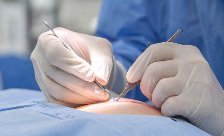
Biopsy
02
Wire Localisation

Wire localisation helps the surgeon identify a small or non-palpable abnormality that cannot be easily felt during surgery, most commonly in the breast. Using mammography, ultrasound, or MRI guidance, a radiologist positions a fine wire in or close to the abnormality before surgery. The wire serves as a guide to the targeted area and is usually removed along with the tissue, which is then examined by a pathologist.

Read More
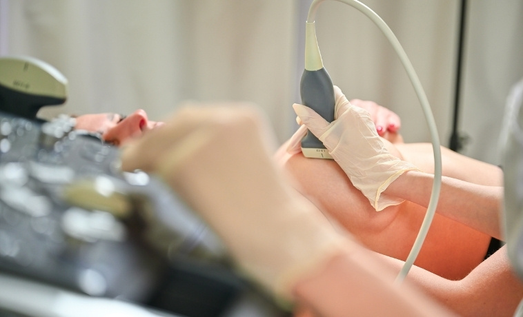
Wire Localisation
03
Clip Placement

Clip placement involves positioning a small marker at the site of an imaging abnormality or biopsy so that the area can be identified during future imaging, localisation, surgery, or treatment planning. It is commonly used in breast care and may also be used at selected tumour or lymph-node sites elsewhere in the body. The clip is placed using image guidance, is designed to remain safely in the body, and does not itself deliver medicine or radiation.

Read More
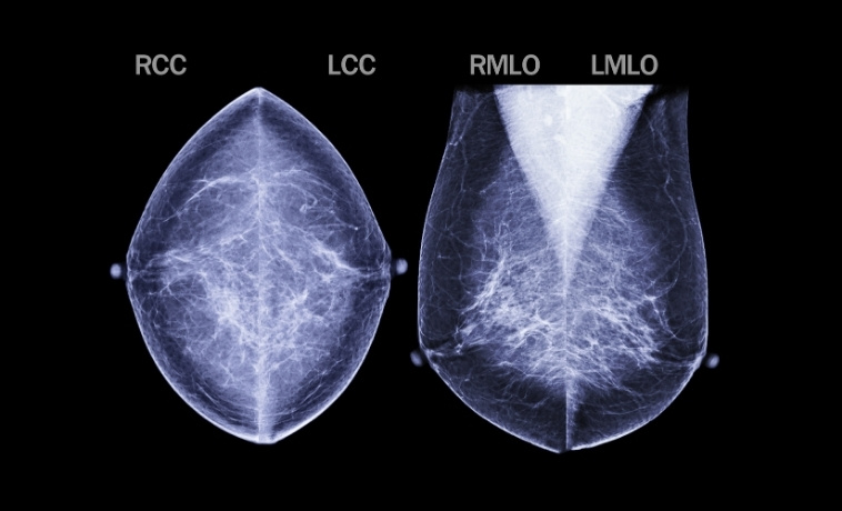
Clip Placement
Key Aspects of Diagnostic Procedures
Selection of the procedure according to the abnormality and clinical question
Image guidance when an abnormality cannot be accurately targeted by touch or direct vision
Collection of an adequate and representative tissue sample
Specialist pathology examination and additional biomarker or molecular testing when appropriate
Correlation of pathology results with imaging and clinical findings
Marker placement when the biopsy site may need to be identified later
Localisation of small or non-palpable abnormalities before surgery
Coordination among radiology, pathology, surgery, and oncology teams
Review of medicines, bleeding risk, allergies, and fitness for sedation or anaesthesia
Clear planning for follow-up when results are inconclusive or do not match the imaging findings
Your Overall Care Plan

How Diagnostic Procedures Fit into Your Overall Care Plan

A diagnostic procedure may be recommended after an abnormality is identified through screening, clinical examination, endoscopy, or imaging. The aim is to obtain enough information to establish or exclude a cancer diagnosis and guide the next step in care.

If a biopsy confirms cancer, the pathology findings are considered together with imaging, laboratory results, and clinical assessment. Additional biomarker or molecular testing may be performed when the results could influence treatment. The case may then be reviewed by a multidisciplinary tumour board to determine the appropriate treatment plan.

A marker clip may be placed so that the biopsied site can be located during follow-up imaging or treatment, particularly if the abnormality is small or expected to change after systemic therapy. If surgery is recommended for an abnormality that cannot be felt, wire localisation may help the surgeon identify the correct area. The tissue removed during surgery is then examined by pathology.

An inconclusive biopsy does not automatically mean that cancer is absent. Repeat biopsy, a different biopsy method, surgical removal, or imaging follow-up may be recommended when the sample is inadequate or the pathology result does not match the clinical or imaging findings.

Frequently Asked Questions
01 Does being advised to have a biopsy mean I have cancer?
icon icon
No. A biopsy is recommended when more information is needed about an abnormality. Many biopsies show benign or non-cancerous changes. The tissue must be examined by a pathologist before a diagnosis can be made.
02 Can a biopsy cause cancer to spread?
icon icon
Biopsy is an established part of cancer diagnosis, and the risk of tumour cells spreading along the biopsy path is extremely low with standard techniques. The biopsy route and method are planned carefully, particularly for certain tumour types where the needle path may influence future surgery.
03 How is the type of biopsy selected?
icon icon
The choice depends on the location and size of the abnormality, the imaging method through which it is best seen, the amount of tissue required, and whether additional biomarker or molecular testing may be needed. Your specialist will recommend the safest method likely to provide an adequate sample.
04 What happens if the biopsy result is inconclusive?
icon icon
The doctor will review whether the sample was adequate and whether the pathology result matches the imaging findings. Repeat biopsy, another biopsy method, surgical removal, or imaging follow-up may be recommended.
05 Can a marker clip move or cause problems?
icon icon
Marker clips are designed to remain at or close to the intended site. Significant movement is uncommon, although a small degree of displacement can occasionally occur. The clip is generally not felt and does not usually cause pain or interfere with daily activities.
06 What is the difference between clip placement and wire localisation?
icon icon
A marker clip is usually placed at a biopsy or tumour site so that the area can be identified during future imaging or treatment. It may remain in the body unless removed during surgery. A localisation wire is placed shortly before surgery to guide the surgeon to a specific abnormality and is usually removed with the targeted tissue.
07 Will the clip or wire interfere with an MRI?
icon icon
Marker clips used in current clinical practice are generally designed to be MRI-compatible or MRI-conditional, but the exact device should be verified before an MRI. A localisation wire is temporary and is usually removed during surgery. Always tell the imaging team about any marker or device in your body.
08 How long will biopsy results take?
icon icon
The time varies according to the tissue obtained and whether special stains, biomarker tests, molecular analysis, or an additional specialist review is required. Your care team will tell you when and how the results will be discussed.
image image
Book an Appointment
Please Enter Your Name
Please Enter Your Mobile Number
Please Enter OTP

