Head and Neck Cancer
Head and neck cancers develop in the mouth, throat, voice box, nose, sinuses, salivary glands, and other structures within the head and neck. Most begin in the squamous cells lining these areas and are therefore called squamous cell carcinomas.
These cancers can affect structures involved in breathing, speaking, swallowing, hearing, taste, and facial appearance. Treatment therefore focuses not only on controlling the cancer but also on preserving these functions wherever this can be achieved safely.
Thyroid and parathyroid cancers occur within the head and neck but behave differently from conventional head and neck squamous cell cancers. They require separate diagnostic, staging, and treatment pathways. Care often involves head and neck surgical oncologists, medical oncologists, radiation oncologists, radiologists, nuclear medicine specialists, pathologists, dental specialists, and reconstructive surgeons.
Types of Head and Neck Cancer
Head and neck cancer is not a single disease. The site where the cancer begins, its cell type, HPV or EBV status, and its stage influence treatment and prognosis.
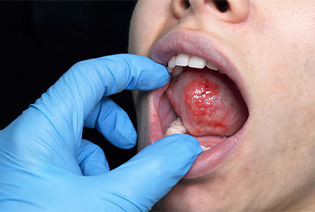
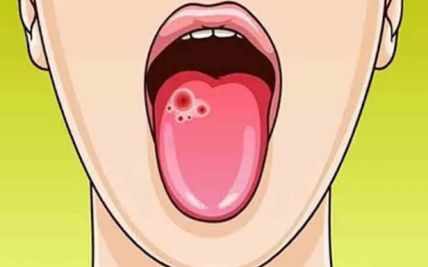
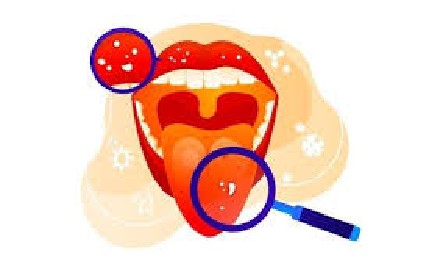
Oral cavity cancer develops in the lips, tongue, gums, inner cheeks, floor or roof of the mouth, or the area behind the wisdom teeth. Most are squamous cell carcinomas.Possible signs include a mouth ulcer that does not heal, a red or white patch, a lump, unexplained bleeding, loosening of teeth, or difficulty moving the tongue or jaw.
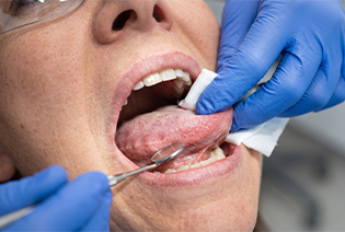
Oropharyngeal cancer develops in the middle part of the throat, including the tonsils, base of the tongue, soft palate, and side and back walls of the throat. Some are caused by persistent high-risk HPV infection, particularly HPV16. HPV-associated oropharyngeal cancers differ biologically from tobacco-associated cancers and have a separate staging system.
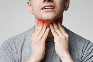
Nasopharyngeal cancer develops in the upper part of the throat behind the nose. It may present with a neck lump, blocked nose, nosebleeds, hearing changes, or persistent fullness in one ear. Many nasopharyngeal cancers are associated with Epstein–Barr virus, although infection alone does not mean that cancer will develop.
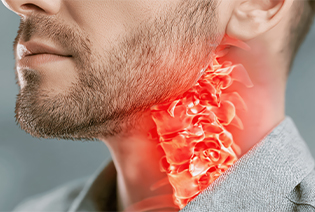
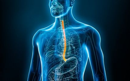
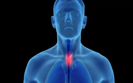
Hypopharyngeal cancer begins in the lower part of the throat surrounding the entrance to the oesophagus. Early symptoms may be subtle. Possible signs include difficulty or pain while swallowing, persistent throat discomfort, a change in voice, ear pain, weight loss, or a neck lump.
Laryngeal cancer develops in the voice box. It may arise above, at, or below the vocal cords. Persistent hoarseness is a common symptom of cancers involving the vocal cords. Other symptoms may include difficulty swallowing, noisy breathing, a neck lump, or persistent throat or ear pain.

These cancers develop in the nasal passages or air-filled sinuses around the nose. They include squamous cell carcinoma, adenocarcinoma, melanoma, olfactory neuroblastoma, and other rare tumours. Symptoms may include persistent one-sided nasal blockage, repeated nosebleeds, facial pain or swelling, numbness, dental changes, double vision, or a bulging eye.
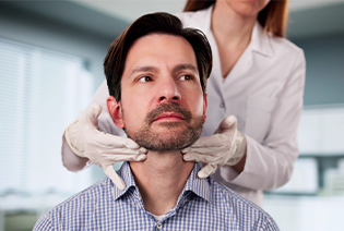
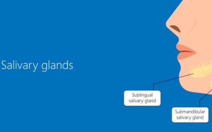
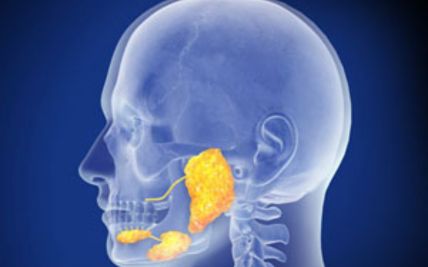
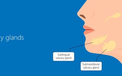
Salivary gland cancer may develop in the major salivary glands near the ear, under the jaw, or beneath the tongue, or in smaller glands throughout the mouth and throat. These cancers include several subtypes with different patterns of growth and spread. A painless lump is common, while pain, numbness, or facial weakness may suggest nerve involvement.
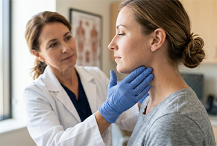
Sometimes cancer is found in a neck lymph node, but the original tumour cannot initially be identified. Examination, imaging, endoscopy, HPV or EBV testing, and targeted biopsies are used to locate the primary site. Treatment is based on the lymph-node findings, molecular results, and the most likely site of origin.
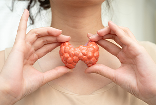
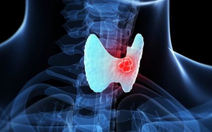
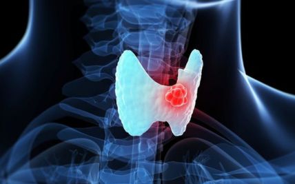
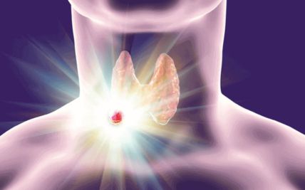
Thyroid cancer develops in the thyroid gland at the front of the neck. The main types are papillary, follicular, medullary, and anaplastic thyroid cancer. Most differentiated thyroid cancers behave differently from head and neck squamous cell cancers. Treatment may involve surgery, radioactive iodine, thyroid hormone therapy, targeted therapy, radiation, or other systemic treatments.
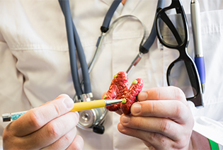
Parathyroid cancer is a rare cancer arising from one of the small glands behind the thyroid. It often produces excess parathyroid hormone, causing high calcium levels. Possible symptoms include weakness, excessive thirst, frequent urination, constipation, kidney stones, bone pain, or a neck lump. Surgery is the main potentially curative treatment.
Risk factors vary by cancer site. Having a risk factor does not mean that cancer will develop, and some people have no identifiable risk factors.
Modifiable Risk Factors
Smoking: Cigarettes, bidis, cigars, and pipes increase the risk of cancers of the mouth, throat, and larynx.
Smokeless tobacco: Chewing tobacco, gutka, khaini, and other smokeless products substantially increase oral cancer risk.
Areca nut: Areca nut or supari, with or without tobacco, increases the risk of oral precancer and cancer.
Alcohol: Heavy or prolonged alcohol consumption increases the risk of oral, pharyngeal, and laryngeal cancers. Combining alcohol and tobacco increases risk further.
High-risk HPV infection: Persistent oral HPV infection is associated with many oropharyngeal cancers. HPV vaccination can prevent infection with the major cancer-causing HPV types.
Ultraviolet exposure: Long-term sun exposure increases the risk of lip cancer.
Occupational exposure: Exposure to wood dust, leather dust, formaldehyde, nickel, or certain industrial substances is associated with selected nasal and sinus cancers.
Poor oral health: Poor oral hygiene and chronic dental irritation may contribute to risk alongside established factors, although they are not independent causes of every oral cancer.
Non-Modifiable Risk Factors
Increasing age
Male sex for several head and neck cancers
Epstein–Barr virus-associated risk for nasopharyngeal cancer
Previous radiation exposure to the head and neck
A weakened immune system
Certain inherited conditions, including Fanconi anaemia
A personal history of head and neck cancer
A family history of thyroid or selected other cancers
Inherited variants associated with medullary thyroid cancer
Long-standing thyroid disease or a previous thyroid nodule
Geographic, genetic, and dietary factors associated with nasopharyngeal cancer
Avoiding tobacco and areca nut, limiting alcohol, receiving HPV vaccination when eligible, using sun protection, and following workplace safety measures can reduce some head and neck cancer risks.
Signs and Symptoms
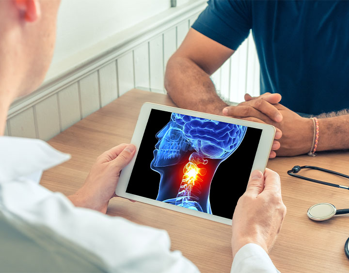
Symptoms depend on the tumour’s location and size. Some early cancers may produce only mild or intermittent changes.
Possible signs and symptoms include:
These symptoms may also result from infection, dental disease, reflux, thyroid nodules, or other non-cancerous conditions. However, a persistent mouth ulcer, neck lump, voice change, swallowing difficulty, one-sided nasal obstruction, or unexplained bleeding should be assessed.
Diagnosis begins with a review of symptoms, tobacco and alcohol use, HPV-related risk, occupational exposure, previous illnesses, and family history. The specialist examines the mouth, tongue, throat, nose, face, skin, thyroid, salivary glands, and neck lymph nodes. Flexible nasoendoscopy or laryngoscopy may be used to inspect areas that cannot be seen directly. Imaging determines the tumour’s extent, while biopsy and laboratory testing establish the cancer type.
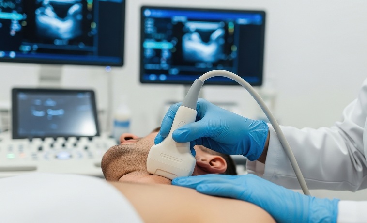
Ultrasound uses sound waves to assess neck lumps, lymph nodes, thyroid nodules, and salivary glands. It can distinguish between solid and fluid-filled abnormalities and guide needle sampling. Ultrasound is particularly useful for thyroid cancer and enlarged cervical lymph nodes but cannot assess every deep structure in the head and neck.
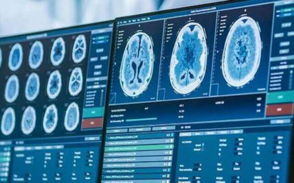
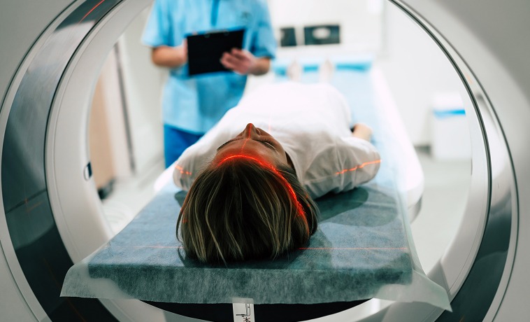
A contrast-enhanced CT scan provides detailed images of the primary tumour, lymph nodes, airway, bones, and surrounding tissues. It is commonly used for cancers of the mouth, throat, larynx, sinuses, salivary glands, and thyroid. CT of the chest may also be performed to assess the lungs or distant spread.


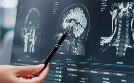
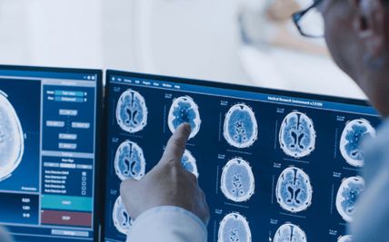
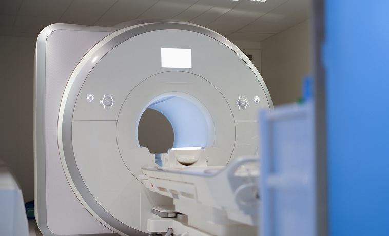
MRI provides detailed images of soft tissues, nerves, the skull base, brain, bone marrow, and spaces deep within the head and neck. It is particularly useful for assessing tongue, salivary gland, nasopharyngeal, sinus, skull-base, and nerve-related tumour involvement.
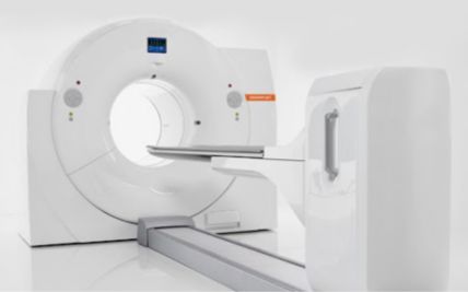
PET-CT combines metabolic and anatomical imaging. It may be used to assess lymph nodes and distant spread, identify an unknown primary tumour, plan radiation, evaluate treatment response, or investigate suspected recurrence. PET-CT is not required for every early head and neck cancer.


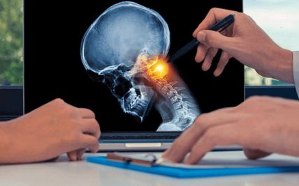
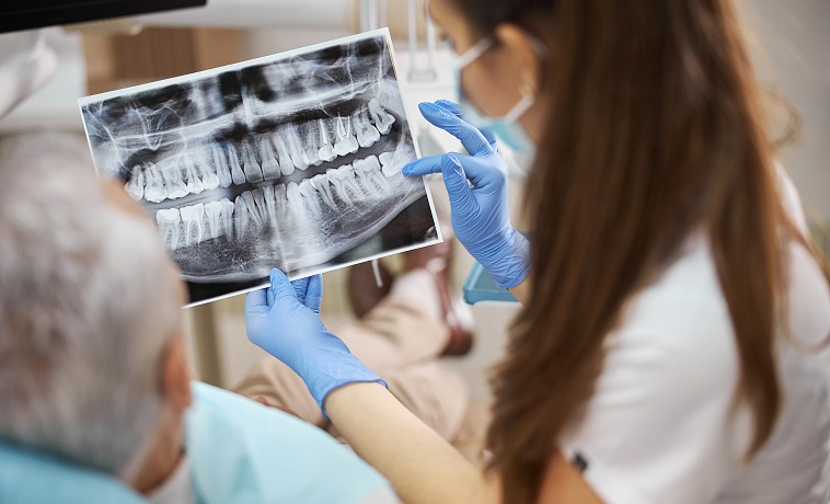
Dental X-rays or specialised scans may be used when a tumour involves the jaw or when dental treatment is required before radiation therapy. Dental assessment before radiation can identify infection, damaged teeth, or other problems that may complicate treatment.
A radioactive iodine or technetium thyroid scan may be used in selected thyroid conditions to assess how thyroid tissue takes up the tracer. It is not the main test used to determine whether a thyroid nodule is cancerous.
After treatment for selected differentiated thyroid cancers, a radioactive iodine scan may be used to identify remaining thyroid tissue or cancer that has taken up iodine.
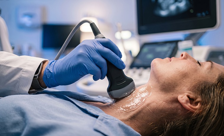
Fine-needle aspiration cytology, or FNAC, uses a thin needle to collect cells from a neck lymph node, thyroid nodule, or salivary gland mass. Ultrasound guidance may be used to improve accuracy. FNAC can identify many cancers but may not always provide enough information for complete classification.
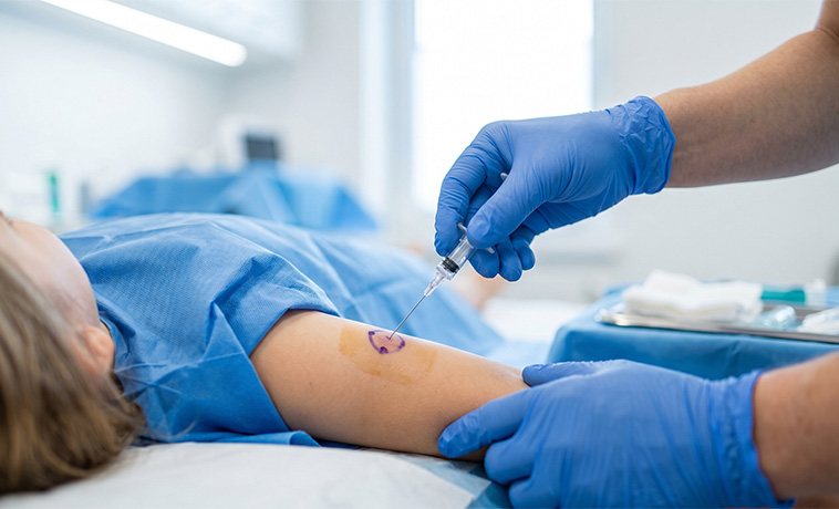
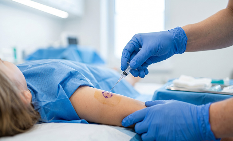
A core needle biopsy removes small cylinders of tissue, usually under ultrasound or CT guidance. It provides more tissue architecture than FNAC. It may be used for selected lymph nodes, salivary gland tumours, thyroid lesions, or deep neck masses when clinically appropriate.
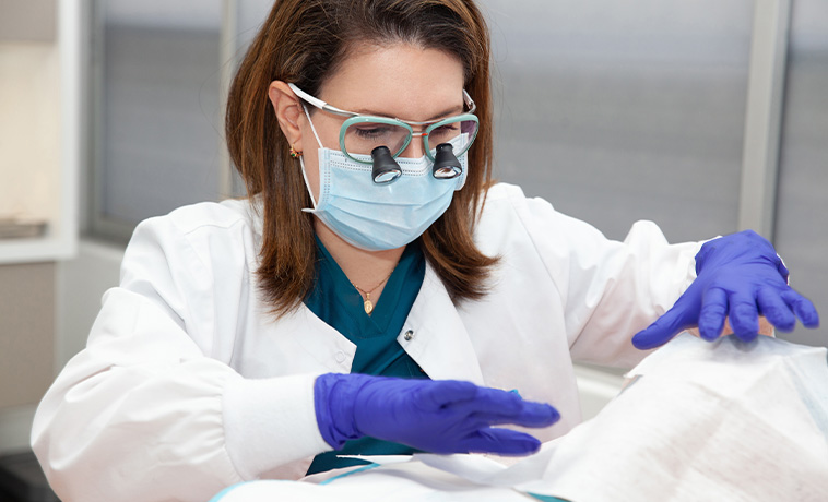
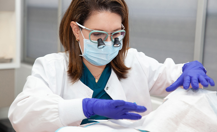
A biopsy may be taken during nasoendoscopy, laryngoscopy, oesophagoscopy, or another endoscopic examination. The procedure allows the specialist to inspect and sample a suspicious area in the nose, throat, larynx, or upper oesophagus. Some biopsies require examination under anaesthesia to assess the tumour fully and obtain adequate tissue.
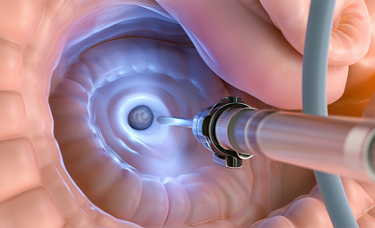
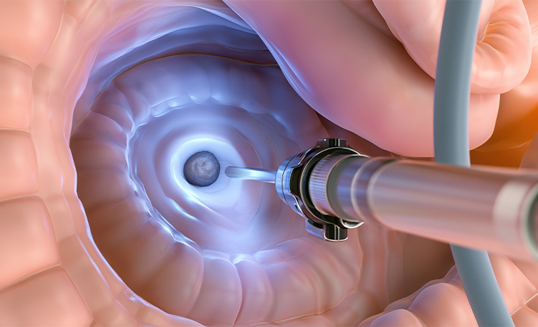
An incisional biopsy removes part of a mouth, skin, or other accessible lesion. It is commonly used when a lesion is too large to remove completely during the diagnostic procedure. The biopsy should be planned so that it does not interfere with later definitive surgery.
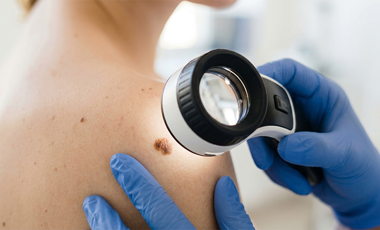
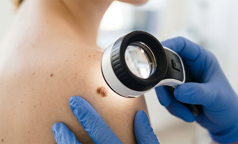
An excisional biopsy removes the entire small lesion or lymph node. It may be used for selected superficial masses when complete removal can be performed safely. An unexplained neck lump should not routinely be removed without appropriate imaging and needle assessment because the surgical approach can affect subsequent cancer treatment.
Sentinel lymph-node biopsy identifies and removes the first lymph node or nodes most likely to receive drainage from the tumour. It may be used in selected early oral cavity, lip, skin, or thyroid cancers according to the clinical situation.
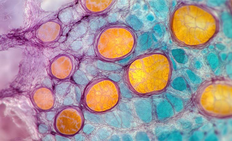
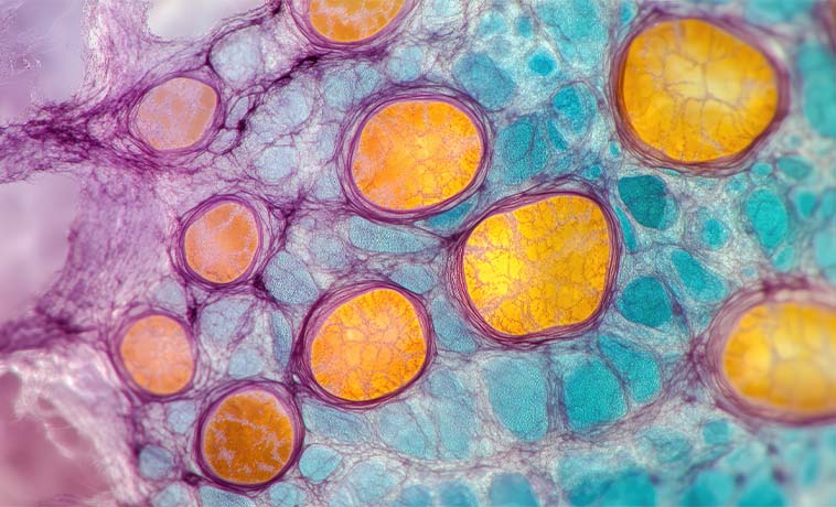
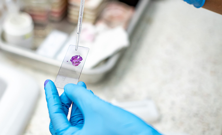
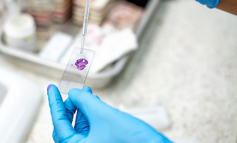
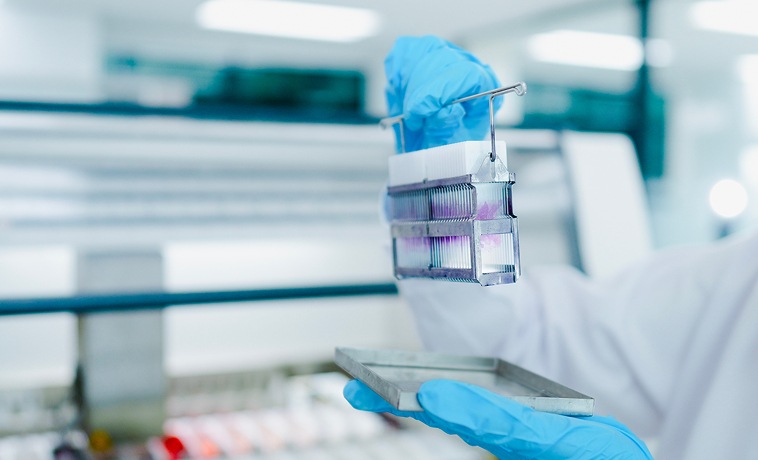
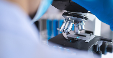
A pathologist examines biopsy or surgical tissue under a microscope. This confirms whether cancer is present and establishes its type, grade, depth of invasion, margins, nerve involvement, blood-vessel or lymphatic invasion, and lymph-node findings.
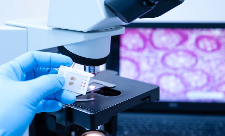
Immunohistochemistry uses specialised stains to identify proteins within tumour cells. It can help distinguish between squamous cell cancer, salivary gland cancer, thyroid cancer, lymphoma, melanoma, and metastatic cancer from another site.

Testing for HPV or the p16 protein may be performed in oropharyngeal squamous cell carcinoma. HPV-associated oropharyngeal cancer has a distinct staging system and generally behaves differently from HPV-negative disease. A positive p16 test is interpreted with the tumour site and other clinical findings.


Epstein–Barr virus testing may be used in nasopharyngeal cancer. Testing can support diagnosis, and plasma EBV DNA may help assess disease burden or monitor selected patients.
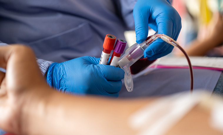
Molecular testing may identify changes that help classify the tumour or guide targeted treatment. Depending on the cancer, testing may include alterations involving BRAF, RET, NTRK, RAS, HER2, androgen receptor, or other relevant markers. Not every head and neck cancer requires broad molecular profiling.


Thyroid-stimulating hormone and other thyroid hormone tests assess thyroid function. They cannot confirm or exclude thyroid cancer but support the evaluation and management of a thyroid nodule.


Calcitonin and carcinoembryonic antigen may be measured when medullary thyroid cancer is suspected or being monitored. Genetic testing for inherited RET variants may also be recommended.
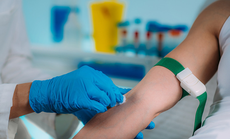

Calcium and parathyroid hormone levels are measured when parathyroid cancer or another parathyroid disorder is suspected. Intraoperative parathyroid hormone testing may be used during parathyroid surgery to assess whether hormone-producing tissue has been adequately removed. It is not a stand-alone test for cancer.


Most head and neck cancers are staged using the TNM system, which considers the primary tumour, regional lymph nodes, and distant spread. The exact definitions vary by tumour site. HPV-associated oropharyngeal cancer, nasopharyngeal cancer, thyroid cancer, and salivary gland cancer have site-specific staging rules.
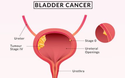
Stage 0: Abnormal cells are confined to the surface lining and have not invaded deeper tissues. This may be called carcinoma in situ.
Stage I: The cancer is generally small and confined to the site where it began, without regional lymph-node or distant spread.
Stage II: The tumour is larger or has grown more deeply into nearby tissue but has limited or no lymph-node involvement.
Stage III: The cancer is larger, has extended into nearby structures, or has spread to a regional lymph node.
Stage IV: The cancer has grown extensively into nearby structures, spread to multiple or advanced regional lymph nodes, or reached a distant organ.
Surgery removes the oral tumour with a margin of surrounding tissue. Depending on the site and extent, this may involve part of the tongue, floor of the mouth, cheek, gum, palate, or jaw. Reconstruction may be performed during the same operation to restore separation between the mouth and neck and support speech, swallowing, and appearance.
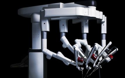
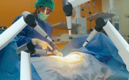
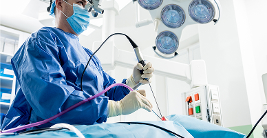
Transoral robotic surgery, or TORS, uses robotic instruments introduced through the mouth to remove selected tumours without an external incision. It may be used for appropriately selected oropharyngeal and other accessible tumours. Suitability depends on tumour size, location, blood-vessel involvement, mouth opening, and whether adequate margins can be achieved.

Transoral laser microsurgery uses a microscope and laser to remove selected tumours through the mouth. It may be used for early laryngeal, pharyngeal, or other accessible cancers. The procedure can preserve surrounding tissue in selected cases, but it is not suitable for every tumour.
Selected early laryngeal cancers may be treated by removing only the affected part of the voice box. Procedures may include endoscopic excision or partial laryngectomy. The choice depends on the tumour’s location and whether safe swallowing, breathing, and voice function can be maintained.
Total laryngectomy removes the entire voice box and separates the airway from the mouth and nose. Breathing then occurs permanently through an opening in the neck called a stoma. Voice can be restored through a voice prosthesis, electrolarynx, or other communication method.
Cancers involving the lower or upper jaw may require removal of part of the mandible or maxilla. The extent depends on whether the tumour involves the surface, marrow, teeth, or surrounding soft tissues. Bone and soft-tissue reconstruction may be performed during the same procedure.
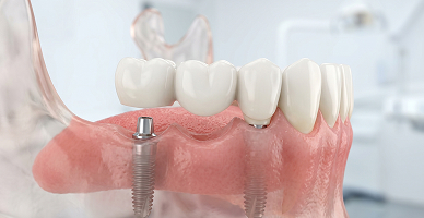
Neck dissection removes lymph nodes and surrounding tissue at risk of containing cancer. It may be performed when lymph-node spread is confirmed or when the risk of microscopic involvement is significant. Modern selective techniques preserve nerves, muscles, and blood vessels whenever this is oncologically safe.
Surgery removes the affected salivary gland and, when required, nearby tissues or lymph nodes. Facial-nerve preservation is attempted when the nerve is not involved by cancer. If part of the facial nerve must be removed, nerve grafting or facial reanimation may be considered.
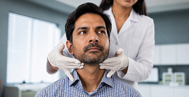
Tumours of the nasal cavity, sinuses, or skull base may be removed through an open, endoscopic, or combined approach. Treatment may require collaboration between head and neck surgeons, neurosurgeons, ophthalmic surgeons, and reconstructive specialists.
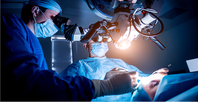
Thyroidectomy removes part or all of the thyroid gland. Lymph nodes may also be removed when cancer has spread or carries a significant risk of nodal involvement. The surgical plan depends on the thyroid cancer type, tumour size, location, genetic findings, and relationship to the recurrent laryngeal nerves and parathyroid glands.
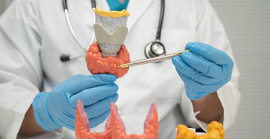
Parathyroid cancer surgery aims to remove the tumour intact, commonly together with the involved parathyroid gland and nearby thyroid tissue. Avoiding tumour rupture is important. Calcium and parathyroid hormone levels are monitored closely during and after surgery.
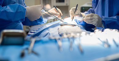
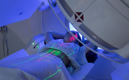
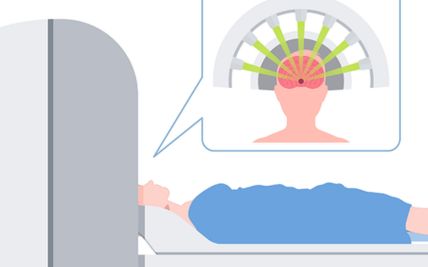
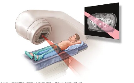
External beam radiation directs radiation towards the tumour and regional lymph nodes. It may be used as the main treatment, after surgery, with chemotherapy, or to relieve symptoms. Its role depends on the tumour site, stage, surgical findings, and expected effect on function.
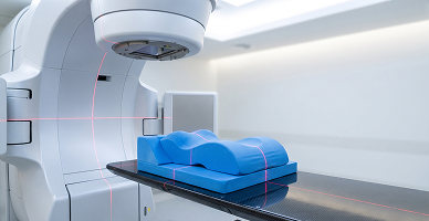
Intensity-modulated radiation therapy and volumetric-modulated arc therapy shape the radiation dose around the tumour while limiting exposure to surrounding tissues. These techniques can help reduce radiation to the salivary glands, swallowing muscles, spinal cord, brainstem, and other nearby structures.
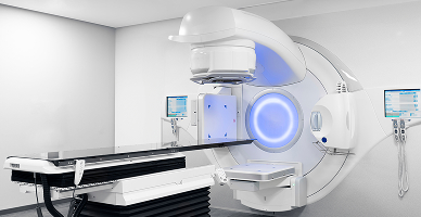
Image-guided radiation therapy uses imaging during treatment to confirm positioning and account for changes in anatomy. This is particularly important in the head and neck, where weight loss or tumour shrinkage can alter the relationship between the target and nearby organs.
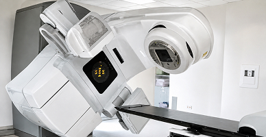
Hypofractionated radiation gives a larger dose during each session over fewer treatments. It may be appropriate for selected early cancers, thyroid cancers, metastatic lesions, or symptom control. It is not a standard shortened option for every curative head and neck treatment.
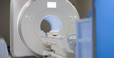
Stereotactic radiation delivers a highly focused dose to a small, well-defined target. It may be used for selected skull-base tumours, limited recurrences, or metastatic lesions. Systems such as Zap-X may be considered for appropriate intracranial or skull-base targets. They are not routinely used for most mucosal head and neck cancers.
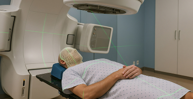
Brachytherapy places radioactive sources within or close to the tumour. It may be used for selected cancers of the lip, tongue, oral cavity, or other accessible sites. Its use depends on tumour size, location, available expertise, and the role of other treatment options.
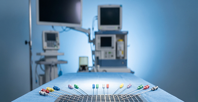
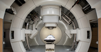
Proton therapy uses proton beams that release most of their radiation within the planned treatment area, with little radiation continuing beyond it. This may reduce radiation exposure to some nearby healthy tissues.
It may be considered for selected skull-base, sinonasal, nasopharyngeal, salivary gland, recurrent, paediatric, or other complex head and neck tumours when comparative planning demonstrates a meaningful advantage. It may be particularly useful when the target lies close to the brain, brainstem, spinal cord, eyes, or other sensitive structures.
Proton therapy cannot guarantee preservation of vision, taste, smell, swallowing, salivary function, or other functions when the tumour involves or lies very close to the structures responsible for them. It is not required for every head and neck cancer.
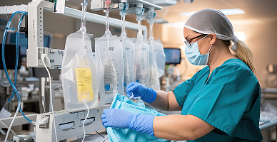
Chemotherapy may be combined with radiation for locally advanced squamous cell cancers or used for recurrent or metastatic disease. It may also be given before another treatment in selected cancers when shrinking the tumour could improve the treatment approach.

Concurrent chemoradiation combines radiation with chemotherapy that makes cancer cells more sensitive to radiation. It is commonly used for selected locally advanced cancers of the oropharynx, nasopharynx, hypopharynx, and larynx. It may also be recommended after surgery when pathology shows high-risk features.
Targeted medicines act on particular proteins or molecular changes within cancer cells. They may be used in selected squamous cell, salivary gland, thyroid, and other head and neck cancers. The relevant target must be identified or supported by the specific diagnosis.

Immunotherapy helps the immune system recognise and attack cancer cells. It may be used for selected recurrent, unresectable, or metastatic head and neck cancers. Suitability may depend on factors such as PD-L1 expression, previous treatment, symptoms, and overall health.

Radioactive iodine is absorbed by thyroid cells and may be used after surgery for selected differentiated thyroid cancers. It can treat remaining thyroid tissue or cancer that has spread but continues to take up iodine. It is not effective for every thyroid cancer and is not used routinely after all thyroid operations.

After total thyroidectomy, thyroid hormone replacement is required. In selected differentiated thyroid cancers, the dose may also be adjusted to suppress thyroid-stimulating hormone, which can encourage some thyroid cancer cells to grow.
Molecular testing may identify treatment options for selected salivary gland, thyroid, squamous cell, or other rare head and neck cancers.
Treatment selection still depends on the cancer type, stage, previous treatment, overall health, and strength of evidence for the identified target.
Microvascular reconstruction transfers skin, muscle, bone, or a combination from another part of the body to the head and neck. Small blood vessels in the transferred tissue are connected to vessels in the neck using microsurgery. The type of flap is selected according to the tissue removed and the function that needs to be restored.
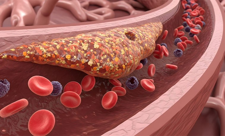
When part of the mandible or maxilla is removed, bone from the leg, shoulder blade, hip, or another site may be used to rebuild the jaw. Plates and computer-assisted planning may help restore facial contour, jaw continuity, and the foundation for dental rehabilitation.
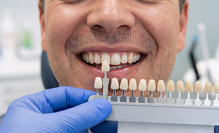
Tissue flaps may be used to reconstruct the tongue, floor of the mouth, cheek, or palate. The reconstruction aims to provide adequate tissue volume and movement for speech, swallowing, and separation of the mouth from the neck or nasal cavity. The approach depends on the size and location of the removed tissue.
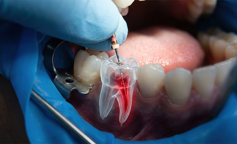
Reconstruction may be required after removal of part of the throat or after total laryngopharyngectomy. A tissue flap or segment of intestine may be used to recreate the passage from the throat to the oesophagus. The method depends on the length and circumference of the defect and previous treatment.
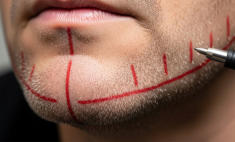
If a tumour involves the facial nerve, reconstruction may use direct repair, a nerve graft, nerve transfer, or muscle transfer. The aim is to restore facial movement, protect the eye, and improve facial symmetry, although complete recovery may not always be possible.
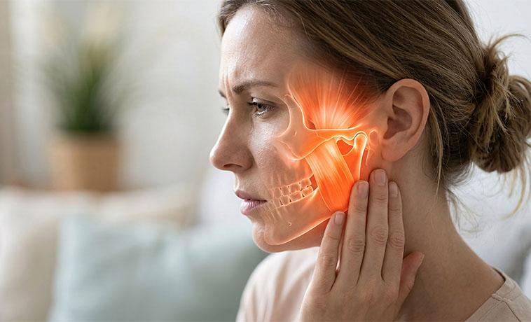
Facial reanimation procedures may be performed when facial movement cannot be restored through direct nerve repair. Tendon or muscle transfers can help restore eye closure, mouth position, and selected facial expressions.
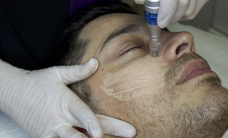
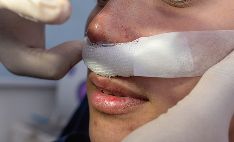
Local flaps, skin grafts, or free flaps may be used to close defects involving the face, scalp, neck, or oral cavity. Reconstruction protects deeper structures and helps restore contour and appearance.
Prognosis for Head and Neck Cancer
The outlook varies according to the cancer site, stage, pathology, and biological features. Many early head and neck cancers can be treated effectively, while locally advanced disease often requires combined treatment.
Factors influencing prognosis include:
The primary cancer site
Tumour size and depth of invasion
Involvement of nearby structures
Number, size, and location of affected lymph nodes
Growth of cancer outside a lymph node
Distant spread
HPV or EBV status where relevant
Cancer type, subtype, and grade
Surgical margins and other pathology findings
Response to radiation or systemic treatment
Tobacco and alcohol use
Nutritional status and overall health
Ability to complete treatment and follow-up
HPV-associated oropharyngeal cancer generally has a more favourable outlook than HPV-negative disease at a comparable clinical presentation, but it still requires appropriate treatment and surveillance.
Thyroid, salivary gland, nasopharyngeal, and other cancers have their own prognostic factors. Prognosis should therefore be discussed according to the individual diagnosis rather than for head and neck cancer as a single group.


Screening for Head and Neck Cancer
There is no universal population screening test for head and neck cancer. Early detection relies mainly on risk reduction, clinical examination of suspicious changes, and prompt evaluation of persistent symptoms.
Oral Examination
A doctor or dental professional can examine the lips, mouth, tongue, gums, and throat for suspicious ulcers, patches, lumps, or other changes. Regular oral examination may be particularly appropriate for people who use tobacco, smokeless tobacco, or areca nut. It does not replace biopsy when a suspicious lesion is found.
HPV Vaccination
HPV vaccination protects against the high-risk HPV types responsible for most HPV-associated cancers. It may reduce the future risk of HPV-related oropharyngeal cancer as well as cervical and other HPV-related cancers. Vaccination does not screen for an existing head and neck cancer, and there is currently no routine screening test for oral HPV infection in people without symptoms.
Why Choose ACC for Head and Neck Cancer Treatment
Multidisciplinary head and neck cancer teams
Organ-specific care for oral, throat, laryngeal, sinonasal, salivary gland, skull-base, thyroid, and related cancers
Transoral robotic surgery and transoral laser microsurgery for selected tumours
Open, endoscopic, minimally invasive, and remote-access surgery selected according to clinical need
Laryngeal, oral, thyroid, salivary, and other function-preserving procedures where oncologically appropriate
Complex neck, skull-base, sinus, and recurrent cancer surgery
Microvascular free-flap, jaw, tongue, pharyngeal, facial-nerve, and soft-tissue reconstruction
Advanced radiation techniques, including IMRT, IGRT, stereotactic radiation, brachytherapy, and proton therapy for selected patients
Comprehensive chemotherapy, targeted therapy, immunotherapy, and precision-oncology services
Radioactive iodine and nuclear medicine services for selected thyroid cancers
HPV, EBV, molecular, thyroid, and parathyroid testing
Multidisciplinary tumour-board planning focused on cancer control and preservation of speech, swallowing, breathing, hearing, and appearance

Health Blogs