icon
icon
Overview

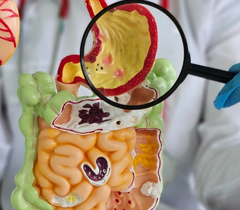
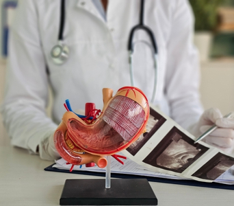
Upper gastrointestinal endoscopy, also called upper GI endoscopy, gastroscopy or esophagogastroduodenoscopy, is a procedure used to examine the lining of the esophagus, stomach and first part of the small intestine, called the duodenum.

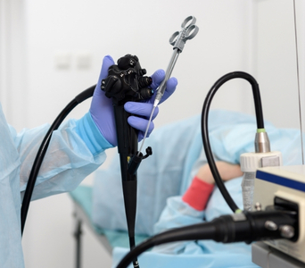
A thin, flexible tube with a light and camera is passed through the mouth, allowing the doctor to view these areas directly. Tissue samples can be collected during the same procedure to help diagnose cancer or other conditions.

Upper GI endoscopy may also be used for surveillance or to perform selected treatments. CT, PET-CT, endoscopic ultrasound or other tests may still be required to determine how deeply a tumour has grown or whether it has spread.
Upper GI Endoscopy
How the Technology Works
Upper GI endoscopy provides a real-time view of the upper digestive tract. Instruments can also be passed through the endoscope to collect tissue or perform selected treatments.
work-icon
Flexible Endoscope
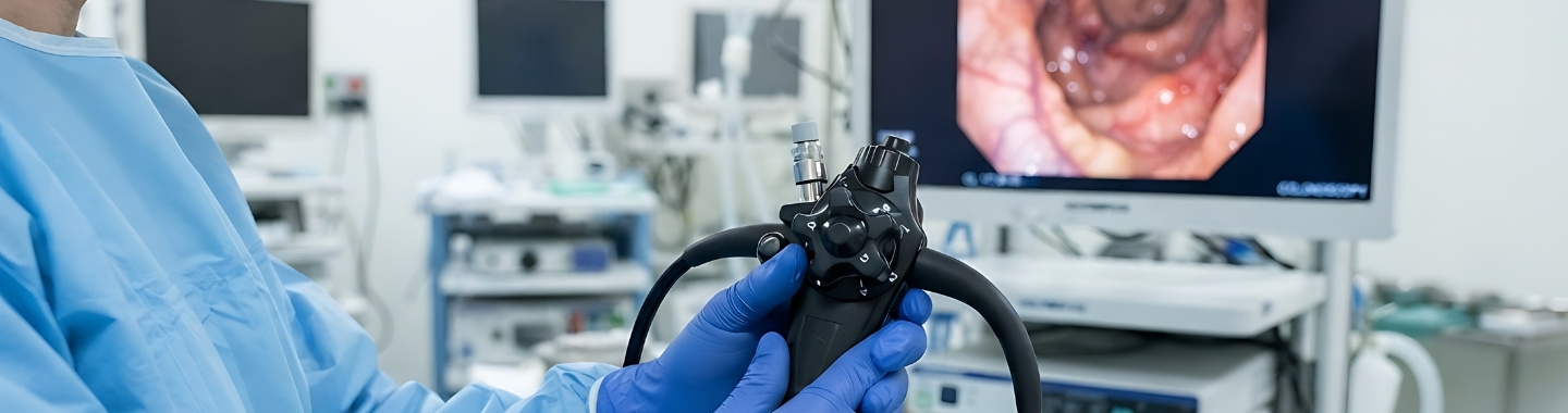
A thin, flexible endoscope is passed through the mouth and guided down the esophagus into the stomach and duodenum. A camera at its tip sends magnified images to a monitor.
work-icon
Direct Visualisation
The doctor examines the lining for ulcers, growths, narrowing, bleeding, inflammation or other abnormal changes. Air or carbon dioxide may be introduced gently to expand the digestive tract and improve visibility.
work-icon
Biopsy and Treatment Channels
Small instruments can be passed through channels within the endoscope to collect tissue samples. The same channels may also be used to remove selected lesions, control bleeding, widen a narrowing or place a stent when clinically appropriate.

Role in Cancer Care

Investigating Symptoms
Endoscopy may be recommended for persistent difficulty or pain when swallowing, unexplained vomiting, upper abdominal pain, gastrointestinal bleeding, anemia, loss of appetite or unexplained weight loss. These symptoms can have many non-cancerous causes. Direct examination helps identify abnormalities that may require biopsy or treatment.
Investigating Symptoms
Detecting Abnormalities
Endoscopy can identify suspicious changes in the esophagus, gastro-esophageal junction, stomach and duodenum. It can show the location, surface appearance and approximate extent of an abnormality. Small or flat lesions may sometimes be detected using image-enhancement techniques or specialised stains.
Detecting Abnormalities
Obtaining a Biopsy
Small tissue samples can be taken from suspicious areas during the procedure. A pathologist examines these samples to determine whether cancer is present and, if so, identify its type and relevant characteristics. Several samples may be collected because some abnormalities require tissue from more than one area.
Obtaining a Biopsy
Supporting Staging and Treatment Planning
Endoscopy can help define a tumour’s position and its visible extent along the digestive tract. However, standard endoscopy cannot reliably determine the full depth of tumour invasion or whether cancer has spread outside the organ. Endoscopic ultrasound, CT, PET-CT or other investigations may therefore be required for staging.
Supporting Staging and Treatment Planning
Providing Selected Treatments
Some early or superficial lesions may be removed endoscopically using techniques such as endoscopic mucosal resection or endoscopic submucosal dissection. Endoscopy may also be used to control tumour-related bleeding, dilate a narrowing or place a stent to improve swallowing or the passage of food. Suitability depends on the lesion, cancer stage, patient’s condition and available expertise.
Providing Selected Treatments
Surveillance and Follow-up
Endoscopy may be used to monitor selected conditions associated with an increased cancer risk, such as Barrett’s esophagus or certain precancerous stomach changes. It may also assess the treatment site or investigate new symptoms after cancer treatment. The timing of surveillance is based on pathology, previous treatment and individual risk.
Surveillance and Follow-up

What to Expect Expect

Before the Test 

The stomach must be empty so the doctor can see clearly and reduce the risk of food or fluid entering the airway. You will usually be instructed not to eat or drink for a specified period before the procedure.

Inform the care team if you:

  • Take blood thinners, aspirin or medicines that affect clotting

  • Have diabetes and use insulin or other glucose-lowering medicines

  • Have heart, lung, kidney or liver disease

  • Have swallowing or breathing difficulties

  • Have allergies or have previously reacted to sedation or anesthesia

  • Are pregnant or may be pregnant

  • Have a pacemaker or another implanted device

  • Have loose teeth, dentures, crowns or other dental concerns

  • Take prescription medicines, non-prescription medicines or supplements

Do not stop or adjust any medicine unless specifically instructed. Blood tests or other assessment may be required if a biopsy or treatment is planned.

If sedation will be used, arrange for a responsible adult to accompany you home. You should not drive after receiving sedation.

Before the Test

During the Test

You will usually lie on your left side. A local anesthetic spray may be applied to the throat, and sedation may be given through a vein to help you relax. The type of sedation or anesthesia depends on the planned procedure and your health.

A mouth guard is placed to protect the teeth and endoscope. The doctor then guides the endoscope through the mouth and into the upper digestive tract. The scope passes through the esophagus and does not obstruct the airway.

Air or carbon dioxide may be introduced to expand the area being examined. You may feel pressure, bloating or the urge to burp. Tissue sampling is not usually felt because the lining of the digestive tract does not sense cutting in the same way as skin.

A routine diagnostic endoscopy commonly takes approximately 10 to 30 minutes. A procedure involving lesion removal, bleeding control, dilation or stent placement may take longer.

During the Test

After the Test 

You will be monitored until the effects of sedation have reduced. Temporary bloating, mild nausea or a sore throat may occur.

Wait until swallowing has returned to normal before eating or drinking if throat-numbing medicine was used. Follow the instructions provided about diet, medicines and activity.

If you received sedation, do not drive, operate machinery, drink alcohol, sign important documents or make major decisions for the period specified by the care team. Arrange for someone to stay with you if advised.

The doctor may discuss the visual findings after the procedure. Biopsy results take longer because the tissue must be processed and examined by a pathologist.

Contact the care team or seek urgent medical attention if you develop:

  • Difficulty breathing or swallowing

  • Severe or worsening chest or abdominal pain

  • Fever

  • Persistent vomiting

  • Vomiting blood or material resembling coffee grounds

  • Black, tarry stools or significant rectal bleeding

  • Marked dizziness, weakness or fainting

After the Test

Potential Benefits

Upper GI endoscopy allows direct examination, tissue sampling and selected treatments during the same procedure.
  • Direct visualisation: The lining of the esophagus, stomach and duodenum can be examined in real time.
  • Magnified assessment: Small, flat or subtle abnormalities may be seen more clearly than with some other tests.
  • Tissue diagnosis: Biopsies can be taken directly from suspicious areas.
  • Accurate localisation: The position and visible extent of an abnormality can be documented.
  • Diagnostic and therapeutic use: Selected abnormalities can be assessed and treated during the same procedure.
  • Early-lesion removal: Some superficial precancerous or cancerous lesions may be removed endoscopically.
  • Symptom relief: Bleeding, narrowing or obstruction may be treated in selected patients.
  • Surveillance: Endoscopy can monitor selected high-risk or precancerous conditions over time.
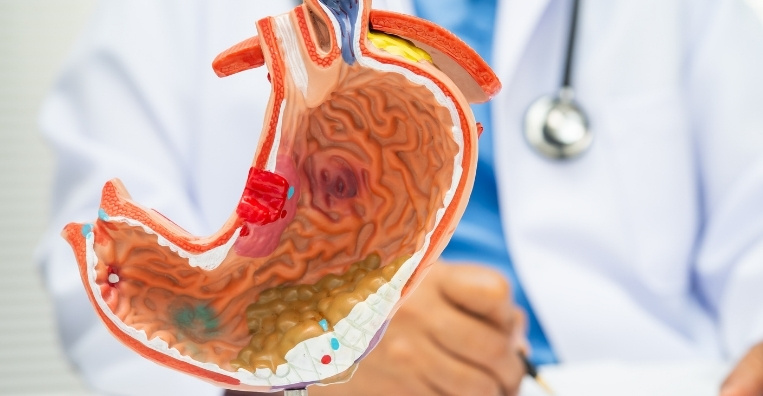
Upper GI Endoscopy
Frequently Asked Questions
01 Is upper GI endoscopy painful?
icon icon
The procedure should not usually be painful, although you may feel pressure, bloating, gagging or temporary throat discomfort. Throat-numbing spray, sedation or anesthesia may be used according to the procedure and individual needs.
02 Will the endoscope affect my breathing?
icon icon
No. The endoscope passes through the esophagus, while breathing occurs through the windpipe. The care team will monitor your oxygen level, breathing, heart rate and other vital signs as appropriate.
03 Is sedation always required?
icon icon
Not always. Some diagnostic endoscopies can be performed using throat-numbing spray alone, while others use conscious or deeper sedation. More complex therapeutic procedures may require deeper sedation or general anesthesia.
04 Can endoscopy confirm upper gastrointestinal cancer?
icon icon
Endoscopy can identify a suspicious abnormality and allow tissue samples to be taken. Cancer is confirmed by pathological examination of the biopsy rather than by appearance alone. If initial samples are inconclusive, repeat or deeper sampling may occasionally be necessary.
05 Can upper GI endoscopy show whether cancer has spread?
icon icon
Standard endoscopy shows the inner surface of the upper digestive tract but cannot fully assess disease outside the organ. Endoscopic ultrasound can help evaluate the depth of a tumour and nearby lymph nodes, while CT, PET-CT or other imaging may be used to assess more distant spread.
06 What are the possible complications?
icon icon
Upper GI endoscopy is generally safe, but complications can include a reaction to sedation, bleeding, aspiration or a tear in the digestive tract. The risk may be higher when a lesion is removed, a narrowing is dilated or another treatment is performed. Your doctor will explain the risks relevant to the planned procedure.
image image
Book an Appointment
Please Enter Your Name
Please Enter Your Mobile Number
Please Enter OTP