icon
icon
Overview

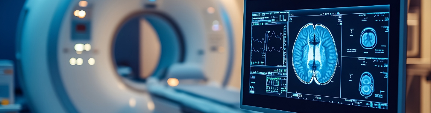
Magnetic Resonance Imaging, commonly called MRI, is a non-invasive imaging test that uses a strong magnetic field, radiofrequency signals and computer processing to create detailed images of organs and tissues. Unlike CT and X-ray, MRI does not use ionising radiation.

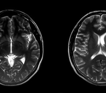
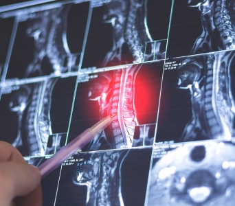
MRI provides particularly detailed views of soft tissues and may be used to assess cancers affecting the brain, spine, head and neck, breasts, liver, pancreas, pelvis, prostate, rectum, female reproductive organs, bones and other parts of the body. Specialised techniques can also provide information about tissue cellularity, blood flow and other tumour characteristics.

MRI scanners are commonly available at magnetic field strengths of 1.5 Tesla and 3 Tesla, abbreviated as 1.5T and 3T. Both can produce high-quality cancer imaging, and the most appropriate scanner depends on the body area, clinical question, imaging protocol, patient factors and compatibility of any implanted devices.
How the Technology Works
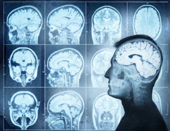
MRI uses a magnetic field and radiofrequency signals to produce detailed images of structures inside the body.
work-icon
Magnetic and Radiofrequency Signals
The magnetic field temporarily aligns hydrogen atoms naturally present in the body. Radiofrequency signals alter this alignment, and the energy released as the atoms return to their usual state is detected and converted into images.
work-icon
1.5T and 3T MRI
Tesla refers to the strength of the MRI scanner’s magnetic field. A 3T scanner can provide a stronger signal, which may support finer detail or faster imaging in selected examinations, while 1.5T MRI provides high-quality imaging across a wide range of applications and may be preferable in certain patients or protocols. One is not universally better than the other.
work-icon
Specialised Sequences and Contrast
MRI uses different sequences to assess tissue structure, fluid, blood flow, cellular density and other characteristics. Some examinations use a gadolinium-based contrast agent to make tumours, blood vessels or patterns of tissue enhancement more visible. Techniques such as diffusion-weighted, dynamic contrast-enhanced, perfusion, spectroscopy or multiparametric imaging may be used when clinically appropriate.

Role in Cancer Care

Detecting and Characterising Abnormalities
MRI may be used to investigate symptoms or further evaluate an abnormality identified on examination or another imaging test. Its detailed soft-tissue images can help show an abnormality’s size, location, internal characteristics and relationship to surrounding structures. Imaging findings may suggest a diagnosis, but a biopsy is often required to confirm cancer.
Detecting and Characterising Abnormalities
Local Staging
MRI can help determine how far a tumour extends within an organ and whether nearby tissues, nerves, blood vessels or other structures are involved. It is particularly useful for local staging in selected cancers of the brain, head and neck, breast, liver, prostate, rectum, female pelvis, bones and soft tissues. These findings may influence treatment selection and surgical planning.
Local Staging
Assessing Spread to Other Areas
MRI may be used to evaluate possible cancer spread to areas such as the brain, spine, liver, bones or bone marrow. Whole-body MRI may be considered in selected cancers or inherited cancer-predisposition conditions, but it is not a routine general cancer-screening test. Other investigations may be needed depending on the cancer and suspected pattern of spread.
Assessing Spread to Other Areas
Planning Treatment
MRI can help surgeons assess whether a tumour can be removed and understand its relationship to structures that may need to be preserved. It may also contribute to radiation therapy planning and guide decisions about systemic or local treatment. In selected cases, MRI can guide biopsy, localisation or minimally invasive treatment.
Planning Treatment
Monitoring Treatment Response
MRI may be repeated during or after treatment to assess changes in tumour size, enhancement, diffusion or other imaging characteristics. These changes can provide information about response to chemotherapy, immunotherapy, radiation therapy or local treatment. Results are interpreted alongside symptoms, clinical findings and other investigations.
Monitoring Treatment Response
Follow-up and Suspected Recurrence
MRI may be used during follow-up to monitor a treated area or investigate possible recurrence. It can help distinguish suspicious tissue from some expected post-treatment changes, although surgery and radiation can sometimes make interpretation difficult. Follow-up timing depends on the cancer type, stage, treatment received and individual care plan.
Follow-up and Suspected Recurrence

What to Expect

Before the Test

Preparation varies according to the body area and imaging protocol. You can usually eat, drink and take regular medicines unless the care team provides different instructions.

Before the scan, inform the team if you:

  • Have a pacemaker, defibrillator, cochlear implant, neurostimulator, medicine pump or other implanted device

  • Have aneurysm clips, vascular stents, surgical clips or metal implants

  • May have metal fragments, bullets or shrapnel in your body, particularly near the eyes

  • Have undergone previous surgery or implantation of a medical device

  • Are pregnant or may be pregnant

  • Are breastfeeding

  • Have kidney disease or reduced kidney function

  • Have previously reacted to an MRI contrast agent

  • Experience claustrophobia or have difficulty remaining still

  • Have tattoos, medicine patches or wearable medical devices

  • Are taking prescription medicines, non-prescription medicines or supplements

Bring information identifying any implant or device, including its model and MRI safety documentation, if available. Many modern implants are MR Conditional, meaning MRI may be performed only under specified conditions, which can include the permitted field strength. The MRI team must confirm compatibility before you enter the scanning area.

If gadolinium contrast is planned, a kidney-function blood test may be required, particularly if you have known kidney disease or relevant risk factors. Remove jewellery, watches, hearing aids, removable dental work, mobile phones, cards and other metallic or electronic items as instructed.

Tell the team in advance if you are anxious in enclosed spaces. Additional preparation, a wider-bore scanner or sedation may be considered when appropriate. If sedation is planned, you will receive fasting instructions and must arrange for someone to accompany you home.

Before the Test

During the Test 

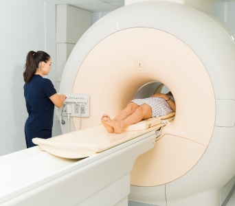
You will lie on a motorised table, and a specialised receiving coil may be placed around or near the body area being examined. Padding and supports may be used to help you remain comfortable and still.

The table moves into the MRI scanner, which is shaped like a tunnel. The machine produces loud knocking, tapping and buzzing sounds while images are acquired. You will be given earplugs or headphones for hearing protection.

It is important to remain still, as movement can blur the images. For some examinations, you may be asked to hold your breath briefly. The technologist will be able to see, hear and communicate with you throughout the scan, and you will be given a call device to request assistance.

If contrast is required, a cannula will be inserted into a vein in the hand or arm. Images may be taken before, during and after the contrast injection.

MRI is painless, although remaining in one position or being inside the scanner may cause discomfort or anxiety. The area being examined may feel slightly warm. Tell the technologist immediately if you experience excessive heating, burning, pain, dizziness, breathing difficulty or another unusual sensation.

Most MRI examinations take approximately 30 to 60 minutes, but specialised or multiparametric studies may take longer.

During the Test

After the Test

If you have not received sedation, no recovery period is usually required. You can generally return to normal activities and resume your usual diet immediately.

If contrast was administered, the cannula will be removed before you leave. You may be advised to drink fluids unless you are on fluid restriction or have received different instructions.

If you received sedation, you will be monitored until it is safe to leave. You must not drive, operate machinery or make important decisions until the effects have worn off, as advised by the care team.

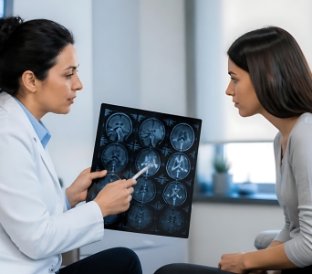
A radiologist will examine the images, compare them with previous studies and prepare a report. The findings will be considered together with pathology results, clinical information and other investigations.

After the Test

Potential Benefits

MRI can provide detailed information about tumours and surrounding tissues without using ionising radiation.
  • Detailed soft-tissue imaging: MRI can distinguish between different types of soft tissue and show tumour boundaries.
  • Multiplanar views: Images can be acquired or reconstructed in different planes without repositioning the patient.
  • No ionising radiation: MRI uses magnetic fields and radiofrequency signals rather than X-rays.
  • Local staging: It can help assess involvement of nearby organs, muscles, nerves, blood vessels and other structures.
  • Specialised tumour assessment: Diffusion, perfusion, contrast-enhanced and multiparametric techniques can provide additional information about tumour characteristics.
  • Treatment planning: MRI may support surgery, radiation therapy, biopsy and selected image-guided treatments.
  • Response monitoring: Changes in tumour size and imaging characteristics can help assess treatment response.
  • Versatile field strengths: Both 1.5T and 3T systems can support high-quality oncology imaging using protocols selected for the individual patient and clinical question.
Potential Benefits
Frequently Asked Questions
01 Is a 3T MRI always better than a 1.5T MRI?
icon icon
No. A 3T scanner provides a stronger signal and may offer finer detail or shorter imaging times for selected examinations. However, it may also be more sensitive to certain artefacts and may not be suitable for every implant or imaging protocol. Both 1.5T and 3T MRI can provide high-quality cancer imaging.
02 Does MRI involve radiation?
icon icon
MRI does not use the ionising radiation used in X-rays, CT, PET-CT or SPECT-CT. It uses a strong magnetic field and radiofrequency signals. MRI safety screening remains essential because the magnetic field can interact with certain metals, devices and implants.
03 Can I undergo an MRI if I have a pacemaker or another implant?
icon icon
Possibly. Many modern devices are MR Conditional and can be scanned only under specified conditions, while some devices may not be suitable for MRI. The exact device model, implantation details, permitted field strength and manufacturer’s conditions must be verified before the examination.
04 Is contrast required for every MRI?
icon icon
No. Many MRI examinations provide useful information without contrast. Gadolinium-based contrast may be recommended when it can better show tumour enhancement, blood vessels, inflammation or treatment-related changes. The decision depends on the body area, clinical question, kidney function and previous reactions.
05 Is gadolinium contrast safe if I have kidney disease?
icon icon
Many patients with kidney disease can undergo contrast-enhanced MRI after appropriate assessment, but the decision depends on kidney function, the contrast agent and the importance of the examination. A rare condition called nephrogenic systemic fibrosis has been associated mainly with certain gadolinium agents in people with severe kidney dysfunction. The radiology team will determine whether contrast is necessary and select the safest appropriate approach.
06 What if I am claustrophobic or cannot remain still?
icon icon
Tell the imaging team before the appointment. Preparation, reassurance, music, a wider-bore scanner or medication for anxiety may help, depending on the examination and facility. Sedation may be considered when necessary, but it requires additional preparation and arrangements for someone to accompany you home.
image image
Book an Appointment
Please Enter Your Name
Please Enter Your Mobile Number
Please Enter OTP