




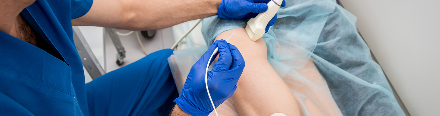
Radiofrequency Ablation (RFA) is a minimally invasive interventional oncology procedure that uses heat to destroy cancer cells. A thin needle-like probe called an electrode is placed within the tumour, and radiofrequency energy passes through it to generate heat. This heats and destroys the targeted tissue.
RFA is usually performed through the skin under ultrasound, CT or other imaging guidance by an interventional oncologist or interventional radiologist. It may also be performed during laparoscopic or open surgery in selected cases. Unlike radiation therapy, RFA does not use ionising radiation to treat the tumour.
RFA is a local treatment generally used for small, well-defined tumours. It may be considered when surgery is not suitable or when preserving as much healthy tissue as possible is important. RFA may be used alone or combined with surgery, systemic therapy, radiation therapy, embolization or other cancer treatments.
- Primary liver cancers, including selected hepatocellular carcinomas
- A limited number of liver metastases
- Selected kidney tumours
- Selected early-stage lung cancers when surgery is not suitable
- A limited number of lung metastases
- Selected adrenal tumours or metastases
- Painful bone tumours or bone metastases
- Small soft-tissue tumours in selected locations
- Residual or recurrent tumours following surgery or other treatment
- Patients who are not suitable for surgery because of other medical conditions or reduced organ function
- Patients for whom surgery would require removal of a significant amount of healthy tissue
- Selected patients requiring local treatment of a limited number of metastatic tumours

How Radiofrequency Ablation Is Performed
Before Treatment
The interventional oncologist or interventional radiologist reviews the diagnosis, biopsy findings, imaging, previous treatments and medical history.
CT, MRI, ultrasound, PET-CT or other imaging is used to evaluate the tumour and plan a safe route for the electrode.
Blood tests are performed to assess blood counts, kidney or liver function and the ability of the blood to clot.
Blood-thinning medicines and certain other medications may need to be temporarily adjusted under medical guidance.
The patient is usually instructed to avoid eating or drinking for a specified period before the procedure.
An anesthesia assessment may be performed when deep sedation or general anesthesia is planned.
Antibiotics may be recommended in selected cases.
The possible need for a biopsy before or during the procedure is discussed.
The treating team explains the expected benefits, possible risks and whether more than one ablation session may be required.

During Treatment
The patient is positioned according to the location of the tumour.
Local anesthesia, sedation or general anesthesia is administered depending on the treatment area and procedure.
Ultrasound, CT or another imaging method is used to locate the tumour.
A small skin puncture is made, and one or more electrodes are guided into the tumour.
The position of the electrode is checked before energy is delivered.
Radiofrequency energy passes through the electrode and generates heat within the target.
The tumour and a planned margin around it are treated where this can be done safely.
Temperature, energy delivery and the patient’s condition are monitored throughout the procedure.
The electrode may be repositioned to treat different parts of a larger or irregularly shaped tumour.
The electrode is removed after the planned ablation is complete, and a dressing is placed over the insertion site.
The procedure may take approximately one to three hours, depending on the tumour’s size, number and location.

After Treatment
The patient is monitored in a recovery area while the effects of sedation or anesthesia wear off.
Pain relief and other medicines are provided when needed.
Follow-up imaging may be performed immediately or shortly after treatment to check the ablation area and identify complications.
Some patients can return home on the same day, while others may need an overnight hospital stay.
Mild pain, tiredness, low-grade fever, nausea or a flu-like feeling may occur for a few days.
The dressing and insertion site should be cared for according to the team’s instructions.
Strenuous activity may need to be avoided for a short period.
Follow-up CT, MRI, ultrasound or PET-CT is arranged to assess the treated area.
Additional ablation or another treatment may be recommended if viable tumour remains or the cancer returns.

Potential Benefits of Radiofrequency Ablation
- Minimally invasive treatment through a small skin puncture
- Destruction of a localised tumour without removing the entire organ
- Preservation of surrounding healthy tissue where possible
- An alternative for selected patients who cannot undergo surgery
- Less pain and a shorter recovery period than major surgery in many cases
- A short hospital stay, with same-day discharge possible for some patients
- The ability to repeat treatment if clinically appropriate
- Treatment of more than one small tumour during the same procedure in selected cases
- The ability to be combined with surgery, embolization, systemic therapy or radiation therapy as part of a multidisciplinary treatment plan
- Local control of selected primary or metastatic tumours
- Relief of pain caused by certain bone tumours or metastases

Side Effects of Radiofrequency Ablation
- Pain, tenderness, bruising or swelling at the insertion site
- Tiredness or weakness
- Low-grade fever, chills or flu-like symptoms after ablation
- Nausea or reduced appetite
- Bleeding or a collection of blood near the treated area
- Infection at the insertion site or within the treated organ
- Injury to nearby nerves, blood vessels or organs
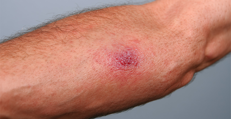
- Skin or tissue burns
- Incomplete tumour destruction or cancer returning at the edge of the treated area
- Collapse of part of the lung or air around the lung following treatment of a lung tumour
- Bleeding into the chest following lung ablation
- Injury to the bile ducts, gallbladder, bowel or diaphragm following liver ablation
- Urine leakage or injury to nearby structures following kidney ablation
- Changes in liver, kidney or other organ function
- Rare blood clots or serious reactions related to anesthesia



