icon
icon
Overview

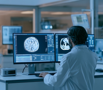
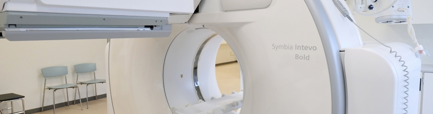
Single-Photon Emission Computed Tomography–Computed Tomography, commonly called SPECT-CT, is a hybrid imaging test that combines functional information from a nuclear medicine scan with detailed anatomical images from CT.

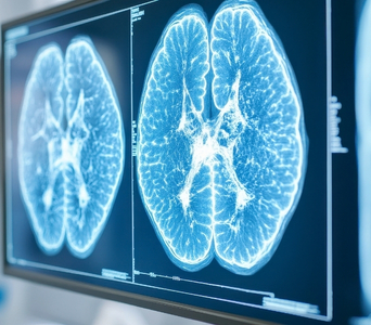
The SPECT component uses a small amount of a radioactive substance called a radiotracer. The tracer is selected according to the organ, tissue or biological process being examined. A gamma camera detects energy released by the tracer and produces three-dimensional images showing where it has accumulated in the body.

The CT component provides detailed structural information. Combining the two scans allows doctors to match areas of tracer uptake with their precise anatomical location. In cancer care, SPECT-CT may be used for selected purposes such as evaluating the bones, localising sentinel lymph nodes, imaging certain tumour types and planning or assessing some radionuclide treatments.

SPECT-CT is not routinely required for every cancer. Its usefulness depends on the cancer type, radiotracer available and specific clinical question.
SPECT-CT
How the Technology Works
SPECT-CT combines functional information from a nuclear medicine scan with detailed anatomical images from CT. The two sets of images are acquired during the same examination and interpreted together.
work-icon
Radiotracer Imaging
A small amount of a radiotracer is usually injected into a vein, although some tracers may be swallowed or inhaled. The tracer accumulates in a particular organ, tissue or cell type according to the biological process being examined.
work-icon
Three-Dimensional SPECT Imaging
Gamma-camera detectors rotate around the body and measure the energy released by the radiotracer. Computer processing uses this information to create three-dimensional images showing how the tracer is distributed.
work-icon
Combined CT Imaging
The CT component uses X-rays to produce cross-sectional images that show the precise anatomical location of tracer uptake. The CT may be performed as a low-dose scan for localisation or, when required, as a diagnostic scan with additional detail or contrast.

Role in Cancer Care

Evaluating Bone Involvement
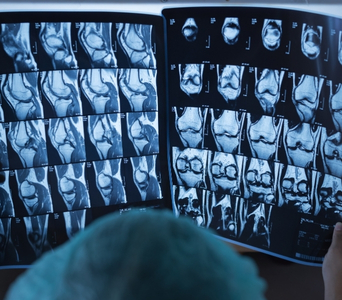
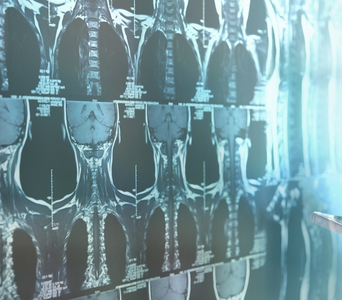
SPECT-CT may be performed as part of a bone scan to evaluate areas of altered bone activity. It can help localise an abnormality to a specific bone or joint and may help distinguish cancer-related changes from fractures, arthritis, infection or other non-cancerous conditions. A bone scan shows changes in bone activity rather than detecting cancer cells directly.
Evaluating Bone Involvement
Sentinel Lymph Node Localisation
SPECT-CT may be used during sentinel lymph-node mapping in selected cancers. A radiotracer is administered near the tumour and travels to the first lymph node or group of nodes likely to receive drainage from that area. The images can help surgeons identify the number and anatomical location of these nodes before surgery.
Sentinel Lymph Node Localisation
Thyroid Cancer Imaging
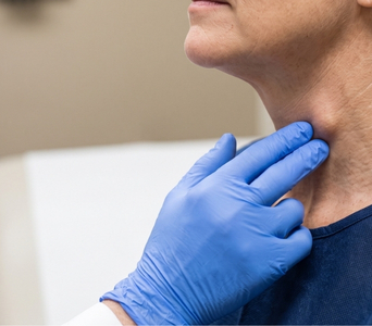
Radioiodine SPECT-CT may be used in selected patients with differentiated thyroid cancer. It can help determine whether radioiodine uptake is located in residual thyroid tissue, lymph nodes, bones or other parts of the body. Imaging may be performed during diagnostic evaluation or after radioactive iodine treatment.
Thyroid Cancer Imaging
Imaging Selected Tumours
Specialised radiotracers may be used to evaluate certain tumours that take up specific substances or express particular molecular targets. These may include selected neuroendocrine tumours, neuroblastoma, pheochromocytoma and paraganglioma. The choice of SPECT-CT depends on the tumour type, tracer availability and whether another test, such as PET-CT, would provide more useful information.
Imaging Selected Tumours
Planning and Assessing Treatment
SPECT-CT may support planning for selected radionuclide therapies or interventional oncology procedures. It can help assess tracer distribution, estimate radiation delivery to tumours and normal tissues, and identify unexpected uptake. Imaging after treatment may also confirm where the therapeutic radiopharmaceutical has accumulated.
Planning and Assessing Treatment
Investigating Possible Recurrence
In selected cancers, SPECT-CT may be used when recurrence is suspected because of symptoms, tumour-marker changes or findings on another scan. It can help localise areas of abnormal tracer uptake and determine whether further evaluation is needed. Its role depends on whether an appropriate radiotracer is available for the cancer being assessed.
Investigating Possible Recurrence

What to Expect

Before the Scan

Preparation varies considerably according to the radiotracer and examination. The nuclear medicine team will provide instructions specific to your scan.

Before the examination, inform the team if you:

  • Are pregnant or may be pregnant

  • Are breastfeeding

  • Have kidney disease or reduced kidney function

  • Have allergies or have previously reacted to CT contrast material

  • Have recently undergone another nuclear medicine test or treatment

  • Have recently had surgery, chemotherapy, immunotherapy or radiation therapy

  • Take prescription medicines, non-prescription medicines or supplements

  • Have difficulty lying still or experience anxiety in enclosed spaces

Some examinations may require fasting, dietary changes, temporary adjustment of certain medicines, blood tests or other preparation. Do not stop or alter any medicine unless instructed.

If contrast-enhanced CT is planned, kidney-function testing or additional preparation may be required. Jewellery and other metal objects may need to be removed. Previous imaging and reports should be provided whenever possible.

Before the Test

During the Scan

The radiotracer is usually administered through a cannula placed in the hand or arm. Depending on the examination, imaging may begin soon afterwards or after an uptake period lasting several hours. Some scans may require delayed images later the same day or on another day.

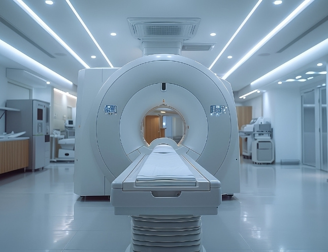
When imaging begins, you will lie on a table while the gamma-camera heads rotate slowly around you. The detectors may come close to the body but should not touch you. It is important to remain still to prevent blurring.

The CT component is usually performed during the same session without changing position. This allows the SPECT and CT images to be accurately combined.

Imaging time varies according to the examination and body area. The scan may take approximately 30 to 60 minutes, although some studies require longer or involve more than one set of images. The technologist can see, hear and communicate with you throughout the scan.

The examination is generally painless apart from the radiotracer injection. Remaining in one position may cause temporary discomfort.

During the Test

After the Scan

You may need to wait briefly while the images are checked to ensure that no additional views are required. The cannula will usually be removed before you leave unless it is needed for another procedure.

Most patients can resume normal activities after the scan. Drinking water may be recommended to help the radiotracer pass out of the body through urine. Follow any hygiene and radiation-safety instructions provided by the nuclear medicine team.

Depending on the radiotracer, you may be advised to limit prolonged close contact with pregnant people and young children for a specified period. Breastfeeding instructions are tracer-specific and must be followed carefully.

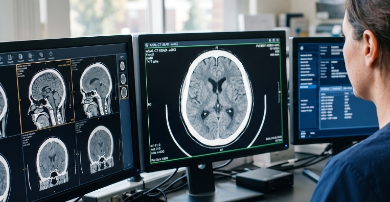
A nuclear medicine physician or appropriately trained radiologist will interpret the SPECT, CT and fused images and issue a report. The findings will be considered alongside previous scans, pathology results and other clinical information.

After the Test

Potential Benefits

SPECT-CT combines functional and anatomical information to provide more precise localisation than either component may provide alone.
  • Functional imaging: SPECT shows how a radiotracer is distributed and provides information about tissue or organ activity.
  • Precise localisation: CT helps identify the exact structure responsible for abnormal tracer uptake.
  • Improved characterisation: Combined images may help distinguish suspicious findings from some non-cancerous causes of tracer uptake.
  • Bone assessment: SPECT-CT can help clarify indeterminate areas identified during bone imaging.
  • Sentinel-node mapping: It can help locate sentinel lymph nodes before selected cancer operations.
  • Tumour-specific imaging: Different radiotracers can target particular organs, cell types or molecular characteristics.
  • Treatment planning: SPECT-CT can support selected radionuclide therapies and interventional oncology procedures.
  • Post-treatment assessment: Imaging can help confirm where a therapeutic radiopharmaceutical has accumulated.
SPECT-CT
Frequently Asked Questions
01 How is SPECT-CT different from PET-CT?
icon icon
Both combine nuclear medicine imaging with CT, but they use different radiotracers and detection systems. SPECT detects gamma rays emitted directly by a single-photon radiotracer, while PET detects paired photons produced after positron emission. PET-CT and SPECT-CT have different clinical applications, and one is not automatically a substitute for the other.
02 Can SPECT-CT detect every type of cancer?
icon icon
No. SPECT-CT is useful only when an appropriate radiotracer and clinical indication exist. Some cancers are better assessed using PET-CT, diagnostic CT, MRI, ultrasound, endoscopy or other tests.
03 Does abnormal tracer uptake always mean cancer?
icon icon
No. Infection, inflammation, fractures, arthritis, healing tissue and normal organ activity can also cause tracer uptake. Areas of reduced uptake can also have several explanations. The findings must be interpreted with the CT images, medical history and other investigations.
04 Does SPECT-CT involve radiation?
icon icon
Yes. Radiation exposure comes from both the radiotracer and the CT component. The amount varies according to the tracer, scan protocol and body area examined. The test is recommended when the expected clinical benefit outweighs the radiation risk. Tell the team if you are pregnant or breastfeeding.
05 Is contrast material always required?
icon icon
No. Many SPECT-CT examinations use a low-dose CT without intravenous contrast for anatomical localisation. Contrast may be used when a diagnostic CT is required. The decision depends on the clinical question, kidney function, allergy history and imaging protocol.
06 Why might the examination take several hours or require delayed images?
icon icon
Radiotracers take different amounts of time to reach and accumulate in their intended target. Some need only a short uptake period, while others require imaging several hours later or on a subsequent day. The nuclear medicine team will explain the schedule for the specific tracer being used.
image image
Book an Appointment
Please Enter Your Name
Please Enter Your Mobile Number
Please Enter OTP