Skin Cancer
Skin cancer develops when abnormal cells grow uncontrollably in the skin. It most commonly occurs on areas exposed to the sun, such as the face, scalp, ears, neck, arms, and hands, but it can develop anywhere, including areas that receive little or no sun exposure.
The three main types are basal cell carcinoma, squamous cell carcinoma, and melanoma. Basal and squamous cell carcinomas are more common and usually remain localised, while melanoma is less common but more likely to spread.
Many skin cancers can be treated effectively when found early. Treatment depends on the cancer type, its depth and location, whether it has spread, and the person’s overall health. Care may involve dermatologists, surgical oncologists, medical oncologists, radiation oncologists, pathologists, radiologists, and reconstructive surgeons.
Types of Skin Cancer
Skin cancers differ in where they begin, how quickly they grow, and their likelihood of spreading.
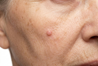
Basal cell carcinoma, or BCC, begins in the basal cells in the lower part of the epidermis. It commonly develops on sun-exposed areas, particularly the face, scalp, ears, and neck. BCC usually grows slowly and rarely spreads to distant organs. However, an untreated tumour can grow into nearby skin, cartilage, bone, or other tissues and may require more extensive treatment.
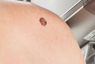
Squamous cell carcinoma, or SCC, begins in the squamous cells that form the outer layers of the skin. It may develop on sun-exposed skin, the lips, ears, scalp, genital region, or within a chronic wound or scar. Most SCCs can be treated successfully when found early. Certain tumours — particularly those that are large, deep, poorly differentiated, located in high-risk areas, or found in people with reduced immunity — have a greater likelihood of recurring or spreading.
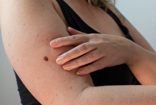
Melanoma begins in melanocytes, the cells that produce skin pigment. It may develop from an existing mole or appear as a new pigmented or non-pigmented growth. Melanoma can spread to lymph nodes and distant organs if it is not diagnosed and treated early. Tumour thickness, ulceration, lymph-node involvement, and distant spread are important in determining its stage and treatment.
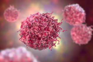
Merkel cell carcinoma is a rare, fast-growing skin cancer. It often appears as a painless, firm, red, purple, or skin-coloured lump that enlarges quickly. Because it has a relatively high risk of spreading to lymph nodes and other organs, treatment commonly involves surgery and radiation therapy. Immunotherapy may be used for advanced disease.
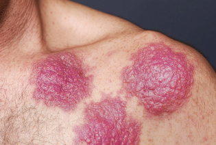
Cutaneous lymphoma begins in lymphocytes, a type of immune cell, and primarily affects the skin. It may cause persistent patches, plaques, lumps, itching, or widespread redness. There are several subtypes of cutaneous lymphoma, each with a different clinical course. Treatment may include skin-directed therapy, radiation therapy, medicines, or systemic treatment.
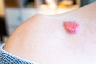
Dermatofibrosarcoma protuberans, or DFSP, is a rare tumour that develops in the deeper layers of the skin. It usually grows slowly but can extend into surrounding tissues. Surgery with careful assessment of the margins is the main treatment. Targeted therapy may be considered for selected advanced or inoperable tumours.
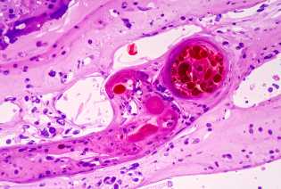
Kaposi sarcoma develops from cells lining blood and lymphatic vessels. It may cause purple, red, or brown patches or nodules on the skin and can sometimes affect internal organs. It is associated with human herpesvirus 8 and occurs more commonly in people with reduced immune function. Treatment depends on the extent of disease and the person’s immune status.
Ultraviolet radiation is the principal preventable cause of many skin cancers. However, skin cancer can affect people of every skin tone, and some tumours arise without a clear preventable cause.
Modifiable Risk Factors
Frequent or prolonged exposure to sunlight
Use of tanning beds or sunlamps
Repeated sunburns, particularly during childhood
Working or spending considerable time outdoors without adequate protection
Inadequate use of protective clothing, hats, shade, and sunscreen
Exposure to arsenic or certain occupational chemicals
Tobacco use, particularly for squamous cell carcinoma of the lip
Non-Modifiable Risk Factors
Increasing age
Fair skin, light-coloured eyes, or a tendency to burn easily
Many moles or unusual-looking moles
A personal or family history of skin cancer
Inherited conditions that increase sensitivity to ultraviolet radiation
Reduced immunity due to illness, organ transplantation, or medication
Previous radiation therapy
Chronic wounds, burns, scars, or long-standing skin inflammation
Certain viral infections, including HPV in selected squamous cell carcinomas
Having one or more risk factors does not mean that skin cancer will develop. People at higher risk may benefit from regular skin examinations and an individualised surveillance plan.
Signs and Symptoms
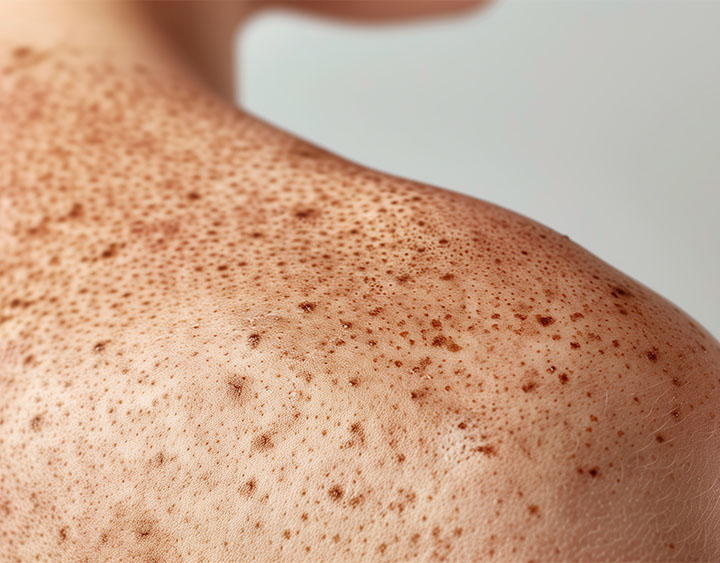
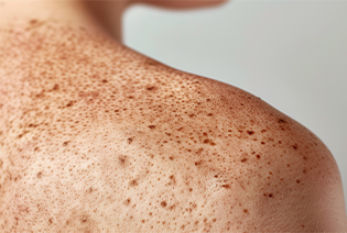
Skin cancer may appear as a new growth or as a change in an existing mole, patch, or sore. It does not always cause pain.
The ABCDE guide may help identify a suspicious mole:
These changes can also be caused by non-cancerous conditions. However, any new, changing, bleeding, or non-healing lesion should be examined by a doctor.
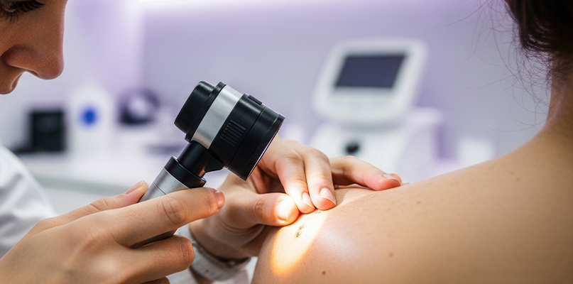
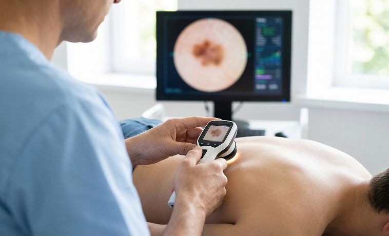
Diagnosis begins with an examination of the suspicious area and the surrounding skin. The doctor may also examine the rest of the skin and nearby lymph nodes, ask how the lesion has changed, and review personal and family history. Dermoscopy can help assess structures and colours beneath the skin surface. A biopsy is required to confirm most skin cancers. Imaging and laboratory tests are generally reserved for melanoma, high-risk tumours, or cancers that may have spread.
Ultrasound uses sound waves to examine lymph nodes near the tumour. It may be recommended when a lymph node feels enlarged or when the cancer has features associated with a higher risk of spread. Ultrasound may also guide a fine-needle aspiration or core biopsy of a suspicious lymph node.
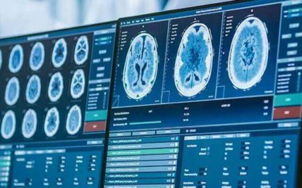
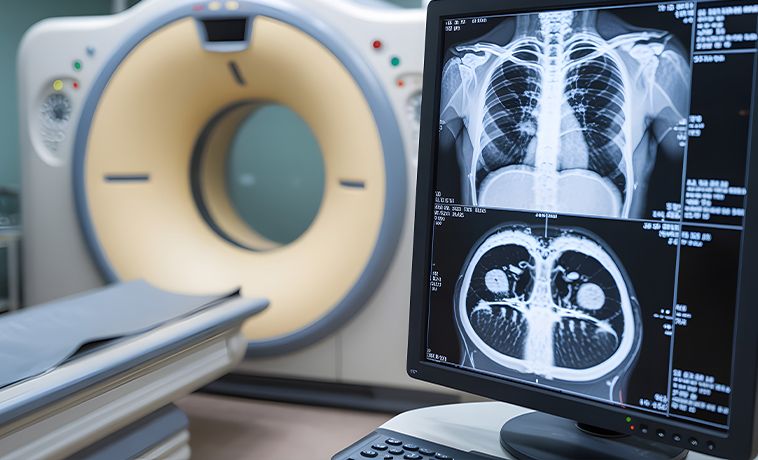
A CT scan uses X-rays to produce detailed images of the body. It may be used to assess lymph nodes, lungs, liver, or other organs when advanced skin cancer is suspected. CT is not routinely required for small, low-risk basal or squamous cell carcinomas.
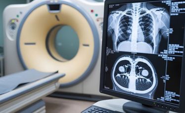
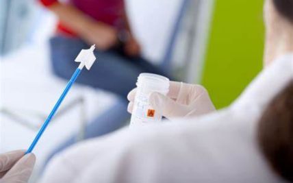
A biopsy is the definitive way to diagnose skin cancer. A sample of the suspicious tissue — or, for small lesions, the entire growth — is removed and examined under a microscope to confirm the presence of cancer and identify its exact type. For many small, early skin cancers, this initial biopsy can sometimes remove the growth entirely.
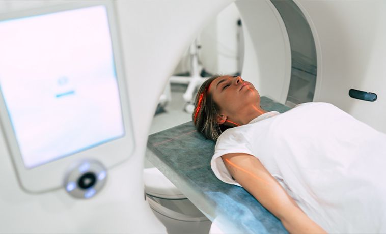
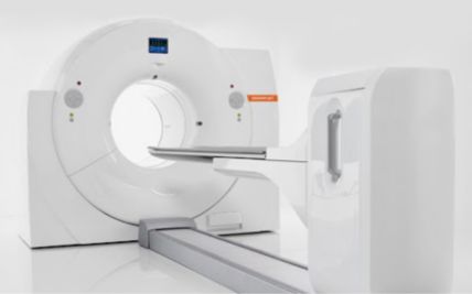
PET-CT combines metabolic and anatomical imaging to identify areas of increased activity in the body. It may be used to assess selected advanced melanomas, Merkel cell carcinomas, or high-risk squamous cell carcinomas. It is not routinely used to evaluate every skin cancer.
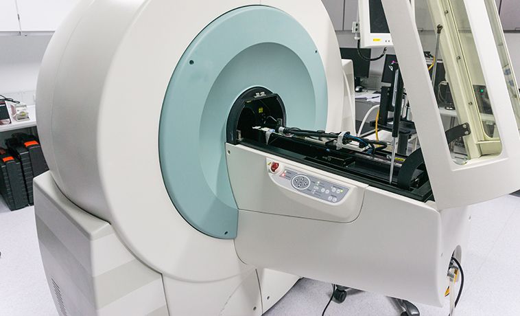
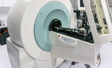
A small amount of radioactive tracer, sometimes combined with a dye, is used to locate the first lymph node or nodes to which the cancer is most likely to spread. These nodes can then be removed through a sentinel lymph-node biopsy. This procedure is primarily used for staging selected melanomas and Merkel cell carcinomas rather than for diagnosing the original skin lesion.
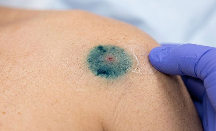
A blade is used to remove the surface layers of a raised or superficial lesion. It may be suitable for suspected basal or squamous cell carcinoma. When melanoma is suspected, the biopsy must obtain enough depth to allow accurate measurement of the tumour. A superficial shave that cuts across the tumour may make staging more difficult.
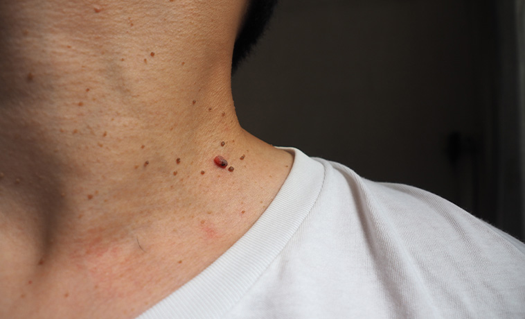
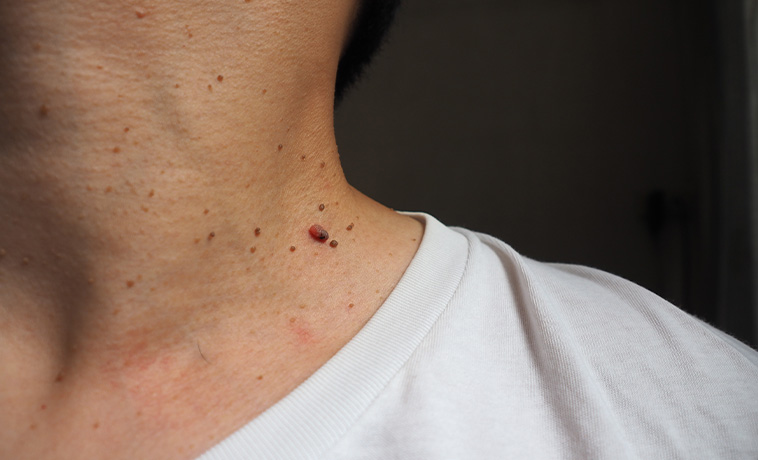
A circular instrument removes a small column of tissue containing the epidermis, dermis, and part of the tissue beneath the skin. It may be used to sample a larger lesion or a condition where the diagnosis is uncertain. Because only part of the lesion may be sampled, the biopsy site should be selected carefully.
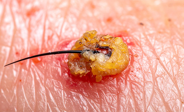
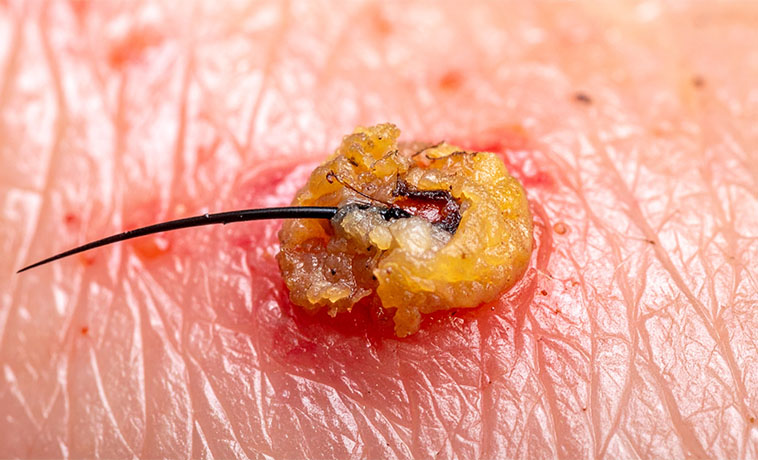
The entire suspicious lesion is removed, usually with a narrow margin of surrounding skin. This is generally preferred when melanoma is suspected and complete removal can be performed safely. The specimen allows the pathologist to assess the cancer type, thickness, ulceration, and other features needed for staging.
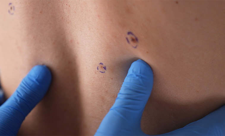
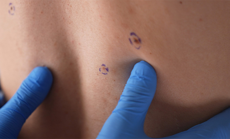
A representative portion of a large lesion is removed when taking out the entire growth during the initial procedure would be difficult or could affect function or appearance. Definitive treatment is planned after the biopsy confirms the diagnosis.
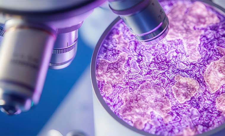
A suspicious lymph node may be sampled using fine-needle aspiration, core needle biopsy, or surgical removal. Sentinel lymph-node biopsy may be recommended for selected clinically node-negative melanomas and Merkel cell carcinomas. The findings help determine whether the cancer has begun to spread beyond the skin.
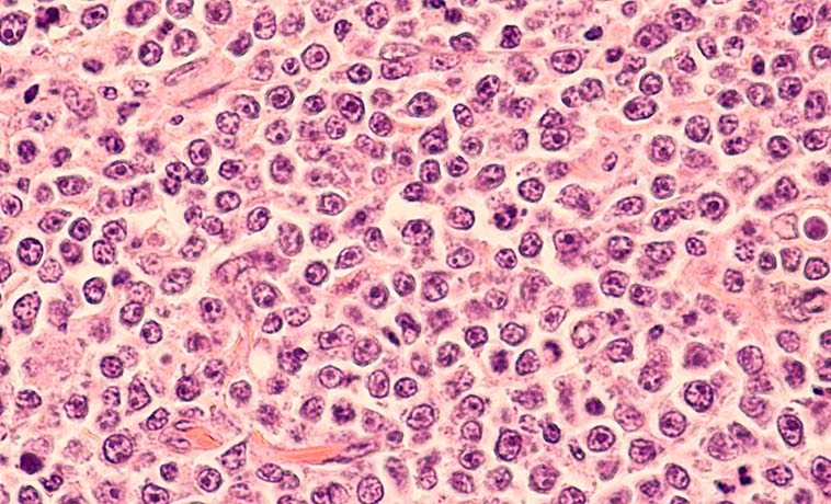
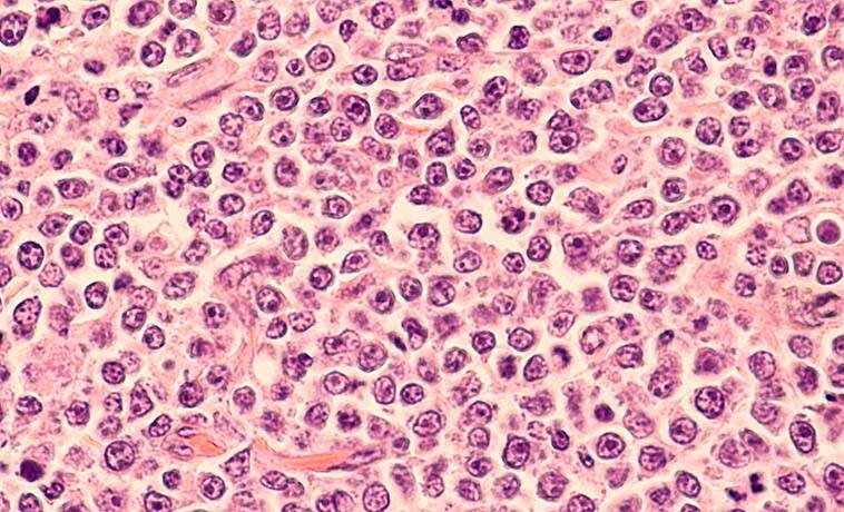
A specialist pathologist examines the biopsy under a microscope to identify the cancer type and features that influence treatment.
For melanoma, the pathology report may include:
Breslow thickness, or how deeply the tumour has grown
Presence or absence of ulceration
Surgical margin status
Mitotic activity and other microscopic features
Involvement of lymphatic or blood vessels, where present
Immunohistochemistry may help distinguish melanoma, carcinoma, lymphoma, sarcoma, and other tumours that can appear similar under the microscope.
Molecular testing may be performed for selected advanced melanomas to identify changes such as BRAF, KIT, or other treatment-relevant alterations. Testing for inherited cancer susceptibility is considered when the medical or family history suggests a hereditary condition.
Blood tests are not used to diagnose an early skin cancer. They may be performed in advanced disease to assess general health, organ function, and—in selected melanoma cases—lactate dehydrogenase levels.
The exact staging system differs between melanoma, basal cell carcinoma, squamous cell carcinoma, Merkel cell carcinoma, and rarer skin cancers. Many small basal cell and squamous cell carcinomas do not require formal stage grouping. The following provides a broad Stage 0–IV framework; the final stage is assigned using the disease-specific system.
Stage 0: The cancer is confined to the epidermis, the outermost layer of the skin, and has not invaded deeper tissue. This is also called carcinoma in situ or melanoma in situ.
Stage I: The cancer is localised to the skin and has no evidence of spread to nearby lymph nodes or distant organs. In melanoma, thickness and ulceration are used to distinguish stage groups.
Stage II: The cancer remains localised but has higher-risk features. Depending on the type, these may include greater thickness, ulceration, deep invasion, involvement of a nerve, or extension into nearby tissues. No regional lymph-node or distant spread is identified.
Stage III: The cancer has spread to regional lymph nodes, nearby lymphatic channels, or tissues close to the original tumour. In melanoma, this may include satellite or in-transit deposits between the primary tumour and nearby lymph nodes.
Stage IV: The cancer has spread to distant lymph nodes, skin, soft tissue, or organs such as the lungs, liver, brain, or bones.
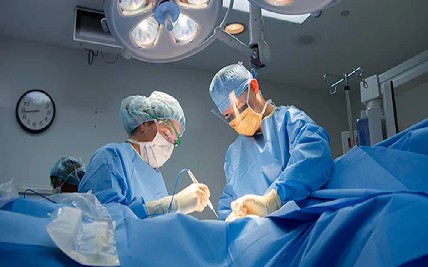
The tumour is removed with a measured margin of apparently healthy skin. The width of the margin depends on the cancer type, tumour thickness, anatomical location, and other risk features. The removed tissue is examined to determine whether the surgical margins are free of cancer.
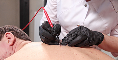
Mohs micrographic surgery removes the tumour one layer at a time. Each layer is examined under a microscope during the procedure, and further tissue is removed only where cancer cells remain. This technique can achieve detailed margin assessment while preserving healthy tissue. It is particularly useful for selected basal and squamous cell carcinomas on the face, ears, eyelids, nose, hands, or other functionally sensitive areas.
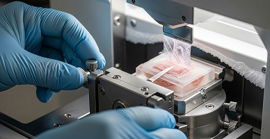
The tumour is scraped away and the treatment area is cauterised using an electric current. The process may be repeated during the same procedure. It may be used for selected small, superficial, low-risk basal or squamous cell carcinomas. It is not suitable for every location or high-risk tumour.
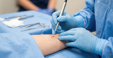
A sentinel lymph-node biopsy may be performed to determine whether melanoma or Merkel cell carcinoma has reached the regional lymph nodes. If lymph-node disease is confirmed, further management may involve observation with ultrasound, surgery, radiation therapy, systemic treatment, or a combination based on the cancer type and clinical situation.
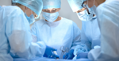
Large or deeply invasive tumours may require removal of involved skin, muscle, cartilage, bone, nerves, or nearby structures. Operations involving the face, scalp, limbs, or genital region may be planned jointly with reconstructive specialists. The aim is to remove the cancer completely while preserving function and appearance wherever possible.
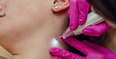
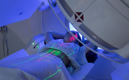
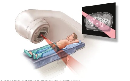
External beam radiation directs treatment at the cancer from outside the body. The dose, treatment area, and number of sessions depend on the cancer type, depth, location, and purpose of treatment. Superficial X-rays or electron beams may be used for cancers close to the skin surface.
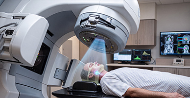
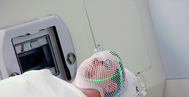
Intensity-modulated radiation therapy shapes and adjusts the radiation dose around the tumour. Image-guided radiation therapy uses imaging during treatment to confirm positioning. These techniques may be useful for irregularly shaped or deeper tumours near sensitive structures.
Proton therapy uses proton beams that release most of their radiation within the planned treatment area, with little dose continuing beyond it. This can reduce radiation exposure to some tissues behind the tumour.
It may be considered for selected complex skin cancers near critical structures, such as certain cancers involving the face, skull base, eye region, or previously irradiated tissues. Proton therapy is not routinely required for skin cancer; suitability is determined by comparing an individual proton plan with other radiation techniques.
Medicines such as imiquimod or 5-fluorouracil may be applied directly to the skin for selected superficial basal cell carcinomas or squamous cell carcinoma in situ. Topical treatment is not appropriate for invasive melanoma or cancers that extend deeply into the skin.

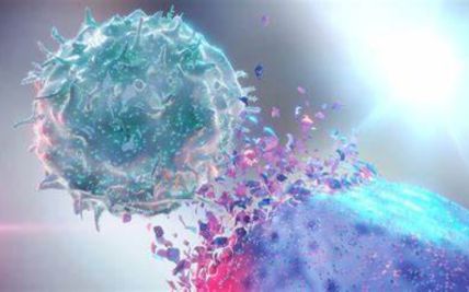
Immunotherapy helps the immune system recognise and attack cancer cells. It may be used before or after surgery, or for unresectable or metastatic disease, depending on the diagnosis. Checkpoint inhibitors have an established role in melanoma, advanced cutaneous squamous cell carcinoma, and Merkel cell carcinoma. Treatment is selected after considering potential benefits and immune-related side effects.
Targeted medicines act on specific pathways involved in cancer growth. BRAF- and MEK-targeted therapy may be used for melanomas with an eligible BRAF alteration. Other targeted treatments include Hedgehog-pathway inhibitors for selected advanced basal cell carcinomas and mutation-directed treatment for certain rare tumours.
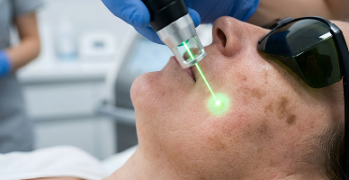
Medication may be injected directly into an accessible tumour. It may be considered for selected melanoma deposits in or beneath the skin and for certain other localised cancers. The choice depends on the number, size, and location of the lesions and whether disease is present elsewhere.

Traditional chemotherapy has a more limited role because immunotherapy and targeted treatment are often more effective for advanced melanoma and certain other skin cancers. It may still be considered for selected advanced cancers, cutaneous lymphomas, Kaposi sarcoma, or situations in which other treatments are unsuitable.
After a small tumour is removed, the wound edges may be brought together directly. This is generally used when closure can be achieved without causing excessive tension or distorting a nearby structure.
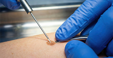
A thin or full-thickness layer of skin is taken from another part of the body and placed over the surgical wound. Skin grafts can cover larger defects when direct closure is not possible.
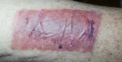
Nearby skin and underlying tissue are moved into the surgical defect while maintaining their blood supply. Local flaps can provide a close match in colour, thickness, and texture, particularly for facial reconstruction.
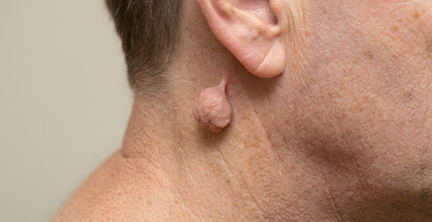
Larger or deeper defects may require tissue transferred from another part of the body. A regional flap remains attached to its original blood supply, while a free flap is reconnected to blood vessels at the reconstruction site using microsurgery.
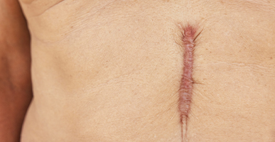
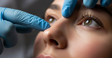
Removal of cancers involving the eyelid, nose, ear, lip, scalp, or jaw may require specialised reconstruction. Treatment is planned to restore coverage, facial contour, and functions such as eyelid closure, breathing, speaking, or eating.
Prognosis for Skin Cance
Most basal cell carcinomas and many squamous cell carcinomas have a favourable outlook when treated early. Melanoma, Merkel cell carcinoma, and other aggressive skin cancers require closer assessment because their outcomes depend strongly on the extent of disease.
Factors that influence prognosis include:
Skin cancer type and subtype
Tumour thickness, depth, and size
Ulceration and other pathological features
Location of the tumour
Whether surgical margins are free of cancer
Lymph-node or distant-organ involvement
Molecular characteristics
Immune status and overall health
Response to treatment
Development of recurrent disease
Regular follow-up is important because some skin cancers can return and people who have had one skin cancer may be at increased risk of developing another.


Why Choose ACC for Skin Cancer Treatment
Multidisciplinary care involving dermatology, surgical oncology, medical oncology, radiation oncology, pathology, and reconstruction
Dermoscopy, specialist pathology, advanced imaging, and molecular testing where clinically appropriate
Surgical options including wide excision, Mohs surgery, lymph-node procedures, and complex tumour removal
Immunotherapy and targeted treatment for eligible advanced skin cancers
Radiation techniques selected according to tumour depth, location, and nearby structures
Reconstructive planning for cancers affecting functionally or cosmetically sensitive areas
Tumour Board review for complex or advanced disease
Individualised follow-up to monitor for recurrence and new skin cancers
Health Blogs