




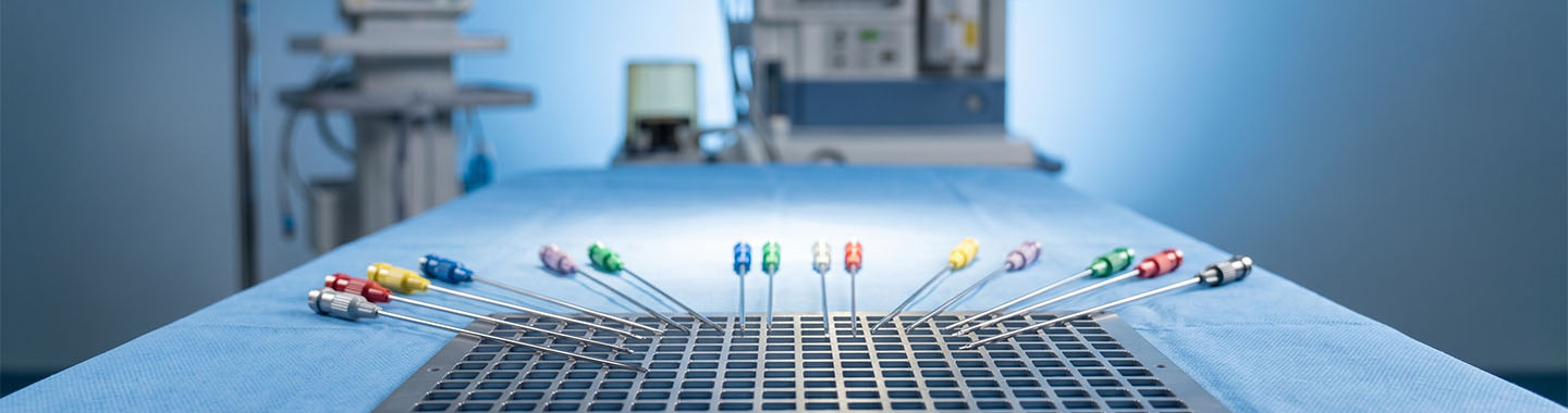
Brachytherapy is a form of internal radiation therapy in which a radioactive source is placed inside or close to a tumour. Because the radiation begins near the treatment area, a concentrated dose can be delivered to the tumour while limiting exposure to more distant healthy tissues.
The radioactive source may be placed within a body cavity, directly into tissue, on the surface of the body or, for certain eye tumours, against the eye. Brachytherapy may be temporary, with the source removed after treatment, or permanent, with small radioactive seeds left in place as their radiation gradually decreases.
Brachytherapy may be used alone or combined with external beam radiation therapy, surgery, chemotherapy or other cancer treatments. The type of implant, radiation dose, number of sessions and need for anesthesia depend on the cancer and treatment area. The National Cancer Institute describes brachytherapy as a local treatment in which a radiation source is placed in or near the tumour.
- Cervical cancer
- Endometrial or uterine cancer
- Vaginal cancer
- Prostate cancer
- Selected breast cancers
- Selected head and neck cancers
- Selected skin cancers
- Eye cancers, including certain ocular melanomas
- Selected oesophageal or airway tumours
- Selected rectal and other pelvic cancers
- Residual or recurrent tumours in an area that can be reached with an applicator, catheter or needle
- Patients who require an additional focused radiation dose after external beam radiation therapy
- Selected patients requiring local treatment while limiting radiation exposure to nearby organs

How Brachytherapy Is Performed
Before Treatment
The radiation oncologist reviews the diagnosis, biopsy findings, imaging, previous treatments and general health.
A physical examination and blood tests may be performed.
CT, MRI, ultrasound, PET-CT or other imaging may be used to assess the tumour and plan treatment.
The treatment team decides whether intracavitary, interstitial, surface or another form of brachytherapy is appropriate.
The type of brachytherapy may be high-dose-rate, low-dose-rate, pulsed-dose-rate or a permanent seed implant.
The patient may require bowel preparation, fasting, bladder preparation or changes to certain medicines.
An anesthesia assessment may be arranged when sedation, spinal anesthesia or general anesthesia is required.
The radiation oncologist, medical physicist and treatment-planning team calculate the dose and determine where the source should be positioned.
The team explains whether the applicator will be removed after each session or kept in place between treatments.
Radiation-safety instructions are discussed if a source will remain in the body temporarily or permanently.

During Treatment
The patient is positioned according to the body area being treated.
Sedation, local anesthesia, spinal anesthesia or general anesthesia may be given, depending on the procedure.
A catheter, needle, tube, plaque or specialised applicator is positioned inside or near the tumour.
Ultrasound, X-ray, CT or MRI may be used to confirm the applicator’s position.
For temporary brachytherapy, a computer-controlled machine transfers the radioactive source into the applicator after it has been positioned.
During high-dose-rate brachytherapy, the source remains in planned positions for a few minutes before returning to the shielded machine.
The patient may be alone in the treatment room while the source is active, but the team can see, hear and communicate with them.
The radioactive source is removed after each temporary treatment session.
In low-dose-rate or pulsed-dose-rate treatment, the source may remain in place for several hours or days, and hospital admission may be required.
For permanent brachytherapy, small radioactive seeds are placed in or near the tumour and remain in the body while their radiation decreases over time.

After Treatment
The applicator, needles or catheters are removed when temporary treatment is complete.
The patient is monitored while recovering from sedation or anesthesia.
Mild pain, tenderness, swelling, spotting or discharge may occur depending on the treatment area.
Pain relief, antibiotics or other medicines may be prescribed when required.
Some patients can return home on the same day, while others may need a hospital stay.
Temporary activity restrictions may be advised while the treated area heals.
No radiation remains in the body after a temporary source has been removed.
Patients with permanent implants may need to temporarily limit close contact with pregnant women or young children.
Individual radiation-safety instructions should be followed until the care team confirms that restrictions are no longer required.
Follow-up examinations, imaging or blood tests are arranged to monitor recovery and assess the treatment response.

Potential Benefits of Brachytherapy
- Delivery of radiation directly inside or close to the tumour
- A concentrated dose to a defined treatment area
- Reduced radiation exposure to more distant healthy tissues
- Protection of selected nearby organs through carefully planned source placement
- Treatment tailored to the shape and location of the tumour
- Use as the main treatment for selected cancers
- Delivery of an additional focused dose following external beam radiation therapy
- Potential preservation of the treated organ and its function in selected patients
- Fewer treatment visits than some external beam radiation schedules
- Outpatient treatment for many high-dose-rate procedures
- An additional local treatment option for certain residual or recurrent tumours

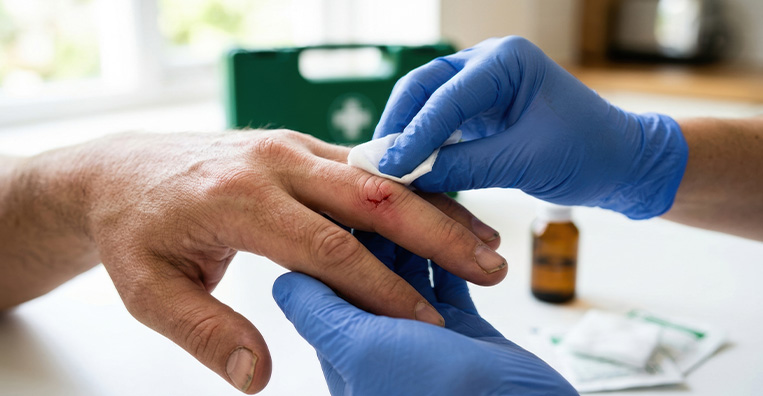
Side Effects of Brachytherapy
- Pain, tenderness, bruising or swelling where an applicator, needle or catheter was placed
- Minor bleeding or discharge
- Infection at the insertion or treatment site
- Tiredness or fatigue
- Temporary skin irritation in or near the treated area
- Urinary frequency, urgency, discomfort or difficulty passing urine following prostate or pelvic treatment
- Diarrhoea, constipation, abdominal cramps or rectal discomfort following pelvic treatment
- Vaginal irritation, dryness, discharge, narrowing or changes in sexual comfort following gynecological treatment
- Breast tenderness, swelling or skin changes following breast treatment
- Mouth soreness, swallowing difficulty or tissue irritation following head and neck treatment
- Temporary changes in sexual function or fertility
- Side effects related to sedation or anesthesia
- Delayed tissue changes, scarring, narrowing or ulceration near the treated area
- Rare injury to nearby organs, nerves or blood vessels
- A small long-term risk of developing another cancer related to radiation exposure



