icon
icon
Overview

Bronchoscopy is a procedure used to examine the trachea and bronchi—the main breathing passages leading into the lungs. It can help investigate abnormal findings, obtain tissue or cell samples and, in selected cases, treat problems affecting the airways.

Most examinations use a thin, flexible bronchoscope passed through the nose or mouth. A rigid bronchoscope may be used under general anesthesia when a larger airway needs treatment, a larger sample is required or significant bleeding or blockage must be managed.
Bronchoscopy
How the Technology Works
Bronchoscopy combines a camera-equipped scope with specialised instruments that allow the clinical team to examine the airways and collect samples.
work-icon
Bronchoscope Insertion
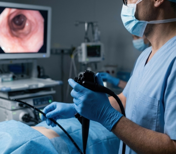
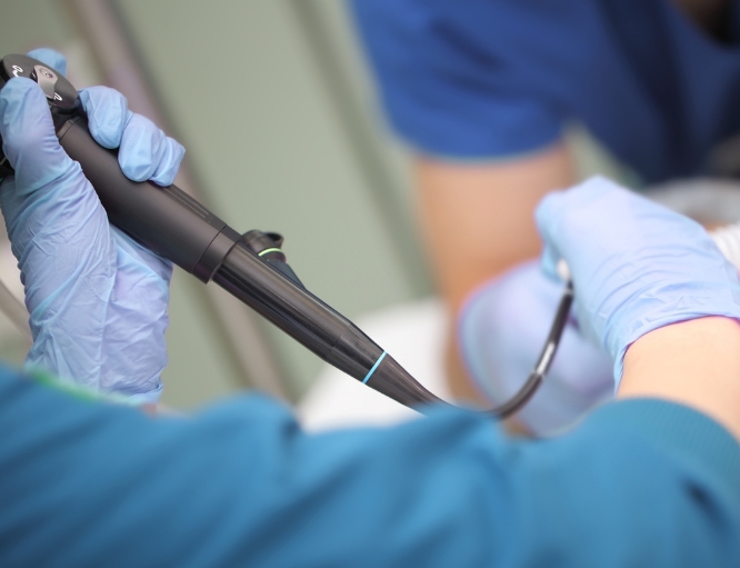
A flexible bronchoscope is passed through the nose or mouth, beyond the vocal cords and into the trachea and bronchi. Local anesthetic helps numb the nose and throat, and sedation is usually provided to improve comfort.
work-icon
Direct Airway Imaging
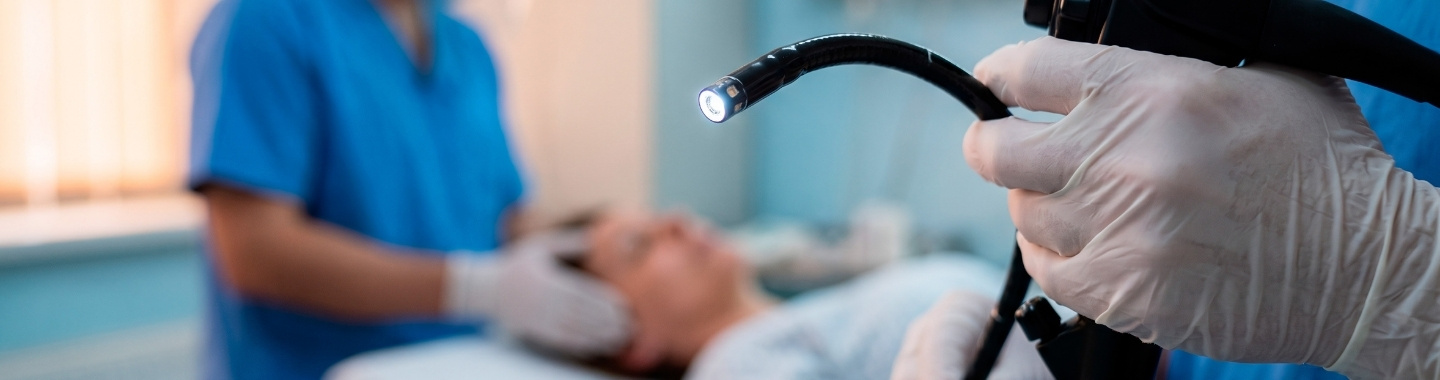
A light and camera at the end of the bronchoscope transmit images to a monitor. This allows the doctor to examine the airway lining and identify narrowing, blockage, bleeding, inflammation or abnormal tissue.
work-icon
Sampling and Treatment Instruments
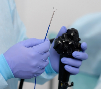
A working channel within the bronchoscope allows saline, brushes, needles, forceps and other small instruments to be introduced. These may be used to collect washings, cells or tissue, or to perform selected treatments such as controlling bleeding, opening a narrowed airway or placing an airway stent.

Role in Cancer Care

Investigating Abnormal Findings
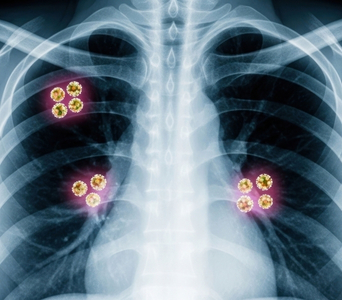
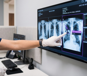
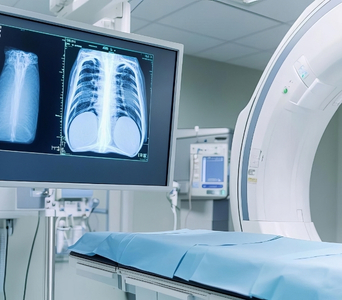
Bronchoscopy may be recommended following an abnormal chest X-ray, CT or PET-CT, or when symptoms suggest an airway problem. These symptoms can include coughing up blood, persistent cough, unexplained breathlessness, recurrent infection or collapse of part of a lung. It can help determine whether an abnormality is caused by cancer, infection, inflammation or another condition.
Investigating Abnormal Findings
Directly Examining a Tumour
The procedure can show a tumour growing within an airway or causing narrowing, obstruction or bleeding. It may also reveal whether an abnormality is pressing on an airway from outside, although imaging is usually needed to define its full extent. Tumours located deeper in the lung may not be directly visible through a standard bronchoscope.
Directly Examining a Tumour
Obtaining Samples for Diagnosis
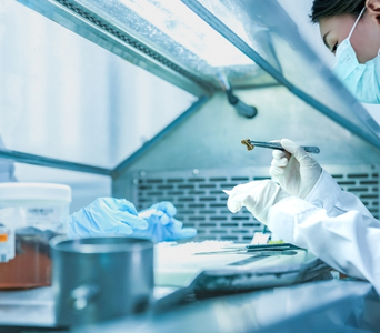
Brushings, washings and biopsies can collect cells or tissue for laboratory examination. Pathology can establish whether cancer is present and identify its type; suitable samples may also undergo biomarker or molecular testing to help guide treatment. If the first sample is insufficient or the abnormality cannot be reached, another biopsy method may be required.
Obtaining Samples for Diagnosis
Assessing the Extent of Disease
Bronchoscopy can assess how far a tumour extends along the visible airways and whether it affects important airway openings. This information may contribute to surgical, radiotherapy and airway-management planning. Bronchoscopy alone does not provide complete cancer staging and is usually considered alongside CT, PET-CT, MRI or other investigations.
Assessing the Extent of Disease
Guiding Samples from Less Accessible Areas
Fluoroscopy, navigational bronchoscopy, robotic systems or a small radial ultrasound probe may be used to help reach selected abnormalities deeper in the lung. Availability and suitability depend on the size and location of the abnormality and the expertise of the centre. Some peripheral lung abnormalities are better sampled using CT-guided needle biopsy or surgery.
Guiding Samples from Less Accessible Areas
Providing Selected Airway Treatments
Therapeutic bronchoscopy may be used to remove or reduce tumour tissue blocking an airway, control bleeding or place a stent to keep an airway open. Heat, laser, freezing or other techniques may be used in specialist centres. These procedures often relieve symptoms or restore airflow but may not treat cancer elsewhere in the body.
Providing Selected Airway Treatments

What to Expect

Before the Test

You will receive instructions about when to stop eating and drinking; the required fasting period depends on the planned sedation or anesthesia. Tell the clinical team about:

  • All medicines and supplements you take, particularly anticoagulants and antiplatelet medicines

  • Diabetes, bleeding disorders, heart or lung conditions and any oxygen requirements

  • Allergies or previous reactions to sedatives, anesthetics or medicines

  • Pregnancy or the possibility of pregnancy

  • Dentures, removable dental work or loose teeth

Do not stop prescribed medicines unless your clinical team instructs you to do so. Blood tests, breathing assessments or recent imaging may be reviewed before the procedure. If you will receive sedation or general anesthesia, arrange for a responsible adult to take you home and follow the instructions about supervision afterwards.

Before the Test

During the Test

Most diagnostic bronchoscopies are performed using a flexible bronchoscope. Local anesthetic is applied to the nose, mouth or throat, and a sedative may be given through a vein; oxygen levels, heart rate and blood pressure are monitored throughout.

The bronchoscope does not usually prevent breathing, although coughing, pressure or a temporary sensation of breathlessness may occur. The doctor may introduce saline to collect cells, brush an abnormal area or take one or more biopsies. The procedure commonly takes approximately 20–60 minutes, although the duration depends on the examinations and treatments required.

Rigid bronchoscopy is performed under general anesthesia and is generally reserved for selected biopsies or treatments involving larger airway tumours, obstruction or bleeding.

During the Test

After the Test

You will be monitored until the sedation has worn off. Do not eat or drink until the throat numbness has resolved and the clinical team confirms that swallowing is safe. A temporary sore throat, hoarse voice, cough or small amount of blood-streaked phlegm may occur, particularly after a biopsy.

If you received sedation, do not drive, operate machinery, drink alcohol, make important decisions or sign legal documents for the period specified by your clinical team. A chest X-ray may be required after certain lung biopsies to check for an air leak around the lung.

The doctor may discuss what was seen shortly after the procedure. Laboratory results from washings, brushings or biopsies take longer and will be reviewed with you when available.

Seek urgent medical advice if you experience:

  • Increasing or severe breathlessness

  • Chest pain

  • Persistent fever or feeling increasingly unwell

  • Heavy or persistent bleeding, or more than a small amount of blood when coughing

  • Fainting, confusion or any rapidly worsening symptoms

After the Test

Potential Benefits

Bronchoscopy may provide several benefits when investigating or managing suspected or confirmed cancer:
  • Direct Visualisation: It allows the doctor to examine the trachea and bronchi directly.
  • Targeted Sampling: It enables the collection of cells, fluid and tissue from selected areas.
  • Diagnostic Confirmation: Samples collected during bronchoscopy can help confirm the cancer type through laboratory analysis.
  • Airway Assessment: It can identify tumour-related airway narrowing, blockage or bleeding.
  • Treatment Planning: Its findings can support decisions about surgery, radiotherapy and other treatments.
  • Symptom Relief: Selected airway obstructions or bleeding may be treated during the same procedure.
  • Minimally Invasive: It accesses the airways through the nose or mouth without requiring a surgical incision.
  • Guided Access: It can be combined with specialised guidance techniques to investigate selected abnormalities deeper in the lungs.
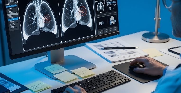
Bronchoscopy
Frequently Asked Questions
01 Will bronchoscopy be painful?
icon icon
Local anesthetic is used to numb the nose and throat, and most people receive sedation to help them relax. You may notice pressure, coughing or an urge to gag, but the team can provide additional local anesthetic, adjust the sedation or pause when necessary. Rigid bronchoscopy is performed under general anesthesia.
02 Can I breathe while the bronchoscope is in place?
icon icon
Yes. A flexible bronchoscope is narrow and does not normally fill the entire airway, so air can pass around it. Your oxygen level and other vital signs are monitored continuously, and additional oxygen can be given if required.
03 Can bronchoscopy confirm that I have cancer?
icon icon
Bronchoscopy can identify suspicious airway changes and obtain samples, but the appearance alone usually cannot confirm cancer. Cells or tissue must be examined by a pathologist to establish a diagnosis. Occasionally, a sample does not contain enough abnormal tissue and another procedure is needed.
04 Can bronchoscopy detect every lung cancer?
icon icon
No. Standard bronchoscopy is most effective for abnormalities within or close to the larger airways. Small or peripheral tumours may be beyond its view or reach and may require navigational bronchoscopy, radial ultrasound guidance, CT-guided biopsy, surgery or another investigation.
05 How is bronchoscopy different from EBUS?
icon icon
Standard bronchoscopy mainly examines the inner surface of the airways and collects samples from visible or reachable abnormalities. Endobronchial ultrasound, or EBUS, adds an ultrasound probe that can show structures beyond the airway wall and guide needle samples from lymph nodes or nearby masses. EBUS is particularly useful for diagnosis and lymph-node staging and is covered separately.
06 What complications are possible?
icon icon
Most bronchoscopies are completed without a serious complication. Possible problems include bleeding, infection, reduced oxygen levels, narrowing of the airways, an abnormal heart rhythm or a reaction to sedation; an air leak around the lung, called a pneumothorax, can occur, particularly after a transbronchial biopsy. Your individual risk depends on your health, the type of sampling or treatment planned and the location of the abnormality, and the clinical team will discuss this before obtaining consent.
image image
Book an Appointment
Please Enter Your Name
Please Enter Your Mobile Number
Please Enter OTP