




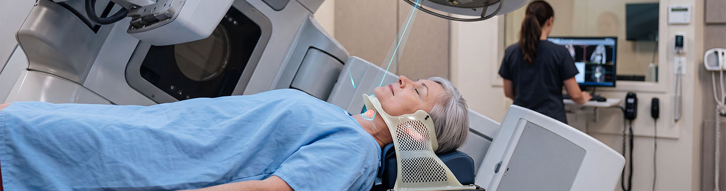
Image-Guided Radiation Therapy (IGRT) uses medical imaging before and, in some cases, during radiation treatment to verify the position of the tumour and surrounding organs. It is not a separate type of radiation. Instead, it is an approach used to improve the accuracy of treatments such as IMRT, proton therapy, stereotactic radiosurgery and SBRT.
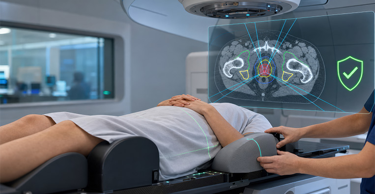
The images taken in the treatment room are compared with the reference images used to create the treatment plan. If the patient or target is not in the planned position, the treatment team can adjust the treatment table, patient position or radiation delivery before proceeding.
IGRT can be particularly valuable for tumours that move with breathing or changes in bladder and bowel filling. It is also useful when the tumour lies close to a sensitive organ and small differences in position could affect treatment accuracy. Imaging may include X-rays, cone-beam CT, standard CT, MRI or ultrasound, depending on the treatment system and body area. RadiologyInfo describes IGRT as medical imaging used to support precise radiation delivery.
- Brain and spinal tumours
- Head and neck cancers located close to sensitive structures
- Lung tumours that move with breathing
- Liver, pancreatic and other abdominal tumours affected by respiratory movement
- Prostate cancer, where the position of the prostate may change with bladder or bowel filling
- Bladder, cervical, uterine and other pelvic cancers
- Tumours located close to the spinal cord, bowel, kidneys, heart, lungs or other critical organs
- Tumours being treated with IMRT, SBRT, stereotactic radiosurgery or proton therapy
- Patients receiving a high radiation dose to a small or precisely defined treatment area
- Tumours requiring small treatment margins around the visible target
- Previously irradiated tumours where another course of radiation is considered safe
- Treatment areas that may change because of weight loss, tumour shrinkage or internal organ movement

How IGRT Is Performed
Before Treatment
The radiation oncologist reviews the diagnosis, imaging, previous treatments and overall health.
A planning or simulation CT scan is performed with the patient in the intended treatment position.
MRI, PET-CT or other imaging may be combined with the planning scan to define the tumour and nearby organs.
A customised mask, body mould, cushion or other immobilisation device may be prepared to help reproduce the same position during each session.
Small reference marks may be placed on the skin or immobilisation device.
Small markers called fiducials may be placed in or near certain tumours to make their position easier to identify. These are not required for every patient.
The movement of tumours in the chest or abdomen may be assessed using four-dimensional CT or other motion-management techniques.
Patients may be given instructions about bladder filling, bowel preparation, fasting or breathing, depending on the treatment area.
Reference images are selected for comparison with the images obtained during treatment.

During Treatment
The patient is positioned on the treatment table using the customised supports and reference marks.
X-ray, CT, cone-beam CT, MRI, ultrasound or another imaging method is used to check the treatment area.
The treatment-room images are compared with the reference images from the planning scan.
The treatment team may adjust the patient’s position or move the treatment table if the target is not correctly aligned.
Additional images may be taken to confirm the corrected position.
For selected tumours, imaging or tracking may continue during radiation delivery to monitor movement.
The patient may be asked to hold their breath or follow specific breathing instructions.
Radiation is delivered only after the required positioning and safety checks have been completed.
Imaging and radiation delivery are painless, although the patient must remain still.
The imaging process may add several minutes to the treatment appointment.

After Treatment
Most patients can return home immediately after each session.
The patient is not radioactive and can safely be around other people.
No special recovery is required from the imaging itself unless fiducial markers were placed as a separate invasive procedure.
Side effects, if they occur, are mainly related to the radiation treatment rather than the image guidance.
The care team reviews the patient regularly and manages any treatment-related symptoms.
If imaging shows a significant change in the tumour, body shape or surrounding organs, the treatment plan may need to be reviewed or repeated.
Follow-up appointments and imaging are arranged according to the cancer and overall treatment plan.

Potential Benefits of IGRT
- Verification of the tumour’s position before radiation is delivered
- Improved consistency of patient positioning across treatment sessions
- Detection of changes in the position of the tumour or surrounding organs
- More accurate treatment of tumours that move with breathing or internal organ filling
- The ability to adjust positioning before each treatment
- Greater confidence when using small treatment margins
- Reduced likelihood of radiation missing part of the intended target
- Reduced radiation exposure to selected nearby healthy tissues
- Support for high-precision treatments such as IMRT, SBRT, stereotactic radiosurgery and proton therapy
- Identification of anatomical changes that may require the treatment plan to be reviewed

Side Effects of IGRT
- Tiredness or fatigue
- Redness, dryness, tenderness or darkening of the skin in the treated area
- Temporary hair loss limited to the treatment area
- Headache or nausea during treatment to the brain
- Mouth soreness, dry mouth or difficulty swallowing during treatment to the head and neck
- Cough, breathlessness or chest discomfort during treatment to the chest
- Nausea, vomiting or abdominal discomfort during treatment to the abdomen
- Diarrhoea or changes in bowel habits during treatment to the abdomen or pelvis
- Increased urinary frequency, urgency or discomfort during treatment to the pelvis
- Temporary discomfort, bruising, bleeding or infection following fiducial-marker placement
- Delayed effects involving tissues or organs close to the treatment area
- A small long-term risk of developing another cancer related to overall radiation exposure



