icon
icon
Overview

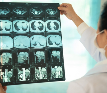
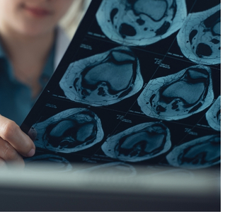
Computed Tomography, commonly called a CT or CAT scan, is an imaging test that uses X-rays and computer processing to create detailed cross-sectional images of the body. Unlike a standard X-ray, which produces a two-dimensional image, CT shows structures in multiple slices and planes. These images can also be reconstructed to provide three-dimensional views when required.

CT can examine almost any part of the body, including the brain, head and neck, chest, abdomen, pelvis, bones and soft tissues. In cancer care, it may be used to identify an abnormality, assess its size and location, determine whether it has spread, plan treatment and monitor response.

A CT finding alone does not always confirm cancer. Depending on the results, additional imaging, endoscopy, laboratory testing or a biopsy may be required to establish the diagnosis.
How the Technology CT Scan
How the Technology Works
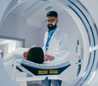
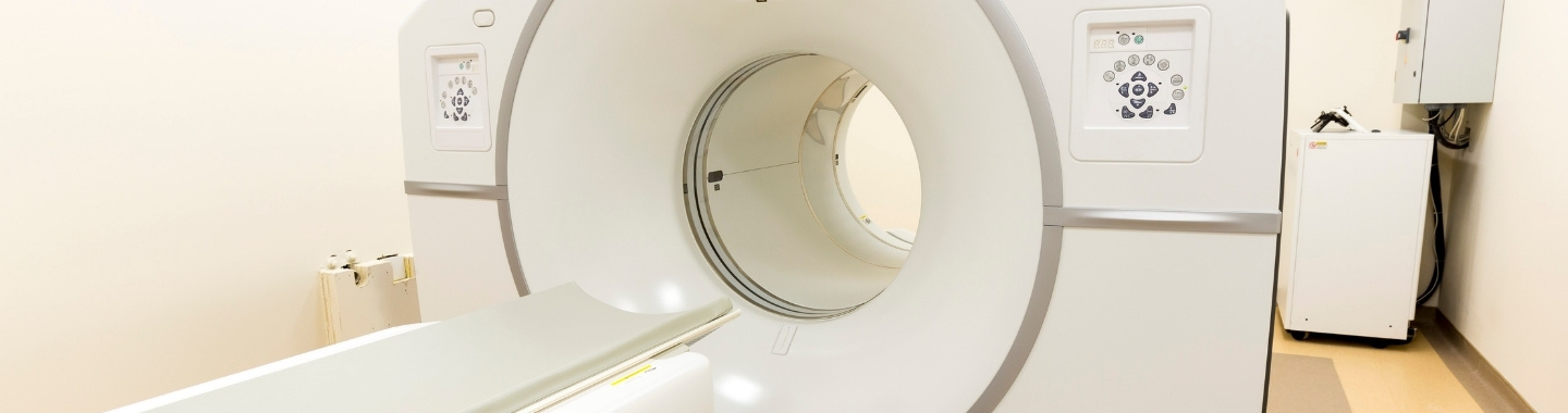
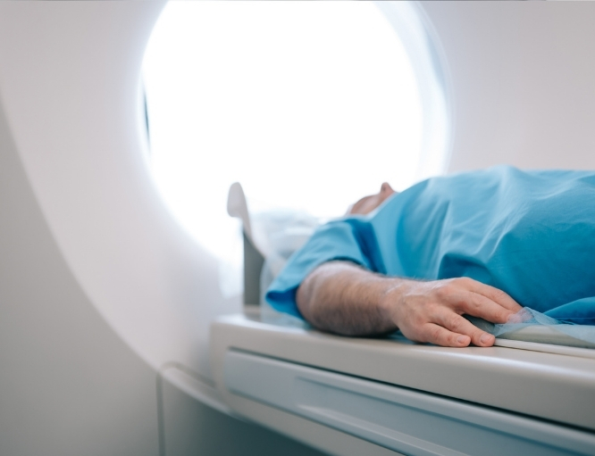
During a CT scan, the patient lies on a table that moves through a large, ring-shaped scanner. An X-ray tube rotates around the body and captures images from different angles.
work-icon
Cross-Sectional Imaging
CT produces thin, cross-sectional images or “slices” of the body. These allow doctors to examine organs, tissues, blood vessels and bones in greater detail than is usually possible with a standard X-ray.
work-icon
Multiplanar and 3D Views
The images can be reconstructed in different planes or combined to create three-dimensional views. This can help define the location of a tumour and its relationship to nearby organs, blood vessels and other structures.
work-icon
Contrast-Enhanced Imaging
Some CT scans use a contrast material to make certain organs, blood vessels or abnormalities more visible. Contrast may be given through a vein, taken orally or, less commonly, administered through the rectum, depending on the part of the body being examined and the clinical question.

Role in Cancer Care

Detecting & Evaluating Abnormalities
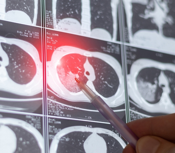
CT may be recommended to investigate symptoms, abnormal examination findings or results from another test. It can help identify masses, enlarged lymph nodes, abnormal fluid collections or changes in organs and tissues.
Detecting & Evaluating Abnormalities
Supporting Diagnosis
CT can show the size, shape, location and imaging characteristics of a suspected tumour. Although these findings may suggest cancer, a biopsy is often required for confirmation.
Supporting Diagnosis
Staging Cancer
Once cancer has been diagnosed, CT may help determine the size and location of the primary tumour, whether nearby tissues or organs are involved, whether lymph nodes appear enlarged or abnormal, and whether the cancer may have spread to other parts of the body. The information is considered together with clinical findings, pathology results and other imaging tests to establish the stage of the cancer.
Staging Cancer
Guiding Biopsies & Other Procedures
CT imaging may be used to guide a needle accurately towards an abnormal area during a biopsy. It may also assist in planning or guiding selected minimally invasive procedures, such as drainage or tumour ablation.
Guiding Biopsies & Other Procedures
Planning Treatment
CT images may help surgeons, medical oncologists, radiation oncologists and interventional oncologists plan treatment. They can provide information about the tumour’s position and its relationship to nearby organs, bones and blood vessels. Specialised CT scans may also be used for radiation therapy planning.
Planning Treatment
Monitoring Treatment Response
CT may be repeated during or after treatment to assess whether a tumour has reduced in size, remained stable or progressed. Doctors compare current images with earlier scans and interpret them alongside symptoms, examination findings and other test results.
Monitoring Treatment Response
Follow-up & Surveillance
After treatment, CT may be used in selected patients to look for recurrence or monitor known areas of concern. The need for follow-up CT and how often it is performed depend on the cancer type, stage, treatment received, symptoms and individual follow-up plan.
Follow-up & Surveillance

What to Expect

Before the Test

Preparation depends on the part of the body being examined and whether contrast is required.

Before the scan, inform the care team if you:

  • Are pregnant or may be pregnant

  • Have kidney disease or reduced kidney function

  • Have diabetes

  • Have previously experienced a reaction to contrast material

  • Have asthma or significant allergies

  • Take prescription medicines, over-the-counter medicines or supplements

  • Have recently undergone another scan involving contrast material

Blood tests may be required to assess kidney function before intravenous contrast is given, particularly in patients with known kidney disease or other relevant risk factors.

You may be asked to avoid food or drink for a few hours before certain contrast-enhanced scans. Do not stop regular medicines unless specifically instructed. Jewellery, metal objects and clothing containing metal may need to be removed, and you may be asked to change into a hospital gown.

Previous scans and reports should be made available whenever possible, as comparison with earlier imaging can help assess changes over time.

Before the Test

During the Test

You will lie on a motorised table, which moves through the centre of the CT scanner. The scanner is open at both ends and does not enclose the entire body like a tunnel.

The technologist will position you according to the area being examined. Supports or straps may be used to help you remain still. You may be asked to hold your breath briefly while some images are taken, particularly during scans of the chest or abdomen.

If intravenous contrast is required, it will usually be given through a cannula placed in the hand or arm. You may briefly experience:

  • A warm sensation through the body

  • A metallic taste in the mouth

  • A feeling as though you need to pass urine

These sensations are usually temporary and pass quickly. Tell the technologist immediately if you develop itching, breathing difficulty, swelling, dizziness or any other unusual symptoms.

The scan itself is painless. Most routine CT examinations are completed within a few minutes, although the overall appointment may take longer when preparation, contrast or additional imaging phases are required. The technologist can see, hear and speak with you throughout the scan.

During the Test

After the Test

Most patients can resume their usual activities immediately after a routine CT scan. If contrast has been given, you may be advised to drink fluids, unless you are on a fluid restriction or have been given different instructions.

The IV cannula will usually be removed after the scan. In some cases, you may be observed briefly, particularly if you have a history of allergies or have experienced symptoms following contrast administration. Seek medical advice promptly if you develop breathing difficulty, facial or throat swelling, widespread rash, severe dizziness or another significant reaction after leaving the imaging centre.

A radiologist will examine the images and prepare a report for the doctor who requested the scan. The findings will be interpreted together with your medical history, examination findings and results from other investigations.

After the Test

Potential Benefits

CT can provide detailed information across many areas of the body and support decisions throughout the cancer care journey.
  • Detailed imaging: CT provides clear cross-sectional views of organs, bones, soft tissues and blood vessels.
  • Rapid examination: Images can be acquired quickly, which can be useful when timely assessment is required.
  • Broad coverage: A single examination can assess large areas of the body, such as the chest, abdomen and pelvis.
  • Accurate localisation: CT can help show the size and position of a tumour and its relationship to nearby structures.
  • Procedure guidance: Real-time or sequential CT images can help guide biopsies and selected minimally invasive treatments.
  • Treatment planning: CT findings may contribute to surgical, radiation and other treatment planning.
  • Response assessment: Scans taken at different points in treatment can be compared to evaluate changes in tumour size and disease extent.
  • Widely applicable: CT can contribute to the evaluation of many cancer types, although its precise role varies according to the organ, tumour and clinical situation.
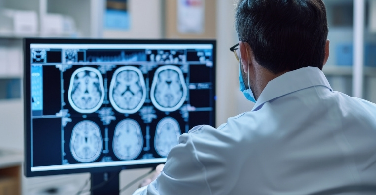
CT Scan
Frequently Asked Questions
01 Does a CT scan expose me to radiation?
icon icon
Yes. CT uses ionising radiation. The radiation dose depends on the body area, scan technique and number of images required. The examination is recommended when the expected diagnostic benefit outweighs the potential radiation risk. The scan does not make you radioactive, and no radiation remains in your body afterwards.
02 Is contrast required for every CT scan?
icon icon
No. Some CT scans are performed without contrast, while others require intravenous, oral or rectal contrast to improve the visibility of specific organs, blood vessels or abnormalities. The decision depends on the area being examined and the information your doctor needs.
03 Can I have a contrast-enhanced CT scan if I have kidney disease?
icon icon
Kidney disease does not automatically mean that contrast cannot be used. Your kidney function and the clinical need for contrast will be assessed. The team may modify the scan, take additional precautions, use a different imaging method or perform the CT without intravenous contrast when appropriate.
04 Is CT safe during pregnancy?
icon icon
Tell your doctor and the imaging team if you are pregnant or could be pregnant. CT may still be performed when it is medically necessary, but the benefits, body area being examined and potential radiation exposure will be considered carefully. Ultrasound or MRI may be used instead when they can provide the required information.
05 How is CT different from MRI and PET-CT?
icon icon
CT uses X-rays to provide detailed anatomical images. MRI uses a strong magnetic field and radiofrequency signals and may provide greater soft-tissue detail in certain areas. PET-CT combines CT images with information about metabolic activity obtained using a small amount of a radioactive tracer. The most appropriate test depends on the cancer type and the clinical question.
06 Can a CT scan confirm that an abnormality is cancer?
icon icon
Not always. CT can identify and characterise an abnormal area, assess its extent and help guide further testing. However, infections, inflammation and non-cancerous conditions can sometimes resemble cancer on imaging. A biopsy may be needed to confirm the diagnosis.
image image
Book an Appointment
Please Enter Your Name
Please Enter Your Mobile Number
Please Enter OTP