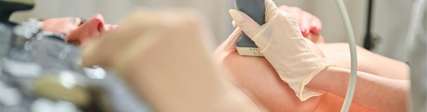
Wire localisation is an image-guided procedure used to mark the position of an abnormal area in the breast that cannot be easily felt during surgery. A thin wire is positioned in or close to the target so that the surgeon can locate and remove the correct area.
The procedure is generally performed shortly before breast surgery using mammography, ultrasound or, in selected cases, MRI guidance. The wire and the targeted breast tissue are removed together during the operation.
Wire localisation does not diagnose or treat the abnormality by itself. It serves as a guide for the planned surgical procedure.
Role in Cancer Care

Localisation Approaches
Mammography-Guided Localisation
Mammography or stereotactic imaging may be used when the abnormality is best seen on a mammogram, such as an area of microcalcification or architectural distortion.
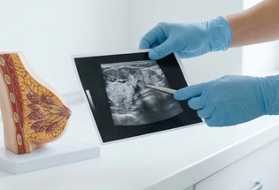
Ultrasound-Guided Localisation
Ultrasound guidance may be used when the lesion, biopsy marker or lymph node can be clearly seen on ultrasound. This approach allows real-time visualisation during wire placement.
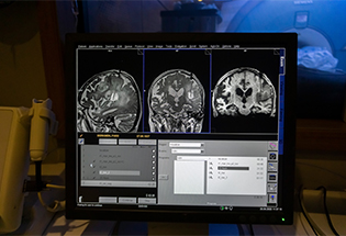
MRI-Guided Localisation
MRI guidance may be considered when the abnormality is visible only on breast MRI and cannot be adequately targeted using mammography or ultrasound.
What to Expect
Before the Procedure
The radiologist reviews the breast imaging and planned surgery to determine the most appropriate guidance method and entry point. Patients should inform the team about allergies, current medicines, previous reactions to local anesthesia and any possibility of pregnancy.
Wire localisation is usually coordinated with the surgery and may be performed on the same day or according to the hospital’s surgical pathway. Instructions about fasting will generally relate to the operation rather than the localisation procedure itself.

During the Procedure
The breast is positioned for mammography, ultrasound or MRI, depending on how the abnormality is best visualised. The skin is cleaned, and local anesthesia is administered.
A fine needle containing the wire is guided into or next to the target. The needle is then removed, leaving the wire in position. Part of the wire may remain outside the breast and is secured with a dressing. Additional images are usually taken to confirm its location.

After the Procedure
The patient is transferred to the surgical area with the wire secured in place. Sudden or strenuous upper-body movements should be avoided while waiting for surgery.
During the operation, the surgeon follows the wire to the target and removes the relevant tissue together with the wire. The specimen may be imaged to confirm that the intended abnormality, marker or calcifications have been removed.

Potential Benefits
- Wire localisation helps the surgeon identify an abnormality that cannot be felt.
- It supports accurate removal of the area identified on breast imaging.
- It may help limit the amount of surrounding healthy breast tissue removed where clinically appropriate.
- It enables coordination between the radiology, surgery and pathology teams.
- Imaging can be used to confirm the position of the wire before surgery.
- Specimen imaging can help verify that the targeted area has been removed.