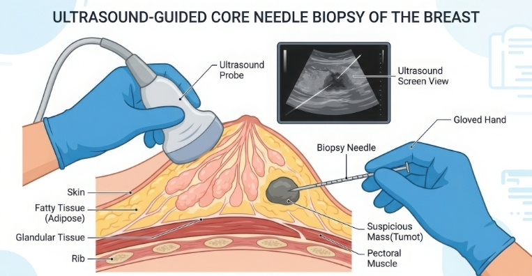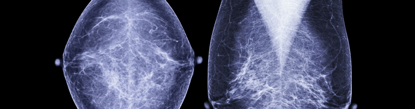
Clip placement is an image-guided procedure in which a very small marker is positioned within the breast or a lymph node to identify a specific site for future imaging or treatment. It is commonly performed at the time of a needle biopsy but may also be carried out as a separate procedure.
The marker can identify the location of a biopsied abnormality even if the lesion becomes difficult to see after sampling or responds to treatment. It may later help with imaging follow-up, surgical localisation or radiotherapy planning.
A marker clip does not treat the abnormality, release medication or emit radiation. It is designed to remain in place unless it is removed with the surrounding tissue during a subsequent operation.
Role in Cancer Care

Placement Sites
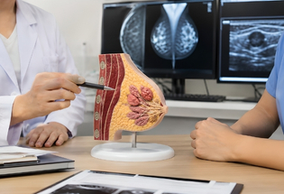
Biopsy Site
A clip may be placed after a breast biopsy to mark the precise location from which tissue was collected. This is especially useful if the imaging abnormality is small or becomes less visible after sampling.

Breast Lesion or Tumour
A marker may be placed within a confirmed breast tumour before neoadjuvant therapy. If the tumour responds well and becomes difficult to identify, the clip continues to mark its original position.
Axillary Lymph Node
A clip may be placed in a suspicious or biopsy-confirmed lymph node. It can help the team identify and assess the same node after systemic therapy or during planned surgery.
What To Expect
Before the Procedure
The radiologist reviews the relevant mammogram, ultrasound or MRI to confirm the site that needs to be marked. Patients should inform the team about allergies, current medicines, anticoagulants, previous procedures and any possibility of pregnancy.
If clip placement is performed during a biopsy, preparation is generally the same as for the biopsy. If it is performed separately, the team will provide procedure-specific instructions.

During the Procedure
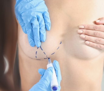
The breast or axilla is positioned according to the imaging method being used. The skin is cleaned and local anesthesia may be given if the clip is not being placed through an existing biopsy needle.
Using mammography, ultrasound or MRI guidance, the doctor advances a needle to the required site and deploys the marker. The needle is then removed. Follow-up imaging may be performed to document the marker’s position.

After the Procedure
Pressure may be applied to minimise bleeding, followed by a small dressing. Mild soreness or bruising can occur, particularly when clip placement accompanies a biopsy.
Most patients can resume routine light activities shortly afterwards, although strenuous activity may need to be avoided for a brief period. The marker usually remains in the body unless it is removed as part of later surgery.

Potential Benefits
- A clip can permanently identify the site of a biopsied abnormality.
- It remains visible even if the lesion becomes smaller or difficult to see.
- It can support accurate follow-up imaging and future comparison.
- It can guide wire or wire-free localisation before surgery.
- It can help identify the original tumour site after neoadjuvant therapy.
- A clipped lymph node can be specifically reassessed or targeted during subsequent treatment.
- Placement usually adds little time when performed during an image-guided biopsy.