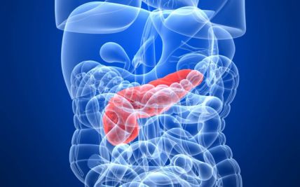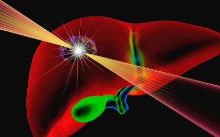
Genitourinary Cancer
Genitourinary cancers develop in the organs of the urinary system and the male reproductive system. They include cancers of the kidneys, renal pelvis, ureters, bladder, urethra, prostate, testicles, and penis.
These cancers differ considerably in their causes, symptoms, behaviour, and treatment. Some are found through screening or imaging before symptoms develop, while others present with blood in the urine, urinary changes, pain, or a lump. Not every urinary symptom indicates cancer, but persistent or unexplained changes should be evaluated.
Treatment often requires close coordination between uro-oncologists, medical oncologists, radiation oncologists, radiologists, nuclear medicine specialists, pathologists, genetic counsellors, and reconstructive surgeons. This helps balance cancer control with the preservation of kidney function, urinary control, sexual function, and fertility wherever possible.
Types of Genitourinary Cancer
Genitourinary cancer is not a single disease. Each cancerrequires an organ-specific approach to diagnosis, staging, and treatment.
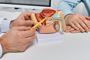
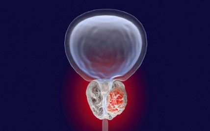
Prostate cancer develops in the prostate gland, which lies below the bladder and surrounds part of the urethra. Most prostate cancers are adenocarcinomas arising from glandular cells. Some prostate cancers grow slowly and may not require immediate treatment, while others are more aggressive. Treatment is based on the cancer’s stage, PSA level, Grade Group, imaging findings, overall health, and individual preferences.

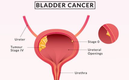
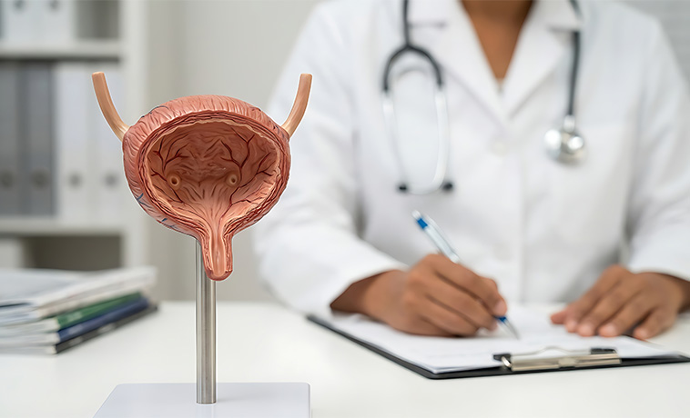
Bladder cancer usually begins in the urothelial cells lining the bladder. The same type of cells also lines the renal pelvis, ureters, and part of the urethra. Bladder cancer is classified as non-muscle-invasive or muscle-invasive according to how deeply it has grown into the bladder wall. This distinction has a major influence on treatment.
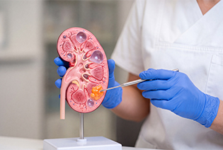
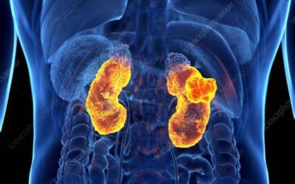
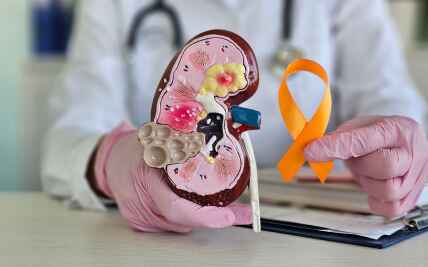
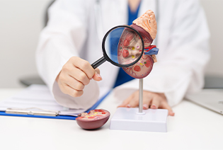
Kidney cancer begins in the kidney. Renal cell carcinoma is the most common type in adults and includes clear-cell, papillary, chromophobe, and several less common subtypes. Some kidney cancers are found incidentally during imaging performed for another reason. Treatment depends on the tumour’s size, location, subtype, kidney function, and whether the disease has spread.

Testicular cancer usually begins in the germ cells that produce sperm. The two main groups are seminoma and non-seminoma, which may require different treatment approaches. It commonly affects adolescents and younger adults, although it can occur at other ages. Testicular cancer is often highly responsive to treatment, including when it has spread, but the outlook depends on the cancer type and extent of disease.
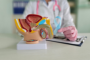
Penile cancer develops in the skin or tissues of the penis. Most cases are squamous cell carcinomas and commonly begin on the glans or foreskin. Human papillomavirus, or HPV, is associated with a proportion of cases. Early assessment may provide more opportunities for organ-preserving treatment.
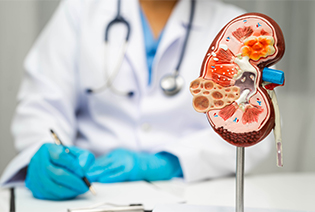
Upper urinary tract cancer develops in the lining of the renal pelvis or ureter. Most are urothelial carcinomas and share some features with bladder cancer. Diagnosis commonly requires specialised imaging and ureteroscopy. Treatment depends on the tumour’s location, grade, extent, and the function of both kidneys.
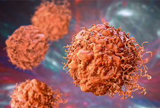
Urethral cancer begins in the tube through which urine leaves the body. It is rare and may include urothelial carcinoma, squamous cell carcinoma, or adenocarcinoma. Treatment depends on where the tumour arises, how deeply it has grown, and whether nearby organs or lymph nodes are involved.
Risk factors differ by cancer type. Having one or more risk factors does not mean that cancer will develop, and some people diagnosed with genitourinary cancer have no identifiable risk factors.
Modifiable Risk Factors
Smoking: A major risk factor for bladder cancer and an established risk factor for kidney cancer.
Excess body weight: Associated with an increased risk of kidney cancer and some aggressive prostate cancers.
High blood pressure: Associated with a greater risk of kidney cancer, although the relationship may also involve other health factors.
Occupational exposure: Long-term exposure to certain chemicals used in dye, rubber, leather, paint, metal, and petroleum-related industries can increase bladder cancer risk.
HPV infection: Persistent infection with high-risk HPV types increases the risk of penile cancer.
Chronic urinary irritation: Long-term catheter use, bladder stones, or recurrent inflammation may increase the risk of certain bladder cancers.
Anabolic steroid misuse: Prolonged use may affect the kidneys and other organs, although it is not a common direct cause of GU cancer.
Non-Modifiable Risk Factors
Increasing age
A family history of prostate, kidney, testicular, or other related cancers
Inherited variants involving genes such as BRCA1, BRCA2, HOXB13, VHL, or genes associated with Lynch syndrome
Inherited conditions such as von Hippel–Lindau syndrome or Birt–Hogg–Dubé syndrome
An undescended testicle, even after corrective surgery
A personal history of cancer in the opposite testicle
Chronic kidney disease or long-term dialysis
Certain congenital abnormalities of the urinary tract
Previous pelvic radiation or selected chemotherapy medicines
Increasing age and male sex for bladder and kidney cancer
Ancestry and inherited background, which may influence prostate cancer risk
People with a strong family history, cancer at an unusually young age, multiple related cancers, or features of an inherited syndrome may be advised to undergo genetic counselling.
Signs and Symptoms
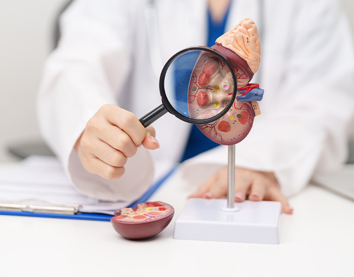
Symptoms depend on the organ involved. Early-stage prostate and kidney cancers may not cause any noticeable symptoms.
Possible signs and symptoms include:
These symptoms may also be caused by urinary infections, kidney stones, benign prostate enlargement, or other non-cancerous conditions. However, visible blood in the urine, a testicular lump, or a persistent penile lesion should be evaluated promptly.
Diagnosis begins with a review of symptoms, urinary and sexual health, medical history, and family history. The examination may include assessment of the abdomen, kidneys, external genitalia, lymph nodes, and prostate where relevant. Imaging, biopsy, and laboratory tests are then selected according to the suspected cancer.
Ultrasound uses sound waves to examine the kidneys, bladder, prostate, testicles, and nearby tissues. It is commonly used to assess a testicular lump, identify a kidney mass, measure urine remaining in the bladder, or guide selected procedures.


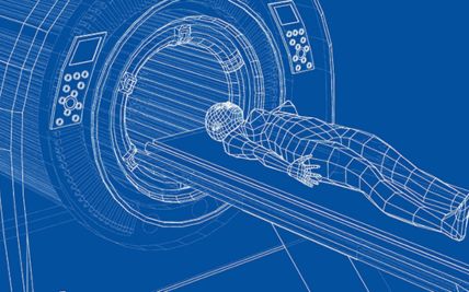
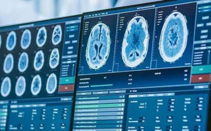
A CT scan produces detailed images of the urinary tract, abdomen, pelvis, and other areas. It is commonly used to evaluate kidney, bladder, ureteric, and testicular cancers and to assess lymph nodes or distant spread. CT urography uses intravenous contrast to examine the kidneys, renal pelvis, ureters, and bladder in people with blood in the urine or suspected urothelial cancer.
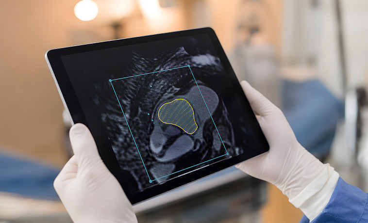
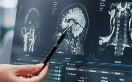
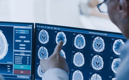
MRI provides detailed images of soft tissues and may be used to assess kidney, bladder, prostate, penile, or urethral cancers. It can help determine whether a tumour has extended into nearby organs, blood vessels, muscles, or other structures.
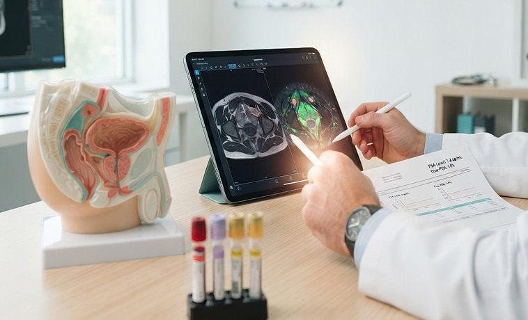
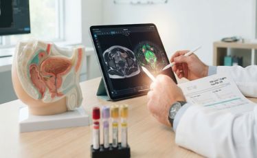
Multiparametric MRI combines different types of MRI images to identify suspicious areas within the prostate. It can help determine whether a biopsy is required, guide targeted biopsy, assess local spread, and support treatment planning. A normal MRI does not completely exclude clinically significant prostate cancer. The result is interpreted together with PSA levels, prostate size, examination findings, and individual risk.


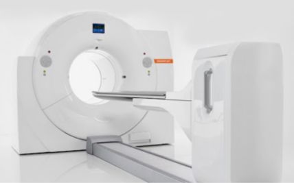
PET-CT uses a radioactive tracer to identify areas of increased biological activity. The type of tracer depends on the suspected cancer. PSMA PET-CT is commonly used in selected prostate cancers to assess disease spread or investigate recurrence. FDG PET-CT and other tracers may be used in selected bladder, kidney, testicular, or penile cancers, but PET-CT is not required for every patient.


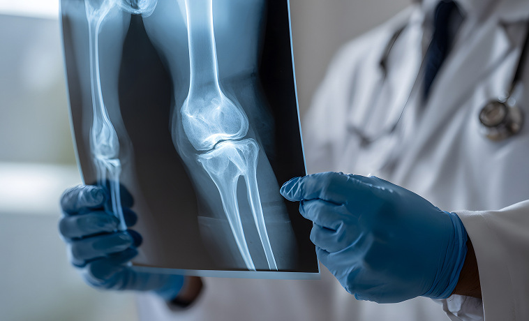
A bone scan may be used to determine whether cancer has spread to the bones, particularly in selected people with prostate cancer. Whether it is required depends on the PSA level, Grade Group, symptoms, stage, and other imaging findings.
Cystoscopy uses a thin camera passed through the urethra to examine the bladder lining directly. It can identify bladder tumours and other abnormalities. Suspicious areas may be sampled or removed during the procedure.
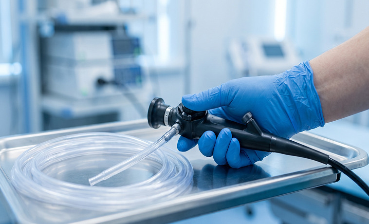
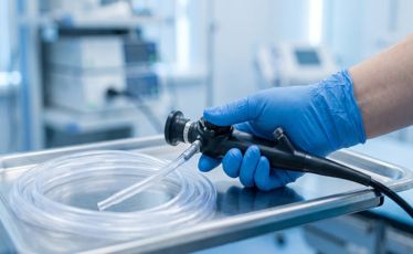
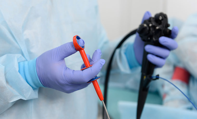
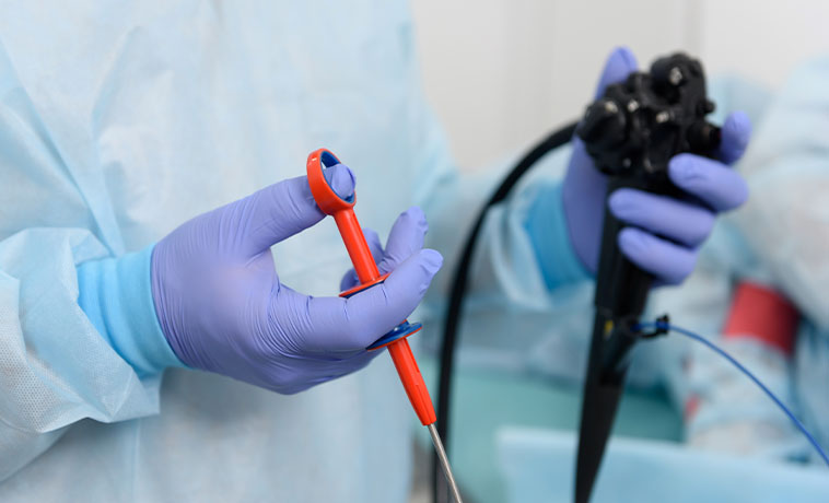
Ureteroscopy uses a narrow camera to examine the ureter and renal pelvis. It may be used to inspect an abnormal area, collect urine samples, or obtain a biopsy when upper urinary tract cancer is suspected.
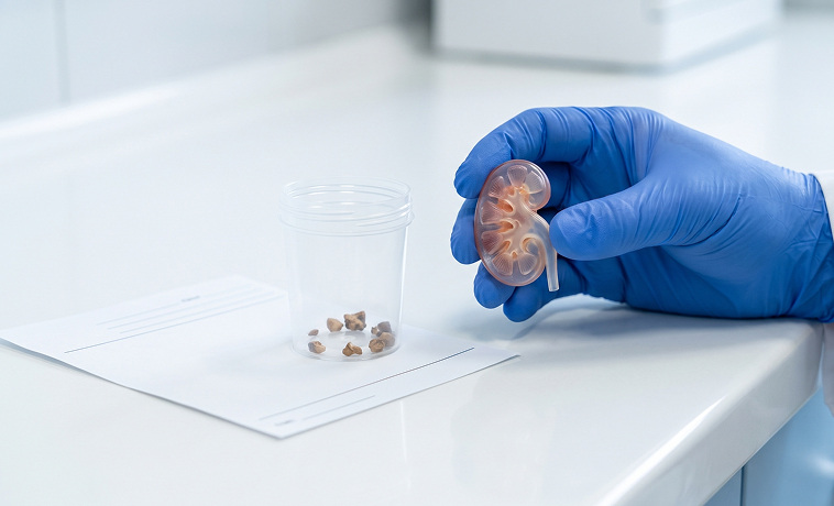
Tissue samples are collected from the prostate using a needle, commonly through the perineum and sometimes through the rectum. MRI findings may be used to target suspicious areas, together with systematic sampling where appropriate. The tissue is assigned a Gleason score and Grade Group, which help indicate how aggressive the cancer is likely to be.
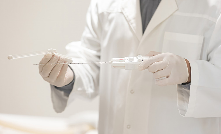
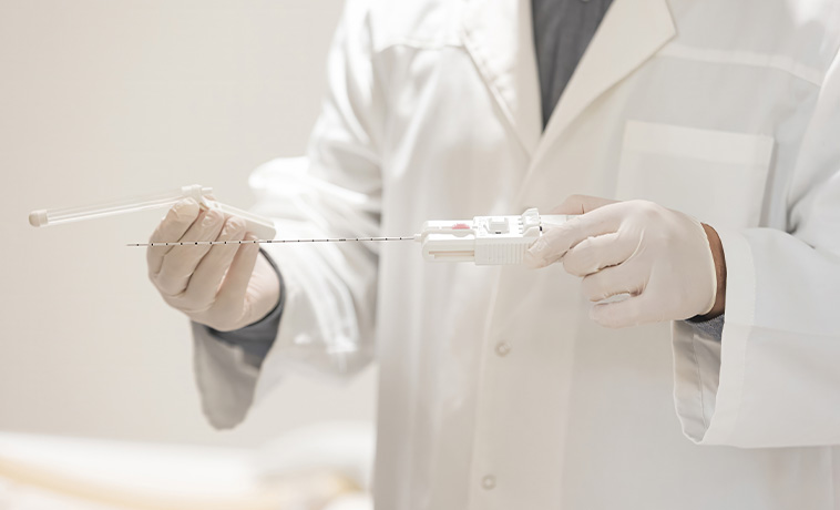
An imaging-guided core needle biopsy may be recommended when the diagnosis is uncertain, before non-surgical treatment, or when the result could change management. It is not required before surgery for every kidney mass.
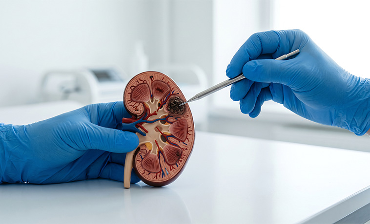
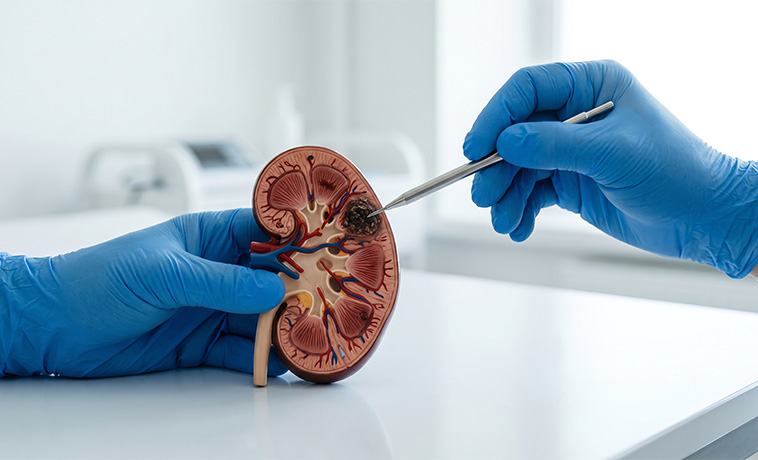
During transurethral resection of bladder tumour, or TURBT, an instrument is passed through the urethra to remove visible bladder tumours and collect tissue. TURBT helps establish the cancer type, grade, and depth of invasion. A repeat procedure may be recommended for selected high-risk or incompletely assessed tumours.
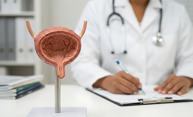
A small tissue sample is collected from a suspicious area in the ureter or renal pelvis during ureteroscopy. The results help determine the cancer’s type and grade and whether kidney-preserving treatment may be appropriate.
When testicular cancer is strongly suspected, the affected testicle is usually removed through an incision in the groin. The tissue is then examined to confirm the diagnosis. A biopsy through the scrotum is generally avoided because it can alter lymphatic drainage and complicate staging and treatment.


A small tissue sample is removed from a penile or urethral lesion. An incisional biopsy may sample part of a larger lesion, while an excisional biopsy may remove a small lesion completely.
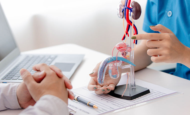
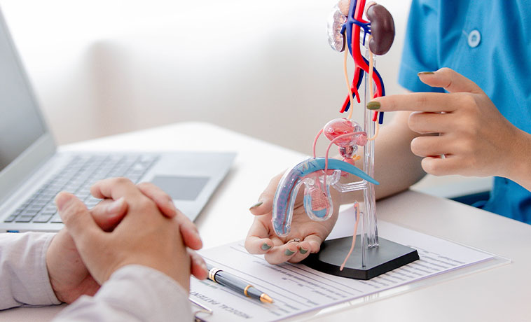
The prostate-specific antigen, or PSA, test measures a protein produced by prostate tissue. A raised PSA can occur with prostate cancer, but it can also result from benign prostate enlargement, inflammation, infection, or recent procedures. The PSA result is interpreted with age, prostate size, previous results, examination findings, imaging, and individual risk.
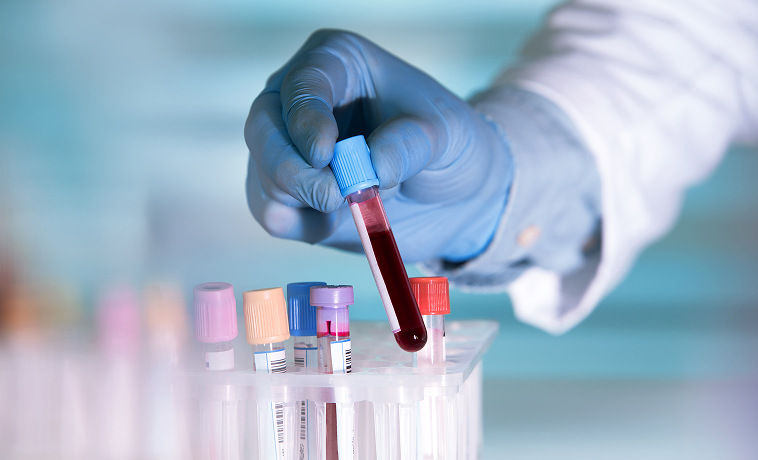
Urinalysis checks for blood, infection, protein, and other abnormalities in the urine. It can help assess urinary symptoms but cannot confirm or exclude cancer by itself.
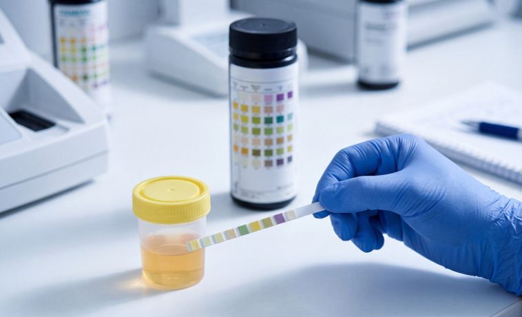
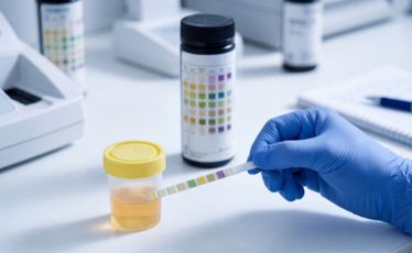
Urine cytology examines urine for abnormal cells shed from the urinary tract. It is more effective at detecting high-grade urothelial cancer than low-grade tumours and is usually interpreted with cystoscopy and imaging.

Blood levels of alpha-fetoprotein, beta-human chorionic gonadotropin, and lactate dehydrogenase may support the diagnosis and staging of testicular cancer. These markers are also measured after surgery and during follow-up. Normal levels do not exclude testicular cancer.
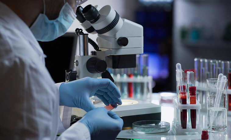
Blood tests assess kidney and liver function before imaging, surgery, systemic treatment, or radiation therapy. They also help determine whether treatment needs to be modified.

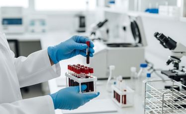
A pathologist examines biopsy or surgical tissue under a microscope to confirm the cancer type, grade, depth of invasion, surgical margins, and other clinically important features.
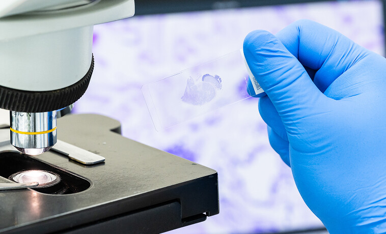
Immunohistochemistry uses specialised stains to identify proteins within tumour cells. It can help establish where the cancer began and distinguish between tumours with a similar microscopic appearance.
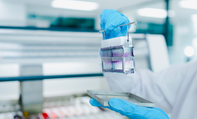
Molecular testing may identify inherited or tumour-specific changes that influence treatment. Examples include DNA-repair gene variants in prostate cancer, FGFR changes in bladder cancer, and hereditary kidney cancer syndromes. Genetic counselling may be recommended when the person’s age, family history, tumour type, or molecular results suggest an inherited cancer risk.
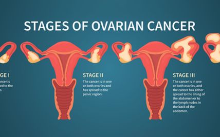
Staging describes the extent of cancer and helps guide treatment and prognosis. Most genitourinary cancers use the TNM system, which considers the primary tumour, lymph-node involvement, and distant spread. The exact definition of each stage differs by organ.
Stage 0: Abnormal or cancerous cells are limited to the surface lining and have not invaded deeper tissues.
Stage I: The cancer is confined to the organ where it began. It has not spread to regional lymph nodes or distant organs.
Stage II: The cancer remains localised but is larger, has grown more deeply, or has higher-risk biological features.
Stage III: The cancer has grown into nearby tissues, major blood vessels, or regional lymph nodes, depending on the organ involved. It has not necessarily spread to a distant organ.
Stage IV: The cancer has spread to distant organs, distant lymph nodes, bones, or other parts of the body.
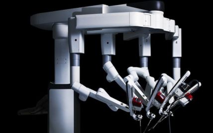
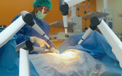
Radical prostatectomy removes the prostate gland and seminal vesicles. Nearby lymph nodes may also be removed in selected patients. The procedure may be performed through an open, laparoscopic, or robotic-assisted approach. Nerve-sparing surgery may help preserve erectile function when the tumour’s location and extent allow it, but this cannot be guaranteed.

Partial nephrectomy removes the kidney tumour with a margin of surrounding tissue while preserving the remaining kidney. It is preferred for many small or suitably located kidney tumours when it can provide effective cancer control. The operation may be performed through an open, laparoscopic, or robotic-assisted approach.
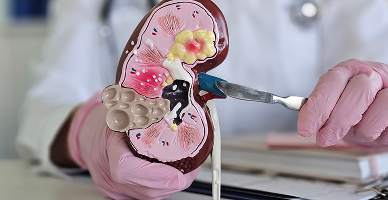
Radical nephrectomy removes the entire affected kidney and, when required, nearby tissue or lymph nodes. It may be recommended for large or complex tumours that cannot be treated safely with partial nephrectomy. Selected advanced kidney cancers extending into major veins may require tumour-thrombus removal as part of surgery.
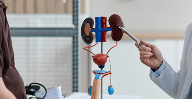
TURBT removes bladder tumours through the urethra without an external incision. It is the main initial treatment for many non-muscle-invasive bladder cancers and an important diagnostic step for muscle-invasive disease. Additional intravesical treatment may be recommended according to the tumour’s grade and risk of recurrence.
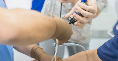
Radical cystectomy removes the bladder and nearby organs or tissues. It may be recommended for muscle-invasive cancer and selected high-risk non-muscle-invasive cancers. A new pathway for urine must be created after the bladder is removed. The appropriate urinary diversion is selected according to the cancer, kidney function, anatomy, overall health, and patient preference.

Radical nephroureterectomy removes the kidney, ureter, and a small part of the bladder around the ureteric opening. It is commonly used for high-risk upper urinary tract urothelial cancer. Selected low-risk tumours may be treated with kidney-preserving ureteroscopic or segmental surgery.
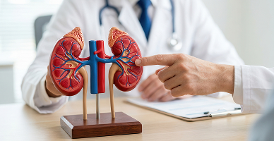
Radical inguinal orchiectomy removes the affected testicle and spermatic cord through an incision in the groin. It is both the main diagnostic procedure and the initial treatment for most suspected testicular cancers. Further treatment depends on the cancer type, stage, tumour-marker levels, and risk of recurrence.
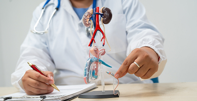
Small or superficial penile cancers may be treated with local excision, glans resurfacing, glansectomy, laser treatment, or other organ-preserving procedures. More extensive disease may require partial or total penectomy. The surgical approach aims to achieve clear margins while preserving urinary and sexual function where possible.
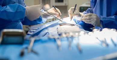
Regional lymph nodes may be removed when there is a significant risk of involvement. This may include pelvic lymph-node dissection for prostate or bladder cancer, retroperitoneal lymph-node dissection for selected testicular cancers, and groin lymph-node dissection for penile cancer.

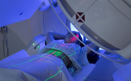
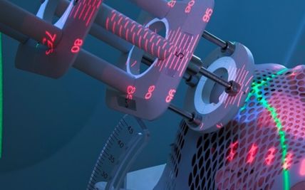
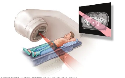
External beam radiation therapy delivers radiation from outside the body. It may be used as a primary treatment, after surgery, or to control symptoms from advanced disease. It has an established role in prostate, bladder, testicular, penile, and urethral cancers and may be considered for selected kidney tumours.
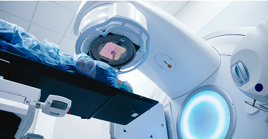
Intensity-modulated radiation therapy shapes the radiation dose around the tumour, while image-guided radiation therapy checks the target’s position during treatment. These techniques are particularly important when treating organs that move or lie close to the rectum, bladder, bowel, kidneys, or other sensitive structures.
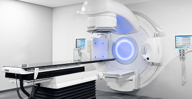
Stereotactic body radiation therapy, or SBRT, delivers highly focused radiation in a small number of sessions. It may be used for selected prostate cancers, kidney tumours, lymph nodes, or limited sites of metastatic disease. Suitability depends on tumour size, location, nearby organs, and the overall treatment plan.
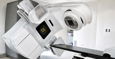
Brachytherapy places a radioactive source within or close to the tumour. It may be used for selected localised prostate cancers, either alone or with external beam radiation. The choice between low-dose-rate and high-dose-rate brachytherapy depends on the cancer’s risk group, prostate anatomy, urinary symptoms, and treatment plan.

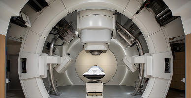
Proton therapy uses proton beams that release most of their radiation within the planned treatment area, with little radiation continuing beyond it. This may reduce radiation exposure to some nearby tissues in selected cases.
It may be considered for carefully selected prostate, bladder, kidney, or paediatric genitourinary tumours when comparative treatment planning demonstrates a meaningful advantage. It has not been shown to be necessary or superior for every person with prostate or another GU cancer.
Suitability is determined after individual assessment and comparison with established photon radiation techniques, surgery, and other appropriate treatments.
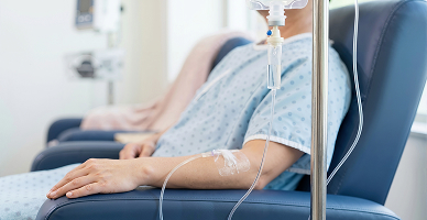
Chemotherapy may be used before or after surgery or to control advanced disease. It has an important role in muscle-invasive and advanced bladder cancer, testicular cancer, and selected penile or urethral cancers. Its role in prostate and kidney cancer is more specific and depends on the disease stage and previous treatments.
Hormone therapy lowers testosterone or blocks its effect on prostate cancer cells. It may be combined with radiation therapy for intermediate- or high-risk localised disease and is a central treatment for metastatic prostate cancer. Additional hormone-pathway medicines may be used according to the stage and response to previous treatment.

Targeted therapies block specific pathways that cancer cells use to grow. They are widely used in advanced kidney cancer and may be considered for prostate, bladder, or other GU cancers with suitable molecular changes.

Immunotherapy helps the immune system recognise and attack cancer cells. It is used in selected kidney and bladder cancers and may be considered for other GU cancers according to their stage, biomarkers, and previous treatments.

Antibody–drug conjugates combine an antibody that recognises a cancer-cell target with a cancer-killing medicine. They may be used for selected patients with locally advanced or metastatic urothelial cancer.
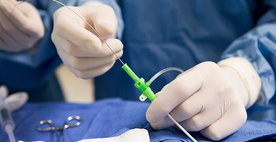
Intravesical therapy delivers medicine directly into the bladder through a catheter. Bacillus Calmette–Guérin, or BCG, and selected chemotherapy medicines may be used after TURBT for non-muscle-invasive bladder cancer. The choice depends on the tumour’s grade, stage, recurrence risk, and previous treatment.

Tumour profiling and inherited genetic testing may identify treatment options for selected patients. Precision approaches are particularly relevant in prostate cancer with DNA-repair changes, selected urothelial cancers, and hereditary kidney cancer.
An orthotopic neobladder is created from a section of intestine after the bladder is removed. It is connected to the urethra so that urine can pass through the usual route. It is suitable only for selected patients and requires adequate kidney function, appropriate tumour location, and the ability to learn a new pattern of bladder emptying.
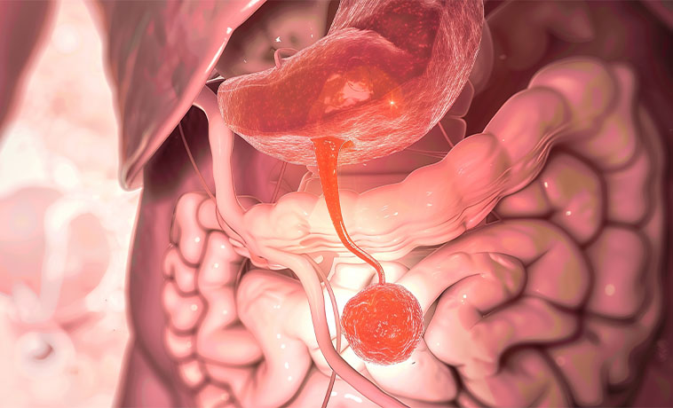
An ileal conduit uses a short segment of intestine to carry urine from the ureters to an opening, or stoma, on the abdomen. Urine drains continuously into an external collection bag. It is a well-established form of urinary diversion and may be suitable for people who are not candidates for a neobladder or continent reservoir.
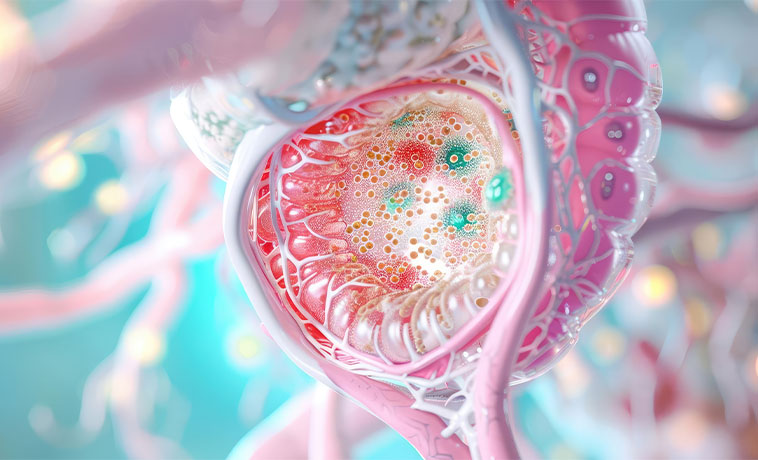
A continent urinary reservoir is an internal pouch created from intestine. Urine is stored inside the body and drained at intervals through a small abdominal opening using a catheter. It avoids the need for a continuously worn external collection bag but requires the ability to catheterise and maintain the reservoir.

When part of a ureter is removed or damaged, urinary drainage may be restored through procedures such as ureteric reimplantation, a Boari flap, or reconstruction using a segment of intestine. The method depends on the length and location of the affected ureter, kidney function, and previous surgery or radiation.

Penile reconstruction may be performed after removal of tissue for penile or urethral cancer. Local flaps, skin grafts, or tissue transferred from another part of the body may be used to restore coverage, urinary function, and appearance. The extent of reconstruction depends on the amount and location of tissue removed.
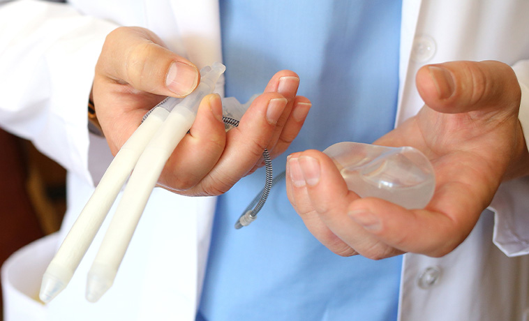
A testicular prosthesis may be placed after orchiectomy to restore the appearance and symmetry of the scrotum. It does not produce sperm or hormones but may be considered for cosmetic or personal reasons. Placement may be performed during cancer surgery or at a later stage, depending on the clinical situation and patient preference.

Prognosis for Genitourinary Cancer
The outlook varies considerably between genitourinary cancers. Many localised prostate, kidney, bladder, testicular, and penile cancers can be treated effectively, but outcomes depend on the disease type and stage.
Factors influencing prognosis include:
The organ in which the cancer began
Cancer type, subtype, grade, and molecular features
Stage at diagnosis
PSA, Grade Group, or testicular tumour-marker levels where relevant
Lymph-node or distant-organ involvement
Kidney function and overall health
Response to treatment
Completeness of surgery where performed
Ability to complete treatment and follow-up
Testicular cancer is often highly responsive to treatment, including in many advanced cases. Some prostate cancers grow slowly and can be monitored safely, while others require prompt combined treatment. Muscle invasion has an important effect on bladder cancer management, and the outlook for kidney cancer depends strongly on whether it remains confined to the kidney.
Regular follow-up is required because recurrence patterns and surveillance tests differ by cancer type and treatment.


Screening for Genitourinary Cancer
Screening recommendations depend on the organ, age, symptoms, family history, and inherited risk.
Prostate Cancer Screening
PSA testing may help detect prostate cancer before symptoms develop, but it can also identify slow-growing cancers that may never cause harm. An elevated PSA does not necessarily mean that cancer is present. The decision to undergo PSA testing should be made after discussing potential benefits, limitations, and individual risk with a doctor. People with a strong family history or other high-risk features may be advised to begin this discussion earlier.
Why Choose ACC for Genitourinary Cancer Treatment
Dedicated multidisciplinary uro-oncology teams
Organ-specific care for prostate, bladder, kidney, testicular, penile, ureteric, and urethral cancers
Robotic, laparoscopic, and open surgical options selected according to clinical need
Kidney-preserving, bladder-preserving, nerve-sparing, and organ-preserving approaches where oncologically appropriate
Advanced imaging, including multiparametric prostate MRI and PET-CT with appropriate tracers
Comprehensive medical oncology, immunotherapy, targeted therapy, and precision-oncology services
Advanced radiation techniques, including IMRT, IGRT, SBRT, brachytherapy, and proton therapy for selected patients
Expertise in urinary diversion and reconstructive urology
Genetic assessment and counselling for suspected hereditary cancers
Treatment planning focused on cancer control and the preservation of urinary, kidney, sexual, and reproductive function

Health Blogs